
Mantidactylus brevipalmatus, AHL, 1929
|
publication ID |
https://doi.org/ 10.1111/j.1096-3642.2010.00667.x |
|
persistent identifier |
https://treatment.plazi.org/id/0E4F87B3-E504-C358-FC86-74A55CAEFD79 |
|
treatment provided by |
Valdenar |
|
scientific name |
Mantidactylus brevipalmatus |
| status |
|
MANTIDACTYLUS BREVIPALMATUS AHL, 1929 View in CoL
The tadpoles of M. brevipalmatus have no obvious external diagnostic characters compared to those of other species of Chonomantis , except for some subtle
M. sp. 59
details of uncertain diagnostic value, and except for M. aerumnalis and M. sp. 59, which can be distinguished by, respectively, their coloration and the presence of rudimentary keratodonts (see accounts on these species for details). Apparently characterized by very long lungs (see below and discussion).
The tadpoles of M. brevipalmatus reported here have been previously described by Blommers-Schlösser (1979) under the name M. aerumnalis ( Vences & Glaw, 2004) and reproduced in Blommers-Schlösser & Blanc (1991) and Glaw & Vences (1992). The description of external morphology is based on one specimen in stage 28, ZMA 7078 (TL 47.3 mm, BL 12.6 mm). Buccopharyngeal features are described based on one tadpole at stage 36, ZMA 6991. These specimens were collected near Manajakatompo in the Ankaratra Massif by R. Blommers in the 1970s (see below) and therefore were not sequenced for molecular identification purposes; however, their identification is reliable because M. brevipalmatus is the only species of Chonomantis in this area ( Vences et al., 2002a).
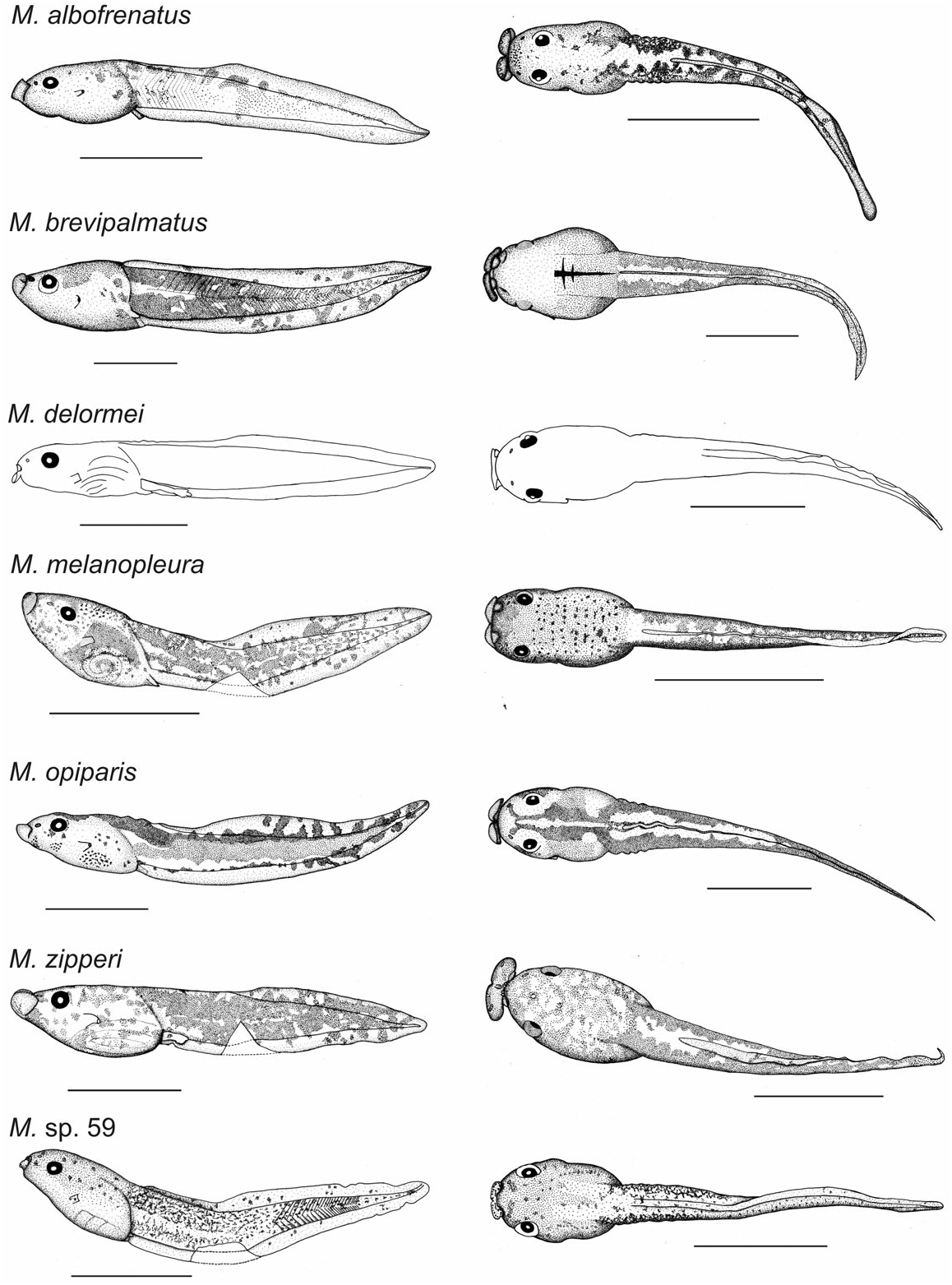
External morphology: In profile ( Fig. 1 View Figure 1 ), BW 117% of BH. In dorsal view ( Fig. 1 View Figure 1 ), body ovoid. Eyes visible in ventral view, directed laterally. A large pineal ocellus present behind eyes. Nares positioned almost dorsally and directed more anteriorly than strictly anterolaterally, aperture directed laterally, RN 49% of NP, NN 61% of PP. Spiracle bulb-like, moderately small, orientated posterodorsally, slightly closer to tip of snout than to end of body, SS 47% of BL; spiracular opening orientated posteriorly. Tail musculature, TMH 69% of BH and 71% of MTH, TMW 58% of BW, proximal quarter roughly parallel then gradually tapering. Tail fins, both UF and LF 29% of MTH, SU 91% of BL, proximal third of upper fin shallow, upper fin horizontal, first half of lower fin straight then curved to reach the tip of tail; TAL 269% of BL, point of maximum height of tail located slightly before mid-point, MTH 97% of BH, tail tip bluntly pointed. Anal tube small, a short tube, directed more posteriorly than posterolaterally, linked to ventral tail fin except its tip. Lateral line organs present on body and tail.
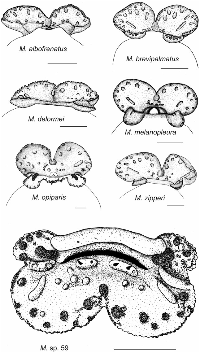
Oral disc ( Fig. 2 View Figure 2 ) ODW 46% of BL and 66% of BW. Upper jaw sheath flat with a very weak median convexity.
Coloration in preservative: Upper side of body and upper part of flanks yellow-brown-grey, upper side of caudal muscle yellow-orange uniformly covered by black dots. Lower part of flanks translucent grey; abdominal area (including the back) covered with black dots, tidily arranged on back and upper part of flanks, forming spots on lower part of flanks. Tissue surrounding intestine uniformly covered by black dots; skin of ventral side translucent grey with some spots anteriorly formed by groups of dots. Caudal muscle yellow-orange uniformly covered by black dots except area of apex of caudal myotomes in anterior quarter. Fins translucent grey, distal half with spots formed by grouping of melanophores.
Variation: Based on the series ZMA 6991, 7078, and 7080, all collected at Manjakatompo by Rose Blommers on 19.ix.1971, 13.iv–3.vi.1972, and 4.xi.1972, respectively. The spiracular opening can be orientated laterally and the cloacal aperture can be strictly posterior. The upper fin can encroach slightly onto body and the tail tip can be finely rounded. The ratios taken on 11 other tadpoles at stages 25–36 (from the batches ZMA 6991 and 7080, TL 28.1 –50.0 mm, BL 8.2–13.6 mm) vary in the following proportions: BW 112–128 % of BH; ED 13–18 % of BL; RN 40–59 % of NP; NN 61–67 % of PP; SS 44–52 % of BL; TMH 58–74 View Materials % of BH; TMH 58–70 View Materials % of MTH; TMW 49–59 % of BW; UF 25–34 % of MTH; LF 30–38 % of MTH; SU 95–160% of BL; TAL 228–292 % of BL; MTH 97–112 % of BH; ODW 39–50 % of BL; ODW 62–74 % of BW.
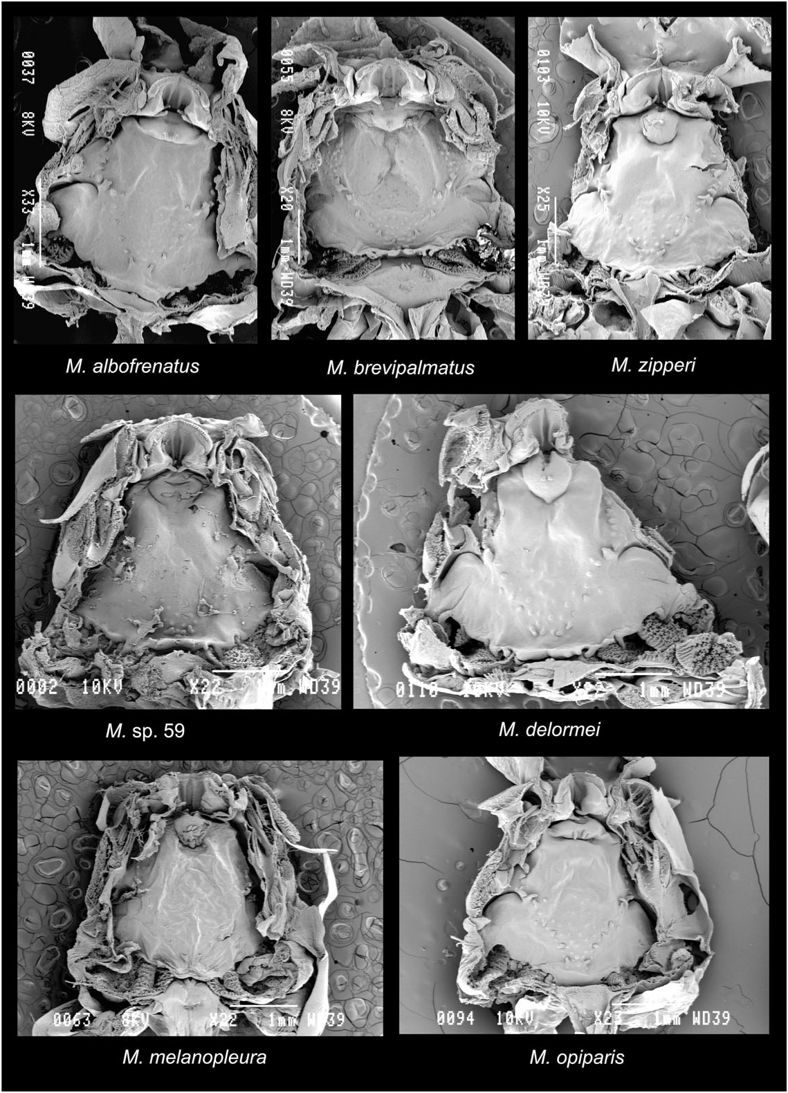
Buccal floor ( Fig. 3 View Figure 3 ): Prelingual arena trapezoidal, two vertical ridges on inner side of lower beak; two pairs of small and simple prelingual papillae on lateral wall of arena, the lowest directed anteromedially, the other, bigger, directed posteromedially. Tongue anlage prominent, roughly rounded; one pair of moderately sized simple lingual papillae orientated posterodorsally. Buccal floor arena rounded delimited by a simple row of about nine small papillae on each side, the biggest, bifid at tip, in front of buccal pocket; interior of arena in a depression with some scarce pustules more densely arranged posteriorly. Buccal pockets relatively wide, slightly curved and almost obliquely orientated, not perforated; a longitudinal cloud of fewer than 20 pustules and prepocket papillae in continuity with the buccal floor arena papillae running up to level of posterior end of tongue anlage. Ventral velum with spicular support, wavy, bearing a row of ten projections medially, close to each other above the first and second filter plates, the median two forming the medial notch; glottis long, fully exposed by medial notch; secretory pits present on margin of velum and projections. Branchial baskets as wide as long; with three filter cavities, filter plates obliquely arranged, filter mesh of low density with tertiary folds. Lungs very long, occupying all length of body.
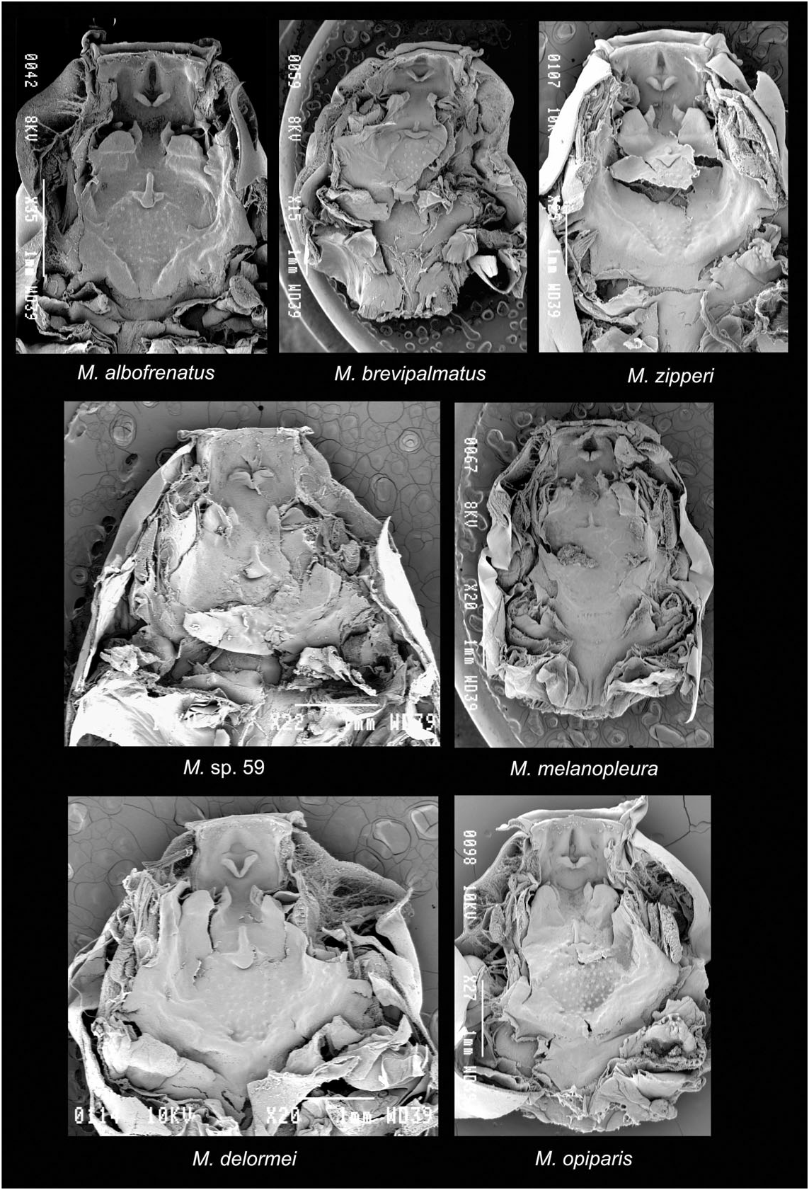
Buccal roof ( Fig. 4 View Figure 4 ): Prenarial arena rectangular, anterior wall with a depression; one pair of obliquely orientated triangular flaps lying on anterior wall of arena and fusing posteromedially. Choanae moderately sized, transverse; anterior wall not elevated, smooth, bearing a small projection on medial half; narial valve slightly elevated forming a triangular flap wrapping medial extremity of choana. A large, flat, and rounded flap behind each choana on a prominence, probably homologous to postnarial papilla. Two abutting pustules in a longitudinal plane in postnarial arena just in front of median ridge. Median ridge curved posteriorly. No lateral ridge papillae. Buccal roof arena diamond-shaped; one or two tiny buccal roof arena papillae on each side anteriorly, the largest anterior; interior of arena with about 40 pustules, the largest anterior. Posterolateral ridge present and prominent, raised, impossible to assess its continuity across the dorsal roof. Glandular zone large, impossible to establish its presence or absence medially, moderately sized secretory pits. Dorsal velum and pressure cushions destroyed during dissection.
No known copyright restrictions apply. See Agosti, D., Egloff, W., 2009. Taxonomic information exchange and copyright: the Plazi approach. BMC Research Notes 2009, 2:53 for further explanation.
|
Kingdom |
|
|
Phylum |
|
|
Class |
|
|
Order |
|
|
Family |
|
|
Genus |