
Lissaclita melaniae, Gomez-Daglio, Liza & Syoc, Robert Van, 2006
|
publication ID |
https://doi.org/ 10.5281/zenodo.171665 |
|
DOI |
https://doi.org/10.5281/zenodo.5676917 |
|
persistent identifier |
https://treatment.plazi.org/id/0A7FCD02-707D-FF80-FEB5-FEB4FCACF939 |
|
treatment provided by |
Plazi |
|
scientific name |
Lissaclita melaniae |
| status |
sp. nov. |
Lissaclita melaniae sp. nov. ( Fig. 1)
Holotype. SEM stubs of the disarticulated wall plates and opercular plates, and two microscope slides of the cirri and mouthparts, California Academy of Sciences ( CASIZ 170229).
Paratypes. SEM stubs of the entire wall and the opercular plates of one individual and one microscope slide of its cirri, California Academy of Sciences ( CASIZ 170230). Two whole specimens from Punta Gorda, Baja California Sur (B.C.S.), Colección Nacional de Crustáceos, Instituto de Biología, Universidad Nacional Autónoma de México ( CNCR 13845 and 13846). Two whole specimens from Bahía de la Paz, B.C.S., Scripps Institution of Oceanography Benthic Invertebrates Collection (SIOBIC C10873).
Other material examined. One specimen from Santa Elena, B.C.S.; two specimens from Coral de los Frailes, B.C.S.; and three specimens from Calerita, B.C.S., México.
Type locality. Punta Gorda ( Fig. 2 View FIGURE 2 ), Baja California Sur, México.
Etymology: Named in honor of Melania Lopez Castro, longtime friend, and her perseverance in turtlebarnacle research.
Distribution. Punta Coyote (24° 35.2 N,110° 26.7 W) to Punta Gorda (23°08.3’ N, 109°58.02’ W), Baja California Sur, México. Attached to rocks, middle and high intertidal zone.
Diagnosis: Shell small, conical to cylindroconical; parietes with two rows of tubes. Radii disparietal, nontubiferous, broadening from opercular orifice and then narrowing again. Plates with transverse striae, with longitudinal color stripes of white and violet. Basis membranous. Base diameter to 1.9 cm.
Description
Shell. Conical to cylindroconical with diametric growth. Carina with two welldeveloped transparietal alae. Rostrum with two radii, radii disparietal, broadening from top until they contact adjacent pariete, then narrowing toward the base. Plates with transverse striae and longitudinal color stripes of white and violet. Parietals with two rows of nonseptal tubes filled with living tissue ( Fig. 1 b). Basis membranous.
Scutum ( Fig. 3 View FIGURE 3 a, b). Exterior concave, triangular with prominent growth ridges; longitudinal brown stripe down middle of valve. Occludent margin with seven to eight oblique teeth formed by extensions from growth ridges. Adductor ridge prominent, rounded, extending about halfway up scutum, not fused with articular ridge. Adductor muscle pit oval, shallow, not well developed; rostral depressor muscle crests eight to nine, short and broad; depressor muscle crests three to four, long, thin, and slight; above these crests, many tiny crests or ridgelets in depressor muscle area. Articular ridge prominent, rounded.
Tergum. ( Fig. 3 View FIGURE 3 c, d). Covered with fine transverse striae; apex rounded, curved to scutal margin. Color white with vertical dark brown broad stripe in middle of valve. Spur moderately wide, about half width of basiscutal margin, elongated with rounded end and with basiscutal margin fused with spur. Depressor muscle crests eight to 10, long, thin, extending up to central part of plate; articular ridge high, very broad and long, with few tiny crests or ridgelets below it; articular furrow projecting down to basiscutal margin.
Labrum ( Fig. 4 View FIGURE 4 a). With deep “V”shaped notch. Crest bullate, with four to six rounded teeth, some specimens with only one or two teeth; crest covered by short setae.
Maxilla I ( Fig. 4 View FIGURE 4 b). Upper margin with two long, prominent spines followed by ten medium spines, inferior angle with four short, slender spines, upper margin covered by long setae (1/3 of total margin), inferior margin with short setae (covering 1/4 of the margin).
Maxilla II ( Fig. 4 View FIGURE 4 c). Distal lobe with three to four rows of long, pinnate setae, covering its outer margin; inner margin with three rows of long setae; proximal lobe covered with a cluster of long pinnate setae.
Mandible ( Fig. 4 View FIGURE 4 d). With five teeth, fourth tooth with four to six subsidiary cusps in margin. Basal comb with four to eight small teeth, inferior angle with two prominent conical spines, inferior margin (1/2 total margin) with long setae, upper margin naked. About 1/4 of mandible surface covered by setae.
Palpus ( Fig. 4 View FIGURE 4 e). Distal margin with five to six long, pinnate setae and several rows of short and medium setae; lower margin naked.
Cirrus I ( Fig. 5 View FIGURE 5 a). Rami unequal in length ( Table 1 View TABLE 1 ), posterior margin of each segment with one or two stout setae, anterior margin with several setae bunched together, distal segments with three long, stout setae, and with pectinate setae covering 1/3 of cirrus.
Cirrus II ( Fig. 5 View FIGURE 5 b). Rami equal in length ( Table 1 View TABLE 1 ), both margins covered by long setae, distal segments with pectinate setae.
Cirrus III ( Fig. 5 View FIGURE 5 c, d). Rami equal in length ( Table 1 View TABLE 1 ), segments wider than long, upper angle of pedicel with cluster of long, thin setae, anterior margin with mediumlong, thin setae. All segments with long pectinate setae on anterior margin, distal segments with stout serrate setae.
Cirrus IV. Rami unequal in length ( Table 1 View TABLE 1 ); with segments wider than long. Anterior margin of pedicel and proximal segments with several rows of small, conical spines. Posterior angle with three setae (two short, erect, and one long, thin), anterior margin with four long and three mediumlong pectinate setae. Distal segments with two long, stout setae each.
Cirrus V. Rami equal in length ( Table 1 View TABLE 1 ), with features resembling those described for cirrus IV.
Cirrus VI. Rami equal in length ( Table 1 View TABLE 1 ); pedicel with one row of small, conical spines, segments wider than long, anterior margin with three to four long setae, posterior margin with two or three erect setae.
Remarks
According to Ross (1969, 1970), tetraclitids have two major evolutionary lines correlated with their shell wall growth pattern (monometric growth being the derived state), and the morphology of their radii (tubiferous being the derived state), with an evolutive tendency to increase the number of rows of tubes in the parietes. The new species and genus described herein retains the ancestral states of solid radii and diametric growth. It lacks either synapomorphy that Ross (1969, 1970) used to define the "tesseroporan" branch (monometric shell growth in Tetraclita, Tesseropora and Tesseroplax , a character which is also present in the Austrobalaninae genera of Austrobalanus and Epopella ) or the "tetraclitellan" branch (tubiferous radii in Tetraclitella , Newmanella and Yamaguchiella ) of the family. Diametric growth is a plesiomorphic character present in the “tetraclitellan” subfamilies Newmanellinae and Tetraclitellinae .
Lissaclita melaniae , lacking either monometric growth (the synapomorphy of the Tetraclitinae and Austrobalaninae ) or tubiferous radii (characteristic of the Newmanellinae and Tetraclitellinae ) does not clearly align itself with any existing subfamily within the Tetraclitidae ; therefore, we place this new genus into this family, but with no subfamily assignment.
Newman and Ross (1976) and Ross and Perreault (1999) defined the four subfamilies of the Tetraclitidae as follows:
(1) Tetraclitellinae : parietal wall tubes in two or more rows; radii tubiferous, broad, and commonly nonseptate; basis membranous or calcareous; growth diametric; scutum lacking rostral and lateral depressor muscle crests; tergum not overlying scutum.
(2) Newmanellinae : parietal wall tubes present in one, two, or several rows; radii tubiferous and broad; basis calcareous; growth diametric; scutum with lateral depressor muscle crests; rostral depressor muscle crests present or not; tergum overlying apical portion of scutum.
(3) Austrobalaninae : four or sixplated ( Epopella , fourplated; Austrobalanus , sixplated); parietes solid or permeated by chitinous rods (not homologous to parietal tubes); radii solid, narrow or obsolete; basis membranous or calcareous; growth monometric; scutum bearing crests for lateral depressor muscles and with or without rostral depressor muscle crests.
(4) Tetraclitinae : shell wall with two or several rows of tubes entirely or partially filled with living tissue or chitinous material, radii solid, narrow or obsolete; basis membranous or calcareous; growth monometric; scutum with lateral and rostral depressor muscle crests.
The other tetraclitids known to the Baja California and Gulf of California region are members three genera which have been assigned to the Tetraclitinae . Their characteristics are contrasted with those of Lissaclita melaniae below.
Tesseroplax has a single species, T. unisemita ( Zullo, 1968) , an extinct species known only from the Pleistocene of Isla Angel de La Guarda, Gulf of California ( Zullo, 1968). This species is the only tetraclitine with transverse septa in the parietal tubes (Table 2). Tesseroplax unisemita lacks lateral depressor muscle crests on the terga and possesses a calcareous basis.
Tetraclita Schumacher, 1817 View in CoL has a worldwide distribution in warm and temperate waters. Tetraclita confinis ( Pilsbry, 1916) and Tetraclita rubescens (Darwin, 1854) are the most common tetraclitids in the northeastern Pacific. According to collection record data at the California Academy of Sciences, Tetraclita confinis ranges from Baja California Sur to Acapulco, Mexico (including the Gulf of California), while T. rubescens extends from Los Cabos, Baja California Sur north to San Francisco, California, U.S.A. Both species are typical members of the Tetraclitinae ( Ross, 1969; Pitombo and Ross, 2002). We examined four small specimens (1 to 2 cm in diameter) of Tetraclita confinis from the Gulf of California. Such very small, young Tetraclita View in CoL resemble L. melaniae in the morphology of the parietal tubes, which arise as the result of splitting of the longitudinal septa intercalated between the inner and outer lamellae of the wall plates. Nevertheless, Lissaclita differs from Tetraclita View in CoL by exhibiting a diametric growth pattern, with a secondary row of tubes in the parietes formed as the primary tubes divide in two near the basis. In T. confinis , the parietal tubes split much closer to the apex, even in very small specimens. Furthermore, T. confinis lacks conical spines on the pedicel of cirrus IV, and the scutum of L. melaniae has small crests above the rostral depressor crests, and also a tergum with longer depressor muscle crests than those of T. confinis . The color and exterior surface texture of the parietes are different, too; T. confinis and T. rubescens have ribbed walls colored gray and reddish respectively, whereas L. melaniae has smooth parietes with transverse fine striae colored in white and purple.
Tesseropora Pilsbry, 1916 View in CoL ranges from the Indopacific and the southwestern Pacific to the North Atlantic ( Bermuda and Azores), and is also known from the Oligocene of Italy ( Newman and Ross, 1977). Only three species are known (Table 2). Pilsbry (1916: 259) defined Tesseropora View in CoL as “ Tetraclita View in CoL with a single row of parietal pores”. Ross (1969) emended this diagnosis to tetraclitids with one row of parietal tubes lacking transverse septa and with a scutum bearing depressor muscle crests.
The parietal tubes of Lissaclita melaniae are generally similar to those found in the three species of Tesseropora View in CoL and suggest an affinity to those taxa. They are especially similar to those in Tesseropora wireni (NilssonCantell, 1921) ; secondary tubes forming within the single row of tubes. However, Lissaclita lacks the incomplete septa on the inner margin of the external lamina exhibited in T. wireni . In addition, the exoskeletal characters differ greatly: L. melaniae has a combination of pectinate and serrate setae on cirrus III, whereas Tesseropora View in CoL species only have bipectinate setae. Also, the number of teeth in the labrum is smaller and the notch shallower in L. melaniae cf. ( Southward, 1998; Young, 1998) (Table 2). All species of Tesseropora View in CoL have a calcareous basis whereas L. melaniae has a membranous basis. Newman and Ross (1977) suggested that the calcareous basis of Tesseropora View in CoL is related to its high intertidal distribution on the rocky shore in as much as a calcareous basis should provide a more secure way to avoid desiccation. One of us (LGD) found L. melaniae attached to rocks in the middle and low intertidal zone, not the upper intertidal.
This new genus and species is probably a relict surviving in the refugium of the Gulf of California and the islands therein, similar to the case of Tesseroplax unisemita . A more thorough understanding of the phylogenetics and biogeography of the Tetraclitidae could help further unravel their evolutionary history.

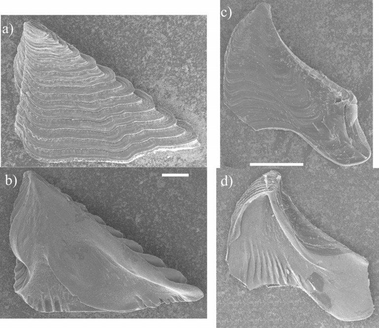


TABLE 1. Number of segments in the cirri of Lissaclita melaniae gen. et sp. nov. (left cirri only).
| I A P | II A P | III A P | IV A P | V A P | VI A P | |
|---|---|---|---|---|---|---|
| Holotype | 7 14 | 8 8 | 6 6 | 8 10 | 14 14 | 15 15 |
| Paratype | 8 16 | 7 8 | 6 6 | 10 12 | 14 14 | 15 15 |
No known copyright restrictions apply. See Agosti, D., Egloff, W., 2009. Taxonomic information exchange and copyright: the Plazi approach. BMC Research Notes 2009, 2:53 for further explanation.
|
Kingdom |
|
|
Phylum |
|
|
Class |
|
|
Order |
|
|
Family |
|
|
Genus |
Lissaclita melaniae
| Gomez-Daglio, Liza & Syoc, Robert Van 2006 |
T. unisemita (
| Zullo 1968 |
Tesseropora wireni (NilssonCantell, 1921)
| Nilsson-Cantell 1921 |
Tetraclita confinis (
| Pilsbry 1916 |
Tesseropora
| Pilsbry 1916 |
Tetraclita rubescens
| Darwin 1854 |
Tetraclita
| Schumacher 1817 |