
Kamabrachys, Constant, 2023
|
publication ID |
https://doi.org/10.5852/ejt.2023.895.2289 |
|
publication LSID |
lsid:zoobank.org:pub:75CAAC73-8100-4D16-B970-4A533DBC7000 |
|
DOI |
https://doi.org/10.5281/zenodo.8407968 |
|
persistent identifier |
https://treatment.plazi.org/id/D3F2D3FC-7031-4A91-B038-6B266B34E020 |
|
taxon LSID |
lsid:zoobank.org:act:D3F2D3FC-7031-4A91-B038-6B266B34E020 |
|
treatment provided by |
Plazi (2023-10-03 09:16:24, last updated 2024-11-29 12:43:31) |
|
scientific name |
Kamabrachys |
| status |
gen. nov. |
Genus Kamabrachys gen. nov.
urn:lsid:zoobank.org:act:D3F2D3FC-7031-4A91-B038-6B266B34E020
Figs 1–5 View Fig View Fig View Fig View Fig View Fig
Type species
Platybrachys signata Distant, 1892 View in CoL by present designation.
Diagnosis
Small to medium sized (6.8–12.6 mm) eurybrachids, brownish grey variegated with black and white, with tegmina flat usually with an anteapical white transverse line. The venation of the tegmina shows veins ScP+RA and RP separated close to base, the first fork of MP very basal, at the level of ScP+RA– RP separation and the first fork of CuA slightly before the apex of the closed clavus. The posterior wings show basicostal area widely, distinctly paler, whitish to yellow orange. The abdomen is bright red. The frons is slightly convex, uniformly coloured and the genae don’t possess subocular spine. The male terminalia show a pygofer with strongly developed lateroventral lobes, the gonostyli fused along most of their length and with a strong dorsal hook (no spoon-shaped process), the anal tube is flat and mostly circular; the connective is very elongate, the aedeagus is bifid with both shafts furcate and with a complex dorsal periandrium showing two lateral processes and one median furcate process. The female terminalia show an excavate membranous fold of gonocoxae VIII delimited dorsally by gonapophysis VIII and ventrally by the posterolateral process and a posterior triangular process of sternite VI.
Differential diagnosis
The females of Kamabrachys gen. nov. differ from those of all other genera by the ventral excavate membranous fold of gonocoxae VIII on the terminalia, delimited dorsally by gonapophysis VIII and ventrally by the posterolateral process and a posterior triangular process of sternite VI.
Among all Australian genera of Eurybrachidae , the males of Kamabrachys gen. nov. can be separated from those of Chewobrachys Constant, 2008 , Fletcherobrachys Constant, 2006 , Hackerobrachys Constant, 2006 , Maeniana Metcalf, 1952 , Nirus Jacobi, 1928 , Olonia Stål, 1862 and Stalobrachys Constant, 2018 by the absence of a spoon-shaped process on the dorsal margin of the gonostyli.
From the remaining genera, males of Kamabrachys gen. nov. can be separated from those of
– Dardus Stål, 1859 , Gelastopsis Kirkaldy, 1906 , Maon Fennah, 1964 and Navorillina Fennah, 1964 by its less broad frons, 1.3–1.5 times as broad as long, as opposed to at least 1.9 times as broad as long in the four other genera and by possessing hind wings with large, differently coloured basicostal area (hind wings uniformly infuscate in the four other genera); Kamabrachys gen. nov. furthermore differs from Dardus by having flat tegmina (convex in Dardus ); it also differs from Gelastopsis and
Navorillina by a uniformly coloured frons (dorsal half of the frons black and modified into fake eyes or tubercles in Gelastopsis and Navorillina ) and from Dardus , Maon and Navorillina by having shorter antennae, not surpassing eye (antennae elongate, surpassing eye in the three other genera).
– Euronotobrachys Kirkaldy, 1906 , Kirkaldybrachys Constant, 2006 , Loisobrachys Constant, 2008 and Platybrachys Stål, 1859 (as defined by P. lanifera (Stål, 1854) , its type species), by possessing hind wings with large, differently coloured basicostal area (hind wings uniformly infuscate in the four other genera); Kamabrachys gen. nov. also differs from Kirkaldybrachys by having a round anal tube in males (anal tube elongate and apically hooked in Kirkaldybrachys ); from Euronotobrachys and Platybrachys by the gonostyli fused along most length (gonostyli fused only basally Euronotobrachys and Platybrachys ) and from Loisobrachys by the flat tegmina and slightly convex frons (strongly convex frons and convex tegmina in Loisobrachys ).
– Gedrosia Stål, 1862 by possessing relatively short antennae, not surpassing eye, while Gedrosia possesses greatly enlarged, subcylindrical antennae, largely surpassing eye, by having elongate and flat tegmina while the tegmina are tuberculate and tapering towards apex in Gedrosia and by the lateral angular projections of the frons while the sides of the frons are round in Gedrosia .
Etymology
The genus name is formed by the combination of ‘ kama ’ and ‘ brachys ’. The former derives from the ‘kamasutra’, an ancient Indian Hindu Sanskrit text on sexuality, eroticism and emotional fulfilment in life and refers to the peculiar copulation position observed in this genus; the latter, ‘ brachys ’ (Greek), means ʻshortʼ and is a common ending of generic names in Eurybrachidae . Gender feminine.
Description
SIZE: ♂: 7.8–11.1 mm; ♀: 8.1–12.6 mm.
COLOURATION. Head, pro- and mesonotum, and tegmina brownish grey with irregular black markings, frons usually darker. Tegmina sometimes with squarish black marking in middle of clavus and/or one or two black transverse lines or bands, more rarely with a pale transverse band; subapical white line or band, sometimes interrupted in middle, nearly always present; apical white spot often present. Posterior wings brown to black with basal third to basal half differently coloured in yellowish brown to orange; sometimes a white marking along posterior margin between veins A1 and CuA; sometimes an apical white spot or narrow band. Legs brown variegated with black to nearly all black; sometimes femora reddish; posterior legs usually paler. Abdomen and ventral face of thorax bright red; genital segments brown to black.
HEAD. Slightly narrower than thorax; vertex 3–4 times as broad as long, concave with all margins slightly carinate; anterior and posterior margins rounded, parallel; frons about 1.3–1.5 times as broad as long, slightly convex, slightly wrinkled to rugulose with peridiscal carina slightly marked; upper margin of frons straight to slightly rounded in perpendicular view of frons; lateral margin slightly incurved above lateral projection of frons, slightly rounded under the projection. Clypeus reaching mesocoxae, elongate, with median carina on anteclypeus prolongated with obsolete carina, sometimes only marked by a dark line, on postclypeus; base of clypeus rounded, rather deeply inserted in frons. Labium reaching metacoxae, with apical segment as long as broad, acuminate, shorter and slightly more slender than penultimate. No infra-ocular spines on genae; ocelli absent; antennae elongate, not surpassing eye, not visible from above; scape about as long as broad, pedicel subcylindrical, elongate, narrowing towards apex.
THORAX. About 1.15–1.35 times as broad as combined length of pro- and mesonotum; mesonotum 2.5– 3.2 times as long as pronotum. Pronotum with disc weakly wrinkled, carina parallel to anterior margin and 2 slightly impressed points on disc. Mesonotum with disc weakly wrinkled, median and peridiscal carinae slightly marked; median carina stopped before scutellum.
TEGMINA ( Fig. 1A View Fig ). Flat, elongate, about 2.5–2.9 times as long as broad; costal margin moderately rounded along basal fifth, then straight to nodal line; apex rounded, slightly sinuate; apical margin obliquely rounded.
VENATION. Veins ScP+RA and RP separated close to base; first fork of MP very basal, at the level of ScP+RA–RP separation; first fork of CuA slightly before apex of clavus; clavus closed; Pcu and A1 fused at about ¾ of clavus length; Pcu+A1 reaching apical angle of clavus; numerous cells along posterior half of costal margin; numerous cross-veins on apical third delimitating small elongate cells.
POSTERIOR WINGS ( Fig. 1B View Fig ). Well developed, as broad as tegmina, about 1.6–1.8 times as long as broad, rounded apically; anal area well developed; sutural margin weakly trilobous; not reaching apex of tegmina at rest. All main veins visible from base, forked at or after nodal line and forming a few closed cells; transverse veinlets delimiting elongate cells on apical ¼; veins A1 and A2 sometimes with 2–3 terminals. Arrangement of secondary veins and veinlets variable between specimens and sometimes between the two wings of the same specimen.
LEGS. Pro- and mesofemora and tibiae dorsoventrally flattened, elongate and slender; metatibiae (PT) with 3 lateral (lSp) and 10 apical spines (aSp); first metatarsomere (MtT1) ventrally with pad of microsetae (PMs) at internoapical angle and two parallel rows of 4 spines (Sp) ( Fig. 1C View Fig ) along outer margin, following the two middle apical spines. Metatibiotarsal formula: (3) 10/4/0.
MALE TERMINALIA ( Figs 2–3 View Fig View Fig ). Pygofer (Py) rather short, as high as long and oblique in lateral view, about twice as wide as high in caudal view; internal basal apodeme (bap) well developed and sclerotized, sinuate in dorsal view; dorsal portion narrow; lateroventral portion strongly developed into a lateroventral lobe (lvl) projecting ventrally; in ventral view, ventral lobe forming posteriorly a round or pointed projection; ventral portion separated from lateroventral lobe by a well-marked depression; ventral portion developed posterad into a ventral lamina (vlp) with anterior and posterior margins parallel in ventral view and posterolateral angles rounded. Anal tube (An) dorsoventrally flattened, round or oval, sometimes with apical margin slightly indented; paraprocts (pa) at basal ¼. Gonostyli (G) fused, attached together by a flexible median membrane; gonostyli developed ventrally into a subtriangular, dorsoventrally flattened, ventral process (vp), and basolaterally into an elongate, laminate, laterodorsal process (ldg) sinuate in caudal view and showing a mediolateral angle (mla) when abruptly curving mesad; laterodorsal process ended with a strong dorsal hook (dhg) curved lateroventrad to anterolaterad. Connective (cv) strongly developed, with two pairs of lateral apodemes; apodemes of anterior pair forming a moderately broad lateral lamina along the connective; apodemes of posterior pair ribbon-like, strongly developed laterally and attached to the inner wall of the pygofer; posterior portion of the connective dorsoventrally flattened and with pair of sclerotized shafts. Aedeagus s. lat. rather complex and symmetrical, generally moderately upcurved. Aedeagus s. str. with pair of elongate apical processes; each process split apically into a simple apicoventral process (avp) smoothly tapering towards the posterior and narrowly rounded apically, and a sinuate apicodorsal process (adp) which is laterally inflated at base, then more or less abruptly tapering towards apex and narrowly rounded at apex. Dorsal periandrium well developed, with two lateral pairs of elongate, spinose processes, one laterodorsal (ldp) and one lateroventral (lvp), one pair of elongate basomedian processes (bmp) apically blunt on each side of elongate median furcate process (mfp) upcurved and bearing a dorsal crest (cfp) on each arm of the apical furca.
FEMALE TERMINALIA ( Fig. 4 View Fig – based on K. signata ). Abdominal sternite VI (As VI) with anterior margin broadly rounded in ventral view ( Fig. 4D View Fig ) and with sides upcurving in caudal view ( Fig. 4B View Fig ); posterior margin in ventral view ( Fig. 4D View Fig ) projecting posteriorly on sides and with median deep, wide, subquadrate emargination accommodating a furca-shaped medioventral process (FAs VI); on each side of emargination, a large triangular process directed posterodorsad; rather deep rounded emargination between triangular process and lateral projection ( Fig. 4B, D, F View Fig ); points of furca of sternite VI diverging and curved posterodorsad ( Fig. 4B,D View Fig ). Sternite VII (As VII) reduced to2 small oblique sclerites. Gonocoxa VIII (Gx VIII) and gonapophysis (Gy VIII) fused, forming (in caudal view – Fig. 4B View Fig ) a horizontally Y-shaped sclerotized structure with dorsal branch, formed by inner end of gonocoxa, joining tergite IX; outer base of the common shaft joining posterolateral projection of sternite VI; common shaft strongly curved in posteroventral view ( Fig. 4D View Fig ) with inner end of gonapohysis VIII tapering towards apex and reaching near triangular process of sternite VI; posterior margin of gonapophysis VIII roundly projecting poterolaterad in ventral view ( Fig. 4D View Fig ). Area between sternite VI and gonapophysis VIII forming a lateroventral oval and excavate membranous fold of gonocoxae VIII ( Fig. 4B, D View Fig – mf in Bourgoin 1993: fig. 4); inner portion of gonapophysis VIII, furca, triangular processes and adjacent part of posterior margin of sternite VI densely covered in short strong setae ( Fig. 4B, D View Fig ). Tergite IX (Tg IX) narrow and curved dorsocephalad with posterior margin projecting posteriorly at base of anal tube ( Fig. 4 A, C View Fig ). Gonoplacs (Gp) unilobed, large and semicircular, mostly hiding the anal tube. Gonocoxal bases IX (Gb IX) projecting laterally. Gonapophysis IX (Gy IX) flattened laterally, strongly reflexed posterodorsad and strongly tapering apically. Anal tube (An) elongate and narrow, curved posteroventrad, v-shaped in cross section beyond anus, lanceolate in dorsal view ( Fig. 4A–B, D View Fig ). Anterior vagina (Av) small and membranous, dorsoventrally flattened, with spermatheca (Sp) attached posterodorsally ( Fig. 4E View Fig ). Posterior vagina (Pv) more strongly sclerified, dorsoventrally flattened, short, wide basally, strongly tapering distally and bearing transverse grooves ( Fig. 4E–F View Fig ). Bursa copulatrix (Bc) attached apically, suboval and tapering distally, much larger than posterior vagina ( Fig. 4A, E–F View Fig ); walls bearing reticulated ornamentation ( Fig. 4E–F View Fig ).
SEXUAL DIMORPHISM. Females about 10% larger than males.
Distribution
Australia: widely distributed throughout the country ( Fig. 5 View Fig ) with the westernmost record in southwest Western Australia (125°48′53″ E) and the southernmost one in northeast New South Wales (29°28′16″ E).
Biology
The species of Kamabrachys gen. nov. seem present more or less all year round. They were recorded from trees in the family Myrtaceae , in the genera Corymbia K.D.Hill & L.A.S.Johnson , Eucalyptus L’Hér. , Gossia N.Snow & Guymer and Melaleuca L. The typical habitat of this genus seems to be open woodland where they are very well camouflaged on the bark of their host tree, usually hiding in the cracks; they are regularly found on partly burnt tree trunks. When disturbed, the specimens tend to walk quickly sideways or backwards, apparently keeping their eyes on the potential ‘enemy’ while escaping, try to hide on the opposite side of the trunk or branch they sit on, out of sight, or move to another nearby hiding place or a high enough spot, out of reach of their presumed ‘aggressor’; if this strategy doesn’t work, they will jump and fly away, their jump being so powerful that it can hardly be detected by the human eye.
The mating behaviour is known in only one species and the male is attached upside down to the female while mating, holding on to the tegmina of the female with its legs, and having its body largely hidden under that of the female; they seem able to quickly separate when disturbed (C. Foelz & A. McDougall pers. com., Apr. 2020).
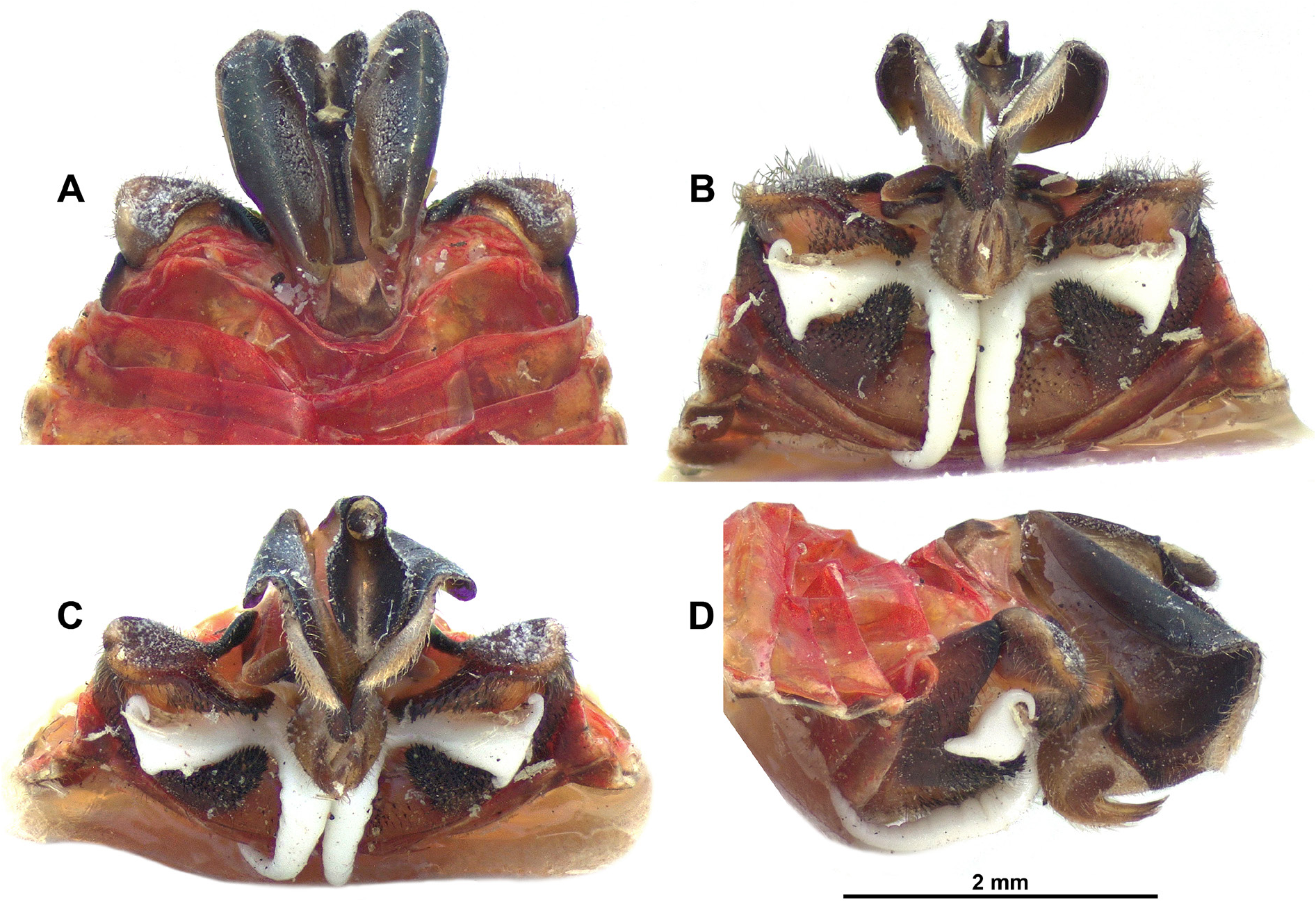
In some females, a symmetrical waxy structure (not made of waxy filaments as on gonoplacs but instead, of white silicone-like wax – see Fig. 45B–D View Fig ), was observed in the area between sternite VI and gonapophysis VIII. It originates under the gonapophysis IX and extends laterally through the excavate membranous fold of gonocoxae VIII in an arm widening towards apex and bearing apical, more or less curved processes; two additional ventral processes, tapering evenly from base to apex, are curved under the body and directed cephalad, diverging slightly towards apex. The formation and role of this secretion remains unknown (see discussion).
Species included (12)
K. andersoni gen. et sp. nov.
K. brennani gen. et sp. nov.
K. campbelli gen. et sp. nov.
K. danielsi gen. et sp. nov.
K. falcata gen. et sp. nov.
K. fasciata gen. et sp. nov.
K. pedemontana gen. et sp. nov.
K. plana ( Kirkaldy, 1906) gen. et comb. nov. K. rieki gen. et sp. nov.
K. signata ( Distant, 1892) gen. et comb. nov. K. v-carinatum gen. et sp. nov.
K. waineri gen. et sp. nov.
Bourgoin T. 1993. Female genitalia in Hemiptera Fulgoromorpha, morphological and phylogenetic data. Annales de la Societe entomologique de France 29: 225 - 244. https: // doi. org / 10.1080 / 21686351.1993.12277686
Constant J. 2008. Revision of the Eurybrachidae (XIII). The new Australian genus Chewobrachys (Hemiptera: Fulgoromorpha). Zootaxa 1898: 41 - 54. https: // doi. org / 10.11646 / zootaxa. 1898.1.3
Constant J. 2018. Revision of the Eurybrachidae XIV. The Australian genera Olonia Stal, 1862 and Stalobrachys gen. nov. (Hemiptera: Fulgoromorpha). European Journal of Taxonomy 486: 1 - 97. https: // doi. org / 10.5852 / ejt. 2018.486
Distant W. L. 1892. Contribution to a knowledge of the homopterous family Fulgoridae. Transactions of the Entomological Society of London 1892: 275 - 286. https: // doi. org / 10.1111 / j. 1365 - 2311.1892. tb 02971. x
Fennah R. G. 1964. Three new genera of Eurybrachidae (Homoptera: Fulgoroidea) from West Africa and Australia. Proceedings of the Entomological Society of London (B) 33 (9 - 10): 157 - 162. https: // doi. org / 10.1111 / j. 1365 - 3113.1964. tb 01633. x
Jacobi A. 1928. Results of Dr E. Mjoberg's Swedish Scientific Expeditions to Australia 1910 - 1913. Rhynchota Homoptera. 1. Fulgoridae und Cercopidae. Arkiv for Zoologi 19 A (28): 1 - 50.
Kirkaldy G. W. 1906. Leafhoppers and their natural enemies. Bulletin of the Hawaiian Sugar Plant Association Division of Entomology 1 (9): 271 - 479.
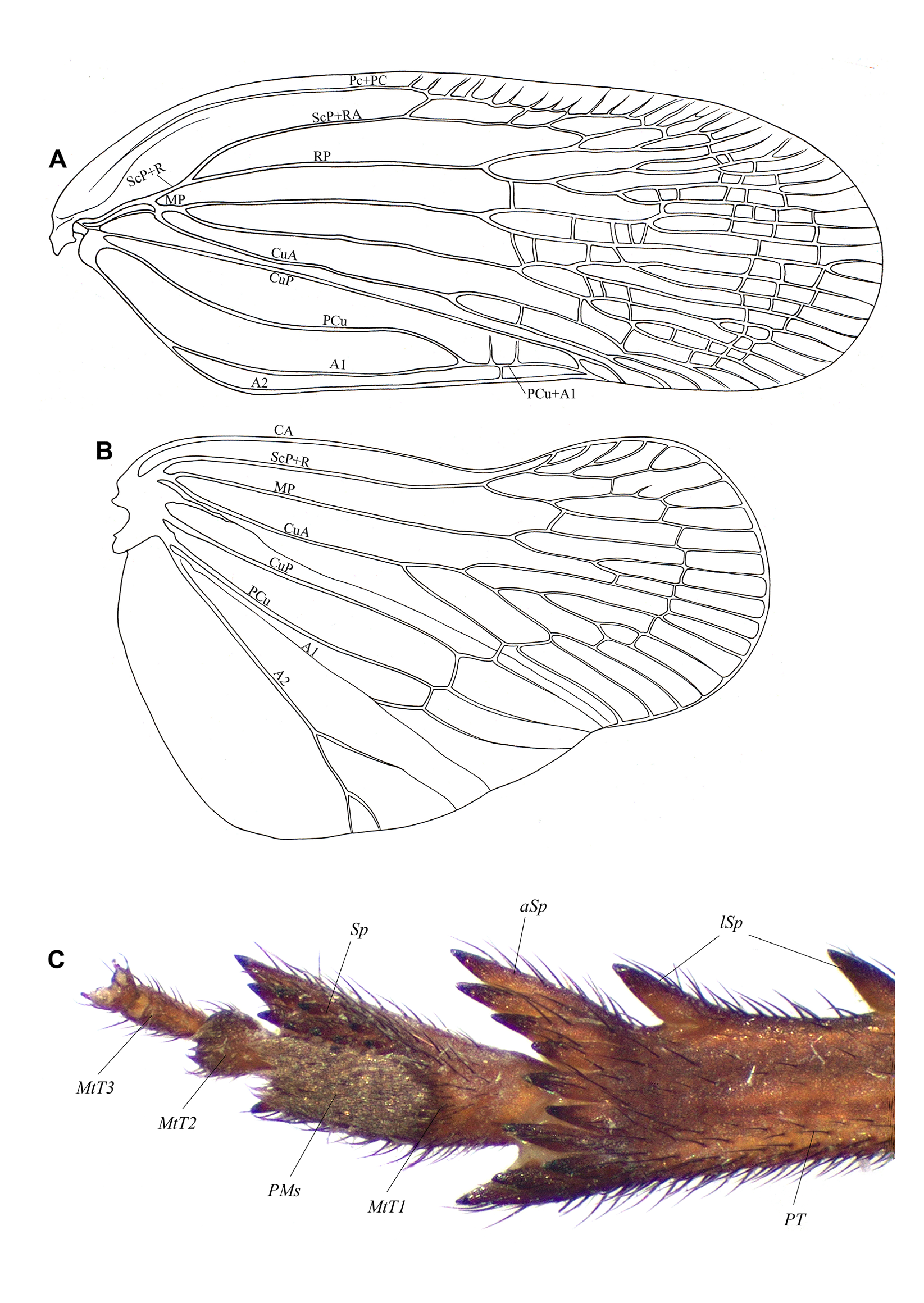
Fig. 1. Kamabrachys signata (Distant, 1892) gen. et comb. nov. A. Right tegmen, venation. B. Right posterior wing, venation. C. Right posterior leg, distal portion, ventral view. Abbreviations: A1 = first anal vein; A2 = second anal vein; aSp = apical spines; CA = costa anterior; CuA = cubitus anterior; CuP = cubitus posterior; lSp = lateral spines; MP = media posterior; MtT1 = first metatarsomere; MtT2 = second metatarsomere; MtT3 = third metatarsomere; Pc+CP = precosta + costa posterior; PCu = postcubitus; PMs = pad of microsetae; PT = posterior tibia; R = radius; RA = radius anterior; RP = radius posterior; ScP = subcosta posterior; Sp = spines.
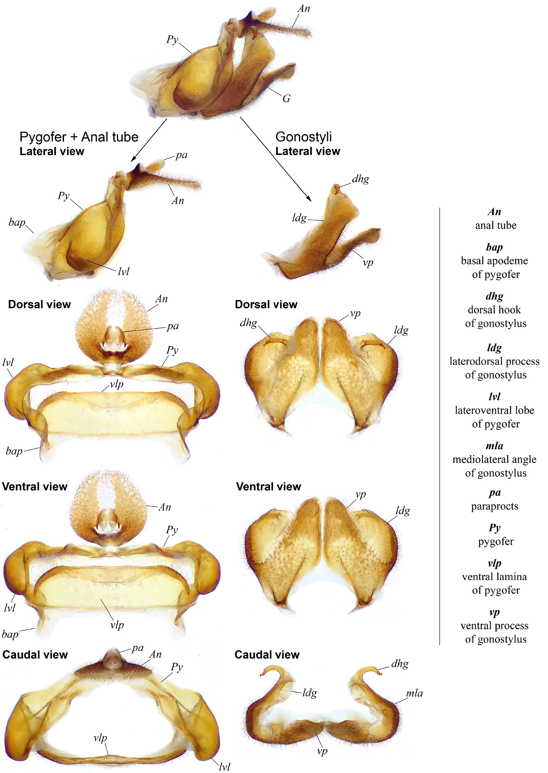
Fig. 2. Kamabrachys signata (Distant, 1892) gen. et comb. nov., male terminalia. Nomenclature used for the description of the pygofer, anal tube and gonostyli.
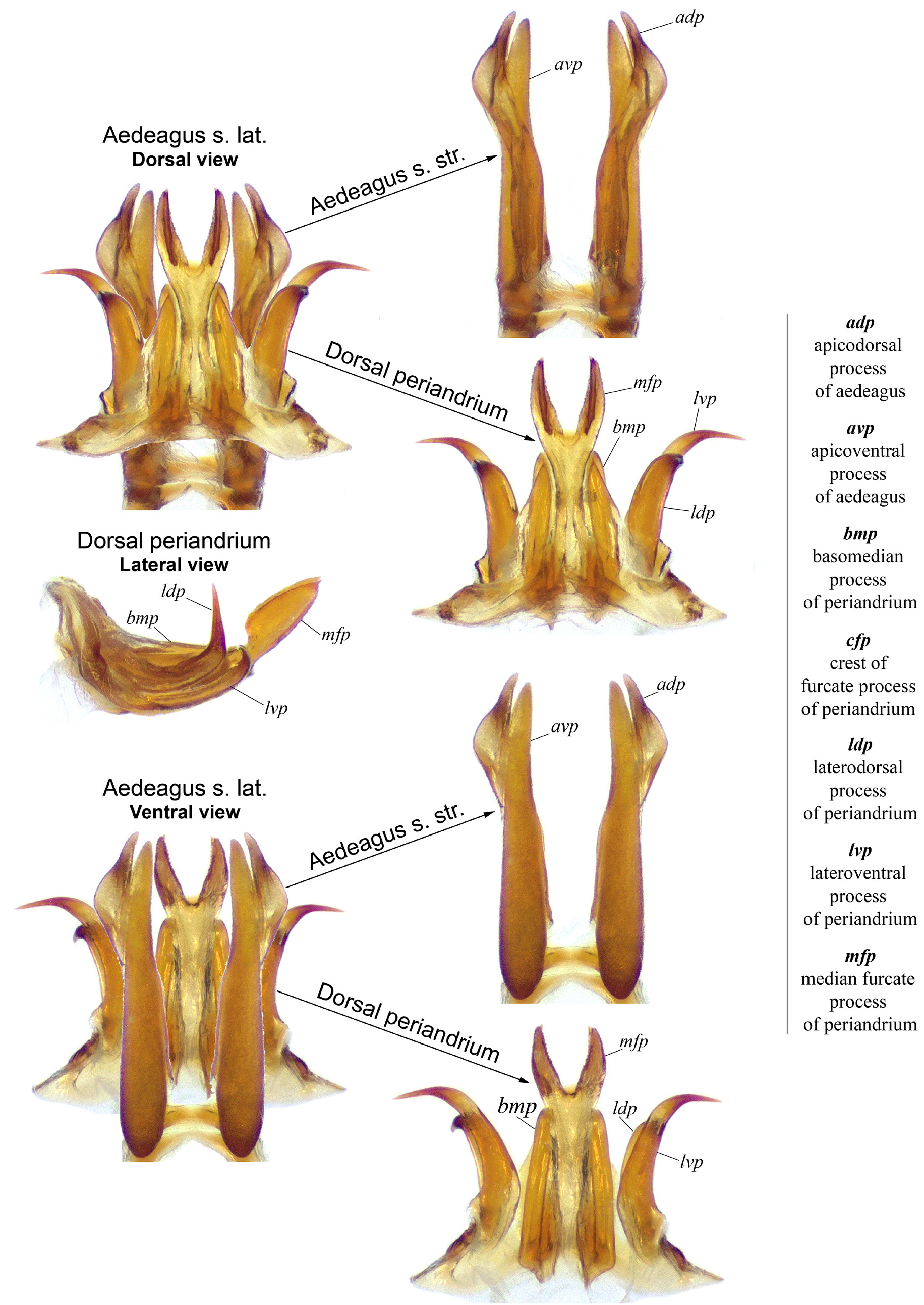
Fig. 3. Kamabrachys signata (Distant, 1892) gen. et comb. nov., male terminalia. Nomenclature used for the description of the aedeagus and periandrium.
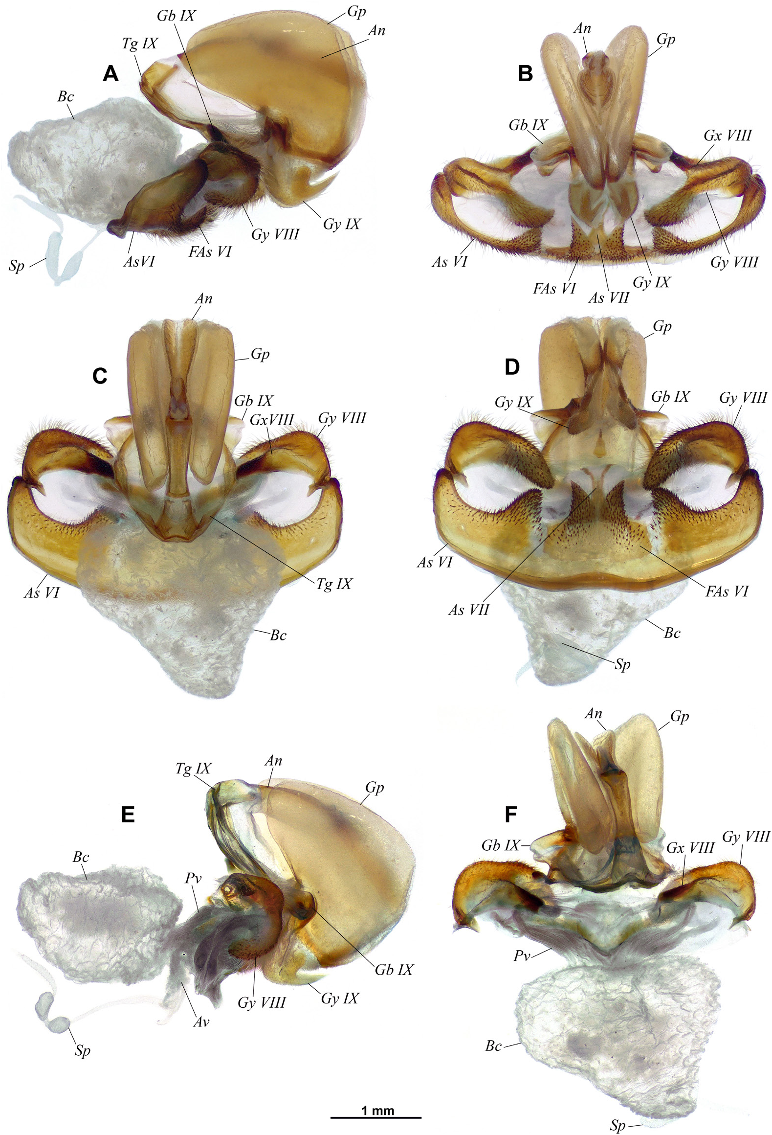
Fig. 4. Kamabrachys signata (Distant, 1892) gen. et comb. nov., female genitalia. A. Left lateral view. B. Caudal view. C. Dorsal view. D. Ventral view. E–F. Sternite VI removed, gonocoxa VIII and gonapophysis VIII detached. E. Lateral view. F. Dorsal view. Abbreviations: An = anal tube; Av = anterior vagina; As VI = sixth abdominal sternite; As VII = seventh abdominal sternite; Bc = bursa copulatrix; FAs VI = furca-shaped medioventral process of sixth abdominal sternite; Gb = gonocoxal base IX;Gp = gonoplac; Gx VIII = gonocoxa VIII; Gy VIII = gonapophysis VIII; Gy IX = gonapophysis IX; Pv = posterior vagina; Sp = spermatheca; Tg IX = ninth abdominal tergite.
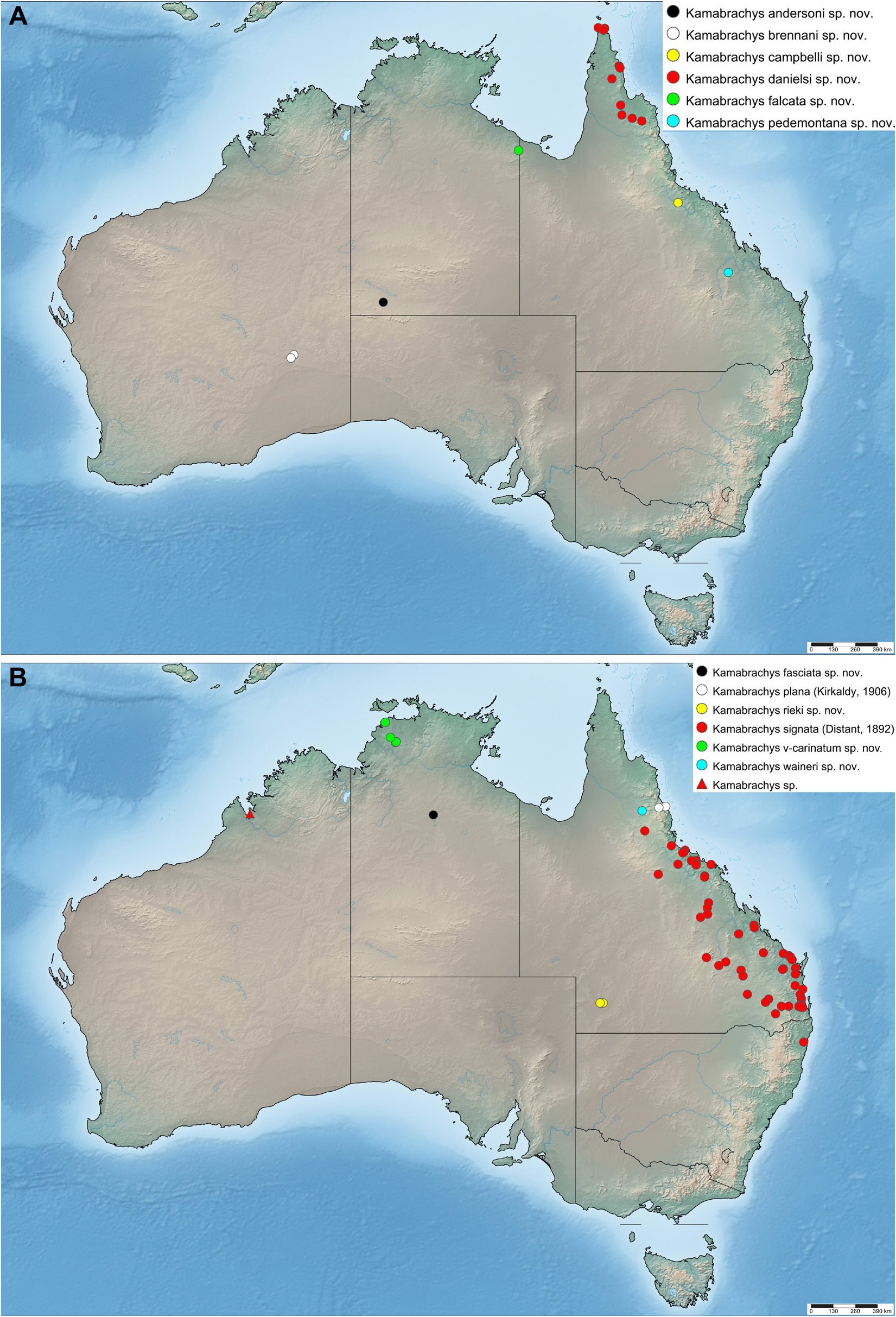
Fig. 5. Kamabrachys spp., distribution maps.A. K. andersoni gen. etsp. nov.,K. brennani gen. et sp. nov., K. campbelli gen. et sp. nov., K. danielsi gen. et sp. nov., K. falcata gen. et sp. nov., K. pedemontana gen. et sp. nov. B. K. fasciata gen. et sp. nov., K. plana (Kirkaldy, 1906) gen. et comb. nov., K. rieki gen. et sp. nov., K. signata (Distant, 1892) gen. et comb. nov., K. v-carinatum gen. et sp. nov., K. waineri gen. et sp. nov., K. sp.
No known copyright restrictions apply. See Agosti, D., Egloff, W., 2009. Taxonomic information exchange and copyright: the Plazi approach. BMC Research Notes 2009, 2:53 for further explanation.
|
Kingdom |
|
|
Phylum |
|
|
Class |
|
|
Order |
|
|
Family |