
Trichomycterus perkos, Datovo, Aléssio, Carvalho, Murilo & Ferrer, Juliano, 2012
|
publication ID |
https://doi.org/ 10.5281/zenodo.213467 |
|
DOI |
https://doi.org/10.5281/zenodo.6174676 |
|
persistent identifier |
https://treatment.plazi.org/id/09268786-296F-8073-A6EF-DCE68CFEFDA1 |
|
treatment provided by |
Plazi |
|
scientific name |
Trichomycterus perkos |
| status |
sp. nov. |
Trichomycterus perkos View in CoL , new species
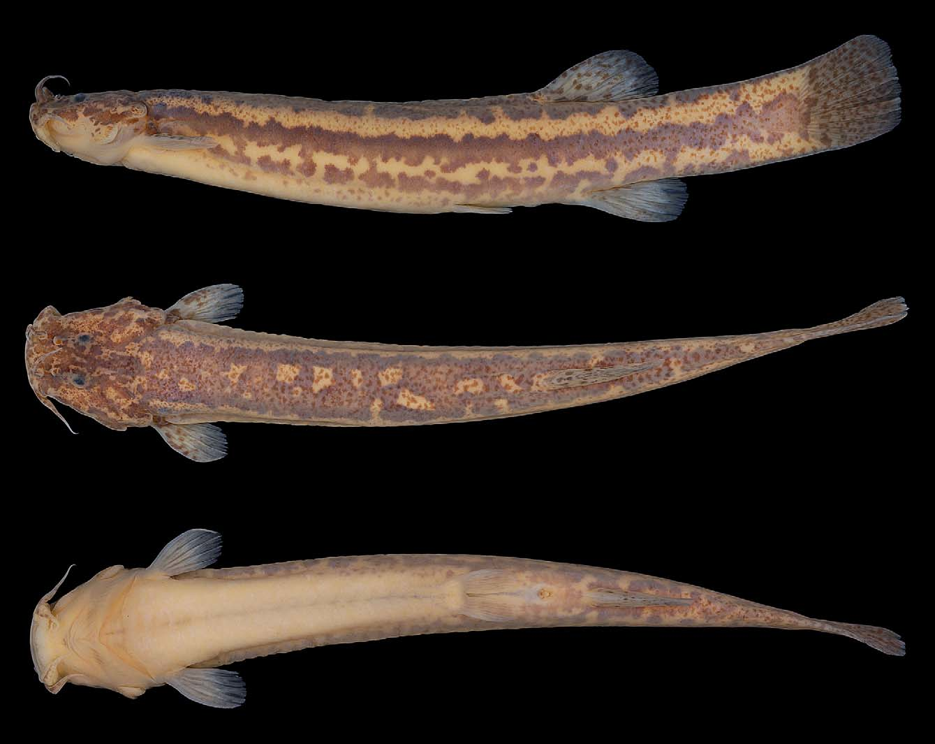
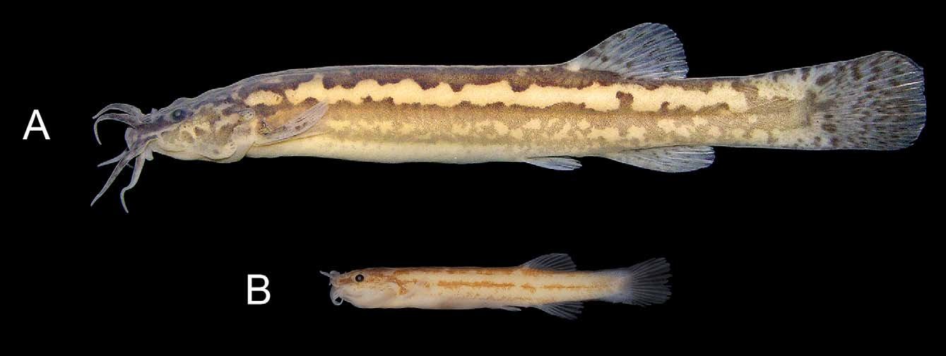
( Figs. 1–2 View FIGURE 1 View FIGURE 2 , Table 1)
Trichomycterus aff. itatiayae: Kantek et al. (2007: 797) View in CoL .
Trichomycterus View in CoL sp. aff. T. itatiayae: Torres et al. (2004: 124) View in CoL .
Trichomycterus View in CoL sp. aff. T. itatiyae [sic]: Sato et al. (2004: 45, 46, 47, Table 1, Table 2).
Holotype. MCP 46679 (93.5 mm SL), XR; Brazil, Rio Grande do Sul State, Passo Fundo Municipality; Uruguai River basin, Passo Fundo River, under the bridge on BR–285 road; approx. 28°14'07"S 52°24'53"W; W. Bruschi Jr. & J. F. P. Silva; 0 3 January 2003.
Paratypes. LIRP 8344, 4 (28.0–93.2 mm SL): 1 CS (28.0 mm SL), 1 MS (35.3 mm SL), 1 XR (28.3 mm SL); Brazil, Rio Grande do Sul State, Passo Fundo Municipality; Uruguai River basin, unnamed stream tributary of Passo Fundo River, about 1 km from BR–285 road; approx. 28°15'14"S 52°21'19"W; W. Bruschi Jr. & J. F. P. Silva; 0 3 January 2003. MCP 31763, 10 (16.2–99.8 mm SL): 1 CS (52.4 mm SL), 1 XR (99.81 mm SL); same data as LIRP 8344. MCP 31764, 2 (17.9–33.1 mm SL; XR); same data as holotype. MCP 31774, 1 (61.2 mm SL); same data as LIRP 8344. MCP 31776, 3 (27.0–43.3 mm SL); Brazil, Rio Grande do Sul State, Passo Fundo Municipality; Uruguai River basin, Passo Fundo River; 28°14'44"S 52°20'53"W; W. Bruschi Jr. & J. F. P. Silva; 0 4 January 2003. MCP 46701, 1 (48.9 mm SL); Brazil, Rio Grande do Sul State, São Valentim Municipality; Uruguai River basin, Passo Fundo River basin, unnamed stream tributary of Erechim River; 27°38'29"S 52°35'51"W; J. F. P. Silva; 20 October 2011. MCP 46711, 1 (69.1 mm SL); Brazil, Rio Grande do Sul State, Campinas do Sul Municipality; Uruguai River basin, unnamed stream tributary of Sepultura Stream, an affluent of reservoir of the Passo Fundo Hydroelectric Power Plant; 27°48'56"S 52°37'51"W; J. F. P. Silva; 20 October 2011. MCP 46715, 1 (32.9 mm SL); Brazil, Rio Grande do Sul State, Campinas do Sul Municipality; Uruguai River basin, unnamed stream affluent of reservoir of the Passo Fundo Hydroelectric Power Plant; 27°51'18"S 52°35'16"W; J. F. P. Silva; 20 October 2011. MCP 46718, 1 (71.4 mm SL); Brazil, Rio Grande do Sul State, Campinas do Sul Municipality; Uruguai River basin, unnamed stream tributary of Cipó Stream, an affluent of reservoir of the Passo Fundo Hydroelectric Power Plant; 27°52'24"S 52°33'35"W; J. F. P. Silva; 20 October 2011. MCP 46721, 1 (50.2 mm SL); Brazil, Rio Grande do Sul State, Campinas do Sul Municipality; Uruguai River basin, Cipó Stream, an affluent of reservoir of the Passo Fundo Hydroelectric Power Plant; 27°52'27"S 52°33'25"W; J. F. P. Silva; 20 October 2011. MZUSP 82372, 1 (72.2 mm SL); Brazil, Paraná State, Castrolanda Municipality; Paranapanema River basin; Tibaji River sub-basin, Iapó River sub-basin, unnamed stream tributary of da Onça River; approx. 24°49'30"S 49°54'28"W; A. L. A. Alves, R. Devid, A. Ferreira, & Y. R. Arvex; 20 March 2002; UFRGS 5922, 1 (41.1 mm SL), XR; Brazil, Rio Grande do Sul State, Condor Municipality; Uruguai River basin, Ijuí River sub-basin, Alegre River sub-basin, Palmeiras River; approx. 28°14'09"S 53°28'01"W; J. A. Anza & J. F. P. Silva; 0 2 November 2002.
Diagnosis. Large-sized specimens (more than 65.8 mm SL), presumably adults, of Trichomycterus perkos are distinguished from all their congeners in having a distinctive coloration with melanophores arranged in two distinct skin layers, forming (1) a freckled pattern, with minute light brown spots scattered on the superficial tegumentar layer of the dorsum and caudal peduncle, and (2) a striped pattern, with three – sagittal, midlateral, and ventrolateral – wide, irregularly bordered, and sometimes interrupted dark brown stripes running along an inner skin layer of the trunk and caudal peduncle ( Fig. 1 View FIGURE 1 ; vs. melanophores in only one skin layer in most other Trichomycterus or melanophores occurring in two layers, but with the inner layer forming blotches in T. brasiliensis Lütken , T. castroi de Pinna, T. crassicaudatus Wosiacki and de Pinna, T. diabolus Bockmann, Casatti , and de Pinna, T. igobi Wosiacki and de Pinna, T. maracaya Bockmann and Sazima , T. mimonha Costa , T. stawiarski (P. Miranda Ribeiro) , and T. tropeiro Ferrer and Malabarba , or forming solely a midlateral stripe in T. giganteus Lima and Costa ; pers. obs.; Bockmann et al., 2004; Bockmann & Sazima, 2004; de Pinna, 1992b; Lima & Costa, 2004; Wosiacki & de Pinna, 2008a, 2008b).
Smaller individuals (less than 43.3 mm SL), presumably juveniles, of Trichomycterus perkos lack the superficial freckled pattern, but already exhibit the three wide dark stripes found in the adults ( Fig. 2 View FIGURE 2 ). This juvenile color pattern, although not identical, may resemble that of some T. duellmani Arratia and Menu-Marque , T. itatiayae A. Miranda Ribeiro , T. nigroauratus Barbosa and Costa , T. pauciradiatus Alencar and Costa , T. reinhardti (Eigenmann) , and T. taenia Kner. Nevertheless , both juveniles and adults of T. perkos unequivocally differ from these congeners in a number of morphological traits:
(1) from T. duellmani in having modally seven pectoral-fin rays (vs. modally eight), two pores in the lateral line (vs. four), distal margin of the adpressed pelvic fin not reaching the anus (vs. distal margin extending posteriorly beyond the anus), modally ten branchiostegal rays (vs. six or seven), and modally 41 post-Weberian vertebrae (vs. 33–36) ( Arratia & Menu-Marque, 1984);
(2) from T. itatiayae by the first pectoral-fin ray not filamentous (vs. filamentous), distal margin of the adpressed pelvic fin not reaching the anus (vs. distal margin extending posteriorly beyond the anus), modally ten branchiostegal rays (vs. eight), and modally 41 post-Weberian vertebrae (vs. 35–37) ( Barbosa & Costa, 2008; Caramaschi & Caramaschi, 1991; A. Miranda Ribeiro, 1906);
(3) from T. nigroauratus by the first pectoral-fin ray not filamentous (vs. filamentous), modally seven pectoral-fin rays (vs. modally eight), modally ten branchiostegal rays (vs. seven or eight), and modally 41 post-Weberian vertebrae (vs. 35–36) ( Barbosa & Costa, 2008);
(4) from T. pauciradiatus in having the first pectoral-fin ray not filamentous (vs. filamentous), modally seven pectoral-fin rays (vs. modally six), five pelvic-fin rays (vs. four), distal margin of the adpressed pelvic fin not reaching the anus (vs. distal margin extending posteriorly beyond the anus), pores i1 and i3 of the infraorbital laterosensory canal lacking (vs. i1 and i3 present), modally ten branchiostegal rays (vs. eight), and modally 41 post-Weberian vertebrae (vs. 36–38) ( Alencar & Costa, 2006);
(5) from T. reinhardti by the first pectoral-fin ray not prolonged as a filament (vs. ray filamentous), distal margin of the adpressed pelvic fin not reaching the anus (vs. distal margin extending posteriorly beyond the anus), absence of the pores i1 and i3 of the infraorbital laterosensory canal (vs. i1 and i3 present), modally ten branchiostegal rays (vs. seven or eight), and modally 41 post-Weberian vertebrae (vs. 38) ( Eigenmann, 1917, 1918; Morris et al., 2006);
(6) from T. taenia by the first pectoral-fin ray not filamentous (vs. filamentous), and distal margin of the adpressed pelvic fin not reaching the anus (vs. distal margin reaching the anus) ( Eigenmann, 1918).
Description. Morphometric data for type series given in Table 1. Refer to Figs. 1–2 View FIGURE 1 View FIGURE 2 for general external aspects.
Body elongate. Dorsal and ventral profiles of trunk ranging from straight to slightly convex; dorsal and ventral profiles of caudal peduncle from straight to slightly concave. Cross section of trunk nearly oval, becoming gradually more compressed posterior to pectoral girdle.
Head wide, depressed, trapezoidal in dorsal view. Larger specimens usually with region lateral to eyes swollen by hypertrophied adductor mandibulae A2A3 muscles. Dorsal profile of head straight; ventral profile ranging from straight to convex. Eyes on dorsolateral region of head, progressively migrating towards more dorsal position with specimen growth. Orbital rim not free. Thin and translucent skin covering eye, not adhered to eyeball surface and forming anteroposteriorly elongated ocular capsule. Anterior nostril surrounded by tubular flap continuous with nasal barbel base; posterior nostril opening slightly smaller than anterior one and with crescent thin flap on anterior border.
Mouth subterminal and slightly curved. Lower lip with lateral fleshy lobes posteromedial to rictal-barbel base. Nasal barbel emerging from lateral region of anterior nostril and reaching center of neurocranium. Maxillary and rictal barbels about same size and usually reaching anterior portion of interopercular patch of odontodes. Branchial membranes thick, united to isthmus only anteriorly and forming small free fold across isthmus. Branchiostegal rays modally ten (one among five CS, MS, and XR specimens with nine); medial most rays hardly visualized through skin.
Opercular patch of odontodes rounded and dorsolaterally placed on head; 8–14 conical odontodes. Interopercular patch of odontodes narrow, posteriorly curved, and placed anterior to opercular patch; 11–17 conical odontodes. Odontodes progressively larger and more curved towards posterior region of both opercular and interopercular patches.
Pectoral-fin rays modally I+6 (four among 27 specimens with I+5); first ray not prolonged as a pectoral filament. Pectoral-fin posterior margin convex. Anterior portion of pectoral-fin base covered by branchial membrane. Axillary pore present.
Pelvic-fin rays I+4, thin pelvic splint parallel to first pelvic ray. Pelvic-fin origin anterior to origin of dorsal fin; posterior margin convex. Bases of pelvic fins contacting to each other. Distal margin of adpressed pelvic fin not reaching urogenital and anal openings.
Dorsal-fin rays i–ii+II+7; eight basal radials distributed between neural spines of 20th–21st and 25th–26th post- Weberian vertebrae. Dorsal fin located on posterior half of trunk; origin approximately at vertical through posterior margin of adpressed pelvic fin; posterior margin convex.
Anal-fin rays i–iii+II+5; six basal radials distributed between haemal spines of 22nd–23rd and 26th–27th post- Weberian vertebrae. Anal-fin origin slightly posterior to vertical through dorsal-fin origin; posterior margin convex.
Caudal-fin rays xiv–xvii+I+5 on dorsal lobe and xii–xiv+I+6 on ventral lobe. Caudal-fin posterior margin slightly convex with rounded dorsal and ventral corners. Two upper hypural plates, presumably hypural 3 (ventral) and compound hypural 4+5 (dorsal); single lower hypural plate (compound hypural 1+2) fused to parhypural (de Pinna & Ng, 2004; Lundberg & Baskin, 1969).
First complete haemal arch on seventh–ninth post-Weberian vertebrae; first complete haemal spine on 15th–16th post-Weberian vertebrae. Post-Weberian vertebrae modally 41 (one specimen with 39 and another with 40 among eight CS and XR specimens). Ribs 12–14.
Laterosensory cephalic canals with simple (non-dendritic) tubes ending in single pores. Supraorbital canal mostly into frontal bone with pores s1, s3 and s6. Infraorbital canal mostly into soft tissue ventroposterior to eyeball and with branches and pores i10 and i11. Otic canal without pores and running through sphenotic-prootic-pterosphenoid. Postotic canal traversing pterotic and posttemporo-supracleithrum, with pores po1 (= preoperculomandibular of Schaefer & Aquino, 2000) and po2 (= pterotic of Schaefer & Aquino, 2000) located anterodorsal to opercular patch of odontodes. Short lateral line canal with pores ll1 and ll2 positioned dorsoposterior to pectoral-fin base.
Coloration in alcohol. Unpigmented body background pale yellow ( Figs. 1 View FIGURE 1 , 2 View FIGURE 2 ). Dark brown melanophores located on inner skin layer and forming three wide body stripes: sagittal, midlateral, and ventrolateral. Sagittal stripe unpaired; midlateral and ventrolateral stripes bilaterally paired. All stripes with notched borders and usually interrupted at some points, thereby occasionally forming few isolated irregular blotches. Stripes borders gradually more notched in larger specimens. Sagittal and midlateral stripes running along entire lengths of trunk and caudal peduncle; sagittal stripe about two times wider than midlateral one. Ventrolateral stripe thinnest and running along posterior part of trunk and caudal peduncle; ventrolateral stripe most often interrupted, sometimes taking form of row of irregular blotches. Dark brown pigmentation from inner tegumentar layer forming highly irregular marks on dorsolateral region of head and small spots on barbel bases. Smallest individuals lacking dark pigmentation on all fins. Median fins and dorsal surface of pectoral fin progressively acquiring increasing number of small spots with specimen growth. Spots on fins also dark brown and located on inner tegumentar layer. Larger individuals (more than 65.8 mm SL), presumably adults, with caudal peduncle and dorsum covered with tiny light brown spots forming a freckled pattern ( Fig. 1 View FIGURE 1 ). Freckled pattern of larger individuals located on outer skin layer and covering part of dark stripes and blotches of inner tegumentar layer. Ventral surface of head and trunk devoid of dark pigmentation ( Figs. 1 View FIGURE 1 , 2 View FIGURE 2 ).
Etymology. From the Greek perkos (περκς), meaning “spotted or streaked with black marks ( Valpy, 1826: 227), in allusion to the color pattern of the new species formed by either dark stripes (small-sized specimens) or dark stripes combined with small spots (larger individuals). An adjective.
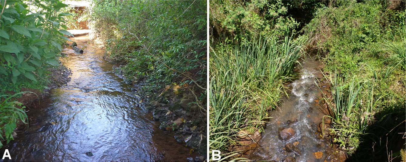
Distribution. A single paratype (MZUSP 82372) is from the Tibaji River basin, a tributary of the Paranapanema River ( Fig. 3 View FIGURE 3 ; see Discussion below). All remaining type series is from two sub-basins of the Uruguai River system, the Ijuí and Passo Fundo drainages ( Fig. 3 View FIGURE 3 ). The Paranapanema is an affluent of the Paraná River, which joins the Uruguai to form the La Plata River/Estuary.
Habitat notes. Ecological data from all localities is unavailable. Most types were collected in clear water streams with 1 to 3 m wide, with bottoms composed of stone, gravel, and sand ( Fig. 4 View FIGURE 4 ). Streamlets were usually surrounded by modified riparian vegetation.

No known copyright restrictions apply. See Agosti, D., Egloff, W., 2009. Taxonomic information exchange and copyright: the Plazi approach. BMC Research Notes 2009, 2:53 for further explanation.
|
Kingdom |
|
|
Phylum |
|
|
Class |
|
|
Order |
|
|
Family |
|
|
Genus |
Trichomycterus perkos
| Datovo, Aléssio, Carvalho, Murilo & Ferrer, Juliano 2012 |
Trichomycterus aff. itatiayae: Kantek et al. (2007 : 797 )
| Kantek 2007: 797 |
Trichomycterus
| Torres 2004: 124 |
Trichomycterus
| Sato 2004: 45 |