
Dendronotus gracilis Baba, 1949
|
publication ID |
https://doi.org/ 10.11646/zootaxa.1960.1.2 |
|
DOI |
https://doi.org/10.5281/zenodo.5242532 |
|
persistent identifier |
https://treatment.plazi.org/id/087D7947-FF91-FFF6-1A86-23E88AADFC5A |
|
treatment provided by |
Felipe |
|
scientific name |
Dendronotus gracilis Baba, 1949 |
| status |
|
Dendronotus gracilis Baba, 1949 View in CoL
( Figures 1E–F View FIGURE 1 , 6C–G View FIGURE 6 )
Material Examined: CASIZ 174950 , Japan, Okinawa, H/S, 3 January 1985, 69 m depth, collected by R. F. Bolland .
Distribution: Thus far, known from Okinawa (present study), Sagami Bay, Japan ( Baba 1949) and New Zealand (Miller pers. comm. in Robilliard 1970).
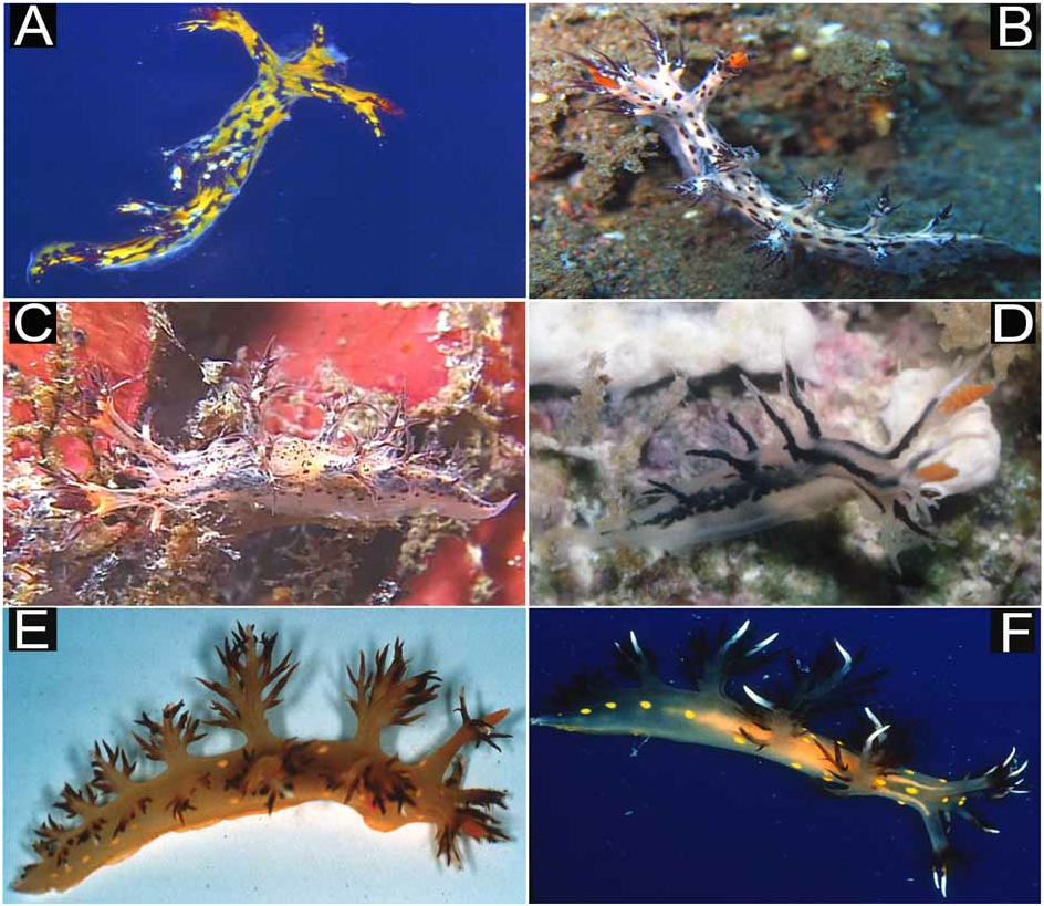
External morphology ( Fig. 1E–F View FIGURE 1 ): The general body shape is limaciform, delicate, and laterally compressed with a short pointed posterior end of the foot. The foot is narrow, rounded along the anterior margin, not sharply marked off from the body, and tapers rapidly to the posterior end of the foot. The lateral margin is not distinct from the dorsum other than for the line of dorsolateral processes. The body surface is smooth and the cardiac elevation is not prominent. The head is rounded with the frontal veil well defined, bearing three branched processes on either side of the mouth. Of these, the two most medial processes are the largest and most branched. The two more lateral processes are shorter but also have several branches. Along the inner end of the veil there is another smaller and simpler papilla. The large, stout rhinophores are inclined anteriorly and are as tall as the first pair of dorsolateral processes. The clavus is conical and perfoliate with about twelve lamellae and is capable of retracting within the campanulate sheath. The sheath bears three marginal processes; the two posterior processes are larger and simple or with few small branches. The anterior process has three branches that are shorter than the posterior ones. There are eight pairs of elaborately arborescent dorsolateral processes along the margins of the back. The distance between pairs decreases towards the posterior end of the foot, as well as the size and number of branches of each dorsolateral process. The dorsolateral processes have a main stalk that extends into at least four elongate papillae, each of which bears a number of shorter subdivisions. The anal opening is along the dorsolateral line about midway between the first and second dorsolateral processes on the right side. The small renal pore is located just above the anal elevation. The reproductive openings are on the right side below the dorsolateral line about midway between the rhinophore and the first dorsolateral process.
The background color of the animal varies from white-translucent ( Fig. 1E View FIGURE 1 ) to orange-brown ( Fig. 1F View FIGURE 1 ). The largest branches of the rhinophoral sheath papillae as well as those of the main branches of each dorsolateral process and the oral tentacles are of the same color as the body but the tips are opaque white. The remaining shorter branches are dark brown. There are a series of small bright yellow spots over the surface forming a line on either side of the body and also several spots in the mid-dorsal line and the frontal velar. The rhinophores are orange.
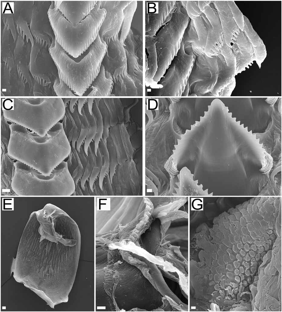
Alimentary Canal: The single specimen was very poorly preserved. None of the internal features could be examined since the internal organs were completely dry and destroyed. Only the buccal bulb and the protruded penis were studied. The radular formula is 37 x 8.1.8. The median tooth is large and strong with its overall length and width about equal ( Fig. 6C View FIGURE 6 ). The dorsal surface is prolonged into a bluntly triangular convex cusp. The lateral margins bear 16 to 19 denticles, similar in size along the margins ( Fig. 6C–D View FIGURE 6 ). The cusp of the preceding median tooth sits in a deep depression of the median basal margin of each median tooth ( Fig. 5C View FIGURE 5 ). There are eight thin lateral teeth on each side with a flat rectangular base. The inner posterior margin is thickened in the innermost six teeth and elongates into a slightly curved cusp at the inner angle. This cusp is longest and strongest in the forth and fifth lateral teeth. Along the outer margin of this cusp there are six to eight sharp denticles increasing in size towards the base of the tooth. The outermost two laterals have a rudimentary or absent cusp ( Fig. 6C View FIGURE 6 ). The mandibles are concave and oval in shape ( Fig. 6E View FIGURE 6 ). The masticatory process begins a short distance below the hinge as a slight elevation terminating in short, low, parallel ridges on the outer face of the process. These gradually increase in size to become well-marked ridge-like denticles ( Fig. 6F View FIGURE 6 ). The lip disk is small and covered by a thin and delicate cuticle with small rodlets on it ( Fig. 6G View FIGURE 6 ). The inner side of this cuticle is continuous with the masticatory process of the mandibles.
Due to poor preservation of this specimen, the only part of the reproductive system that could be examined was the penis because it was extended out of the body wall at the time of preservation. The penis is unarmed, elongate and truncated.
Remarks: Dendronotus gracilis was described from Sagami Bay, Japan, where it was found at a depth of 160 m deep. Sagami Bay is located approximately 1,500 kilometers northeast of Okinawa, Japan. Only external features, the radula, and the jaws were described by Baba (1949). All those features are similar to those of the specimen here described, with a few exceptions. The original specimen of D. gracilis has only four velar processes instead of six. The placement of the spots along the dorsum is irregular in Baba’s specimen instead of in a straight line, although Robilliard (1970) found that two specimens of D. gracilis collected in New Zealand by Dr. Michael Miller also had the spots arranged in a linear fashion. Miller’s specimens were found in dredging from 9 to 24 meters. The radular formula of the specimen described by Baba (1949) is 41 × 8.1.8, which is very similar to the 37 × 8.1.8 formula of our specimen. All of these differences could be due to intraspecific variation, especially since only a few specimens have ever been studied. An interesting note that also supports the identification of this animal as D. gracilis Baba, 1949 is that they both have simple, digitiform processes on the rhinophore sheaths, which is an uncommon trait seen within this genus ( Robilliard 1970, Valdés & Bouchet 1998).
We conclude that our specimen is D. gracilis . This finding increases the range known for the species from temperate to tropical Japan. However, this specimen from Okinawa was collected at a considerable depth (69 m), where waters are colder than near the surface. New specimens from New Zealand should be found and compared with material from Japan to help clarifying the taxonomy of this apparently widely distributed species.

| R |
Departamento de Geologia, Universidad de Chile |
No known copyright restrictions apply. See Agosti, D., Egloff, W., 2009. Taxonomic information exchange and copyright: the Plazi approach. BMC Research Notes 2009, 2:53 for further explanation.