
Paracatonidia, Long, Jian-Kun, Yang, Lin & Chen, Xiang-Sheng, 2015
|
publication ID |
https://doi.org/ 10.11646/zootaxa.4052.2.2 |
|
publication LSID |
lsid:zoobank.org:pub:EEE19165-7EAD-4F71-A776-485CCC969653 |
|
DOI |
https://doi.org/10.5281/zenodo.6121783 |
|
persistent identifier |
https://treatment.plazi.org/id/072787B5-BB6F-926E-FF10-FE78FCFFF8E4 |
|
treatment provided by |
Plazi |
|
scientific name |
Paracatonidia |
| status |
gen. nov. |
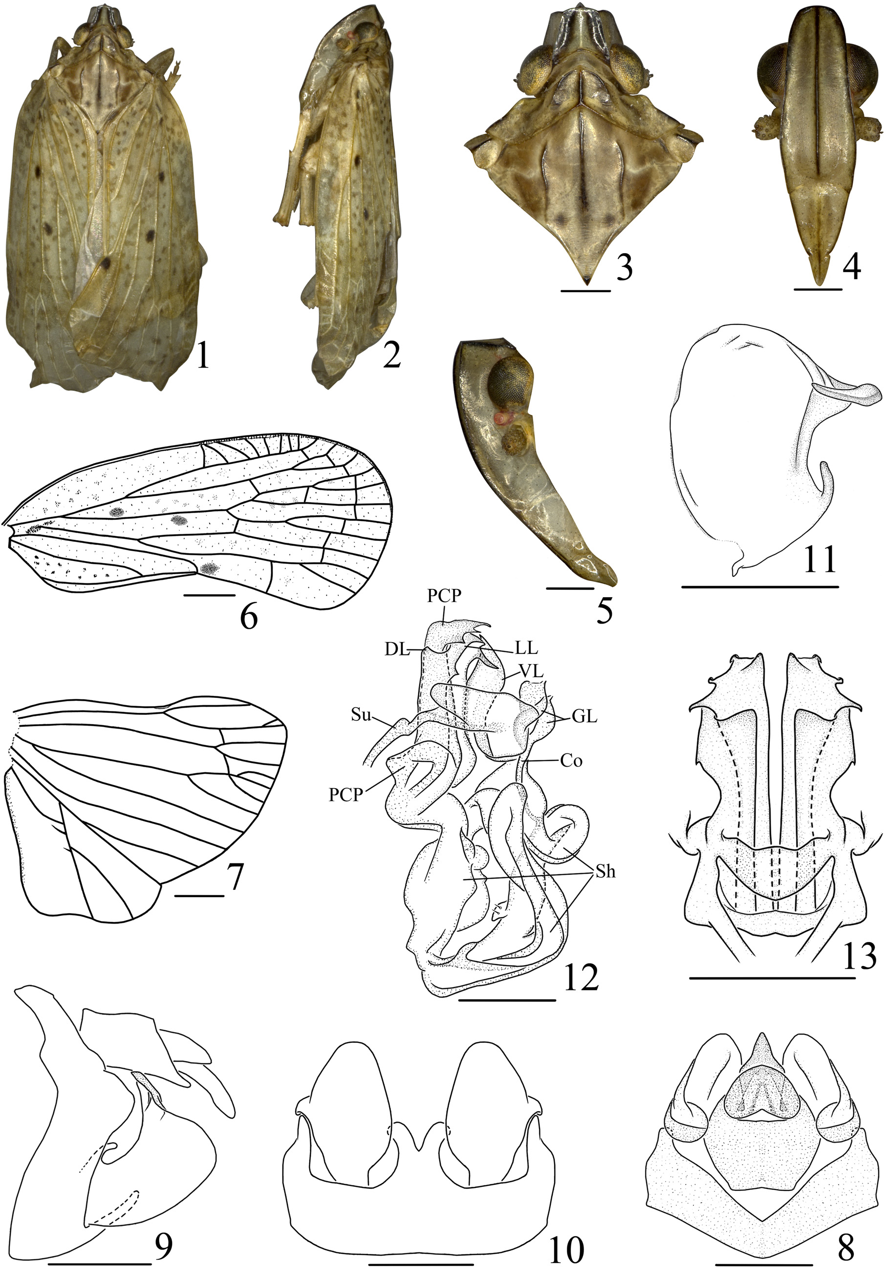
Genus Paracatonidia View in CoL View at ENA gen. nov.
( Figs 1–13 View FIGURES 1 – 13 )
Type species. Paracatonidia webbeda sp. nov., here designated.
Description. Width of head with eyes less than two-thirds width of pronotum. Vertex with disc slightly depressed, median carina protuberant, obsolete apically, anterior margin carinate, subtruncate, lateral margins carinate and straight, diverging basad, posterior margin broadly concave ( Fig. 3 View FIGURES 1 – 13 ). Frons slightly convex in lateral view ( Fig. 5 View FIGURES 1 – 13 ), upper margin roundly concave, median carina percurrent, lateral margin carinate, sinuately diverging to below level of antennae then gradually incurved to suture ( Fig. 4 View FIGURES 1 – 13 ). Clypeus with median and lateral carinae distinct ( Fig. 4 View FIGURES 1 – 13 ). Rostrum slightly exceeding base of post-trochanter, with length of subapical segment equal to apical segment. Antenna subglobose, not sunk in a depression ( Fig. 5 View FIGURES 1 – 13 ). Ocelli separated from eyes ( Fig. 5 View FIGURES 1 – 13 ). Eyes excavate beneath ( Fig. 5 View FIGURES 1 – 13 ), not overlapping pronotum in dorsal view ( Fig. 3 View FIGURES 1 – 13 ). Pronotum with length in midline as long as length behind eyes, anterior margin of disc roundly convex, posterior margin subangulately excavate about 115 degrees; median carina distinct, lateral carinae diverging posteriorly, not reaching hind margin; lateral lobe with a longitudinal carina between eye and tegula ( Fig. 3 View FIGURES 1 – 13 ). Tegula with a longitudinal carina in middle ( Fig. 3 View FIGURES 1 – 13 ). Mesonotum with three carinae more or less obsolete ( Fig. 3 View FIGURES 1 – 13 ). Forewing with costal margin slightly convex; apical margin roundly convex; posterior margin angulately excavate (155 degrees) at apex of clavus; vein Sc+R forking slightly basad of Cu1 forking; vein Sc with several branches, directed towards anterior margin; veins R and M with two and five apical branches, respectively; vein Cu1 forking slightly distad of Sc+R fork, equal to level of union of claval veins, with Cu1b two-branched; clavus terminating at middle of forewing ( Fig. 6 View FIGURES 1 – 13 ). Hindwing with two, two and three branches of veins R, M and Cu1, respectively; vein A2 with blind branches ( Fig. 7 View FIGURES 1 – 13 ). Post-tibiae with one lateral spine.
Male genitalia. Anal style distinctly exceeding apical margin of anal segment ( Fig. 8 View FIGURES 1 – 13 ). Pygofer in lateral view with dorsal margin distinctly shorter than ventral margin ( Fig. 9 View FIGURES 1 – 13 ), medioventral process divided into two branches ( Fig. 10 View FIGURES 1 – 13 ). Genital style with a stout, ear-like process and a finger-like process rising from apical third and near base of dorsal margin, respectively ( Fig. 11 View FIGURES 1 – 13 ). Aedeagus with phallobase sheath-shaped, two dorsal, two lateral and one ventral lobe with anterior portions connected together, protruding anteriorly into body cavity ( Figs 12–13 View FIGURES 1 – 13 ). Suspensoria suspended phallobase with dorsolateral portions of pygofer. Genital lamina sclerotized ( Fig. 12 View FIGURES 1 – 13 ). Phallobasal conjunctival processes exceeding apical margin of phallobase, with apex broadly webbed like a duck foot ( Figs 12–13 View FIGURES 1 – 13 ). Sheath extremely developed, broad, twisted and membranous ( Fig. 12 View FIGURES 1 – 13 ). Connective relatively stout and short ( Fig. 12 View FIGURES 1 – 13 ).
Etymology. The genus name, which is masculine, is a combination of “para-” (similar to) and “ Catonidia ” (name of the related genus), and indicates the new genus is similar to the genus Catonidia Uhler.
Host plant. Unknown.
Distribution. Oriental region (southwestern China).
Diagnosis. The genus is readily distinguished from other known genera in the tribe Achilini by the tegula having a longitudinal carina in middle ( Fig. 3 View FIGURES 1 – 13 ), the medioventral process of the pygofer having two branches ( Fig. 10 View FIGURES 1 – 13 ), the phallobasal conjunctival process with apex broadly webbed duck-foot like ( Figs 12–13 View FIGURES 1 – 13 ), and the sheath extremely developed, broad, twisted and membranous ( Fig. 12 View FIGURES 1 – 13 ).
No known copyright restrictions apply. See Agosti, D., Egloff, W., 2009. Taxonomic information exchange and copyright: the Plazi approach. BMC Research Notes 2009, 2:53 for further explanation.