
Ctenocolum triangulatus Albuquerque & Ribeiro-Costa
|
publication ID |
https://doi.org/ 10.11646/zootaxa.3838.1.1 |
|
publication LSID |
lsid:zoobank.org:pub:1534C775-D28D-470F-9AEC-8BABB3D8FA56 |
|
DOI |
https://doi.org/10.5281/zenodo.6124249 |
|
persistent identifier |
https://treatment.plazi.org/id/03FF87F5-FFC8-FFDF-38AD-FF75FD0E76D7 |
|
treatment provided by |
Plazi |
|
scientific name |
Ctenocolum triangulatus Albuquerque & Ribeiro-Costa |
| status |
sp. nov. |
Ctenocolum triangulatus Albuquerque & Ribeiro-Costa sp. nov.
( Figs. 17 View FIGURES 17 – 21 , 30 View FIGURES 22 – 30 , 42 View FIGURES 35 – 43 , 61 View FIGURES 61 – 65 , 74 View FIGURES 72 – 76 , 86 View FIGURES 85 – 90 , 98 View FIGURES 91 – 99 )
Type material. Holotype deposited in ZMHB, male: “53758” [white label printed in black]; “misotur/ m.” [white label handwritten in black]; “Hist. -Coll. ( Coleoptera )/ Nr. 53758/ Bruchus / aequinoctialis Erichs./ Columb., Moritz./ Zool. Mus. Berlin” [green label with black margin, printed in black]; “ Ctenocolum / SP/ det./ J. M.Kingsolver” [white label handwritten, printed in black]; “♂” [white label printed in black];]; “ HOLOTYPE / Ctenocolum triangulatus / Det. Albuquerque & Ribeiro-Costa” [white label with red margin, printed in black].
Diagnosis. Ctenocolum triangulatus Albuquerque & Ribeiro-Costa sp. nov. is different from C. milelo Albuquerque & Ribeiro-Costa sp. nov. and C. podagricus by having ocular index 4.5 and pygidium oval in both sex; differs from C. martiale by elytral striae 3 and 4 with less conspicuous teeth at base. This species differs from all others by the male genitalia at submedian region with squamous subtriangular-shape sclerite ( Fig. 86 View FIGURES 85 – 90 ).
Description. BL: 2.4 mm; BW: 1.5 mm.
Integument. Dorsum reddish brown and dark brown. Antenna dark brown, first 3 antennomeres paler ( Fig. 61 View FIGURES 61 – 65 ). Pygidium reddish brown and dark brown.Ventral region reddish brown and black. Front and middle femur and tibia brown; hind femur brown, reddish brown to dark brown.
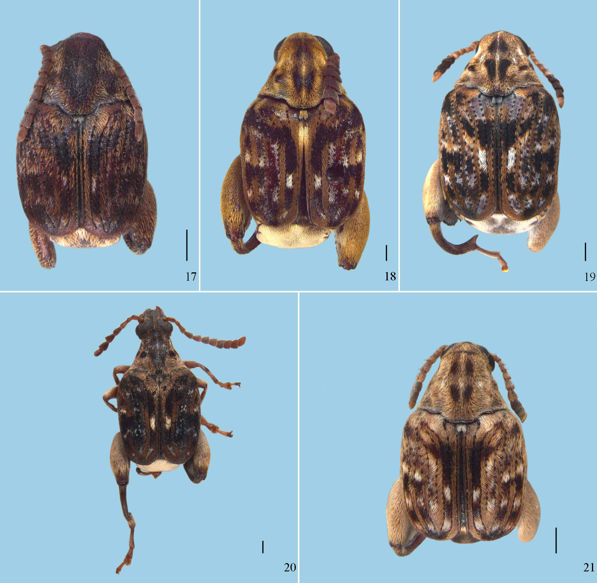
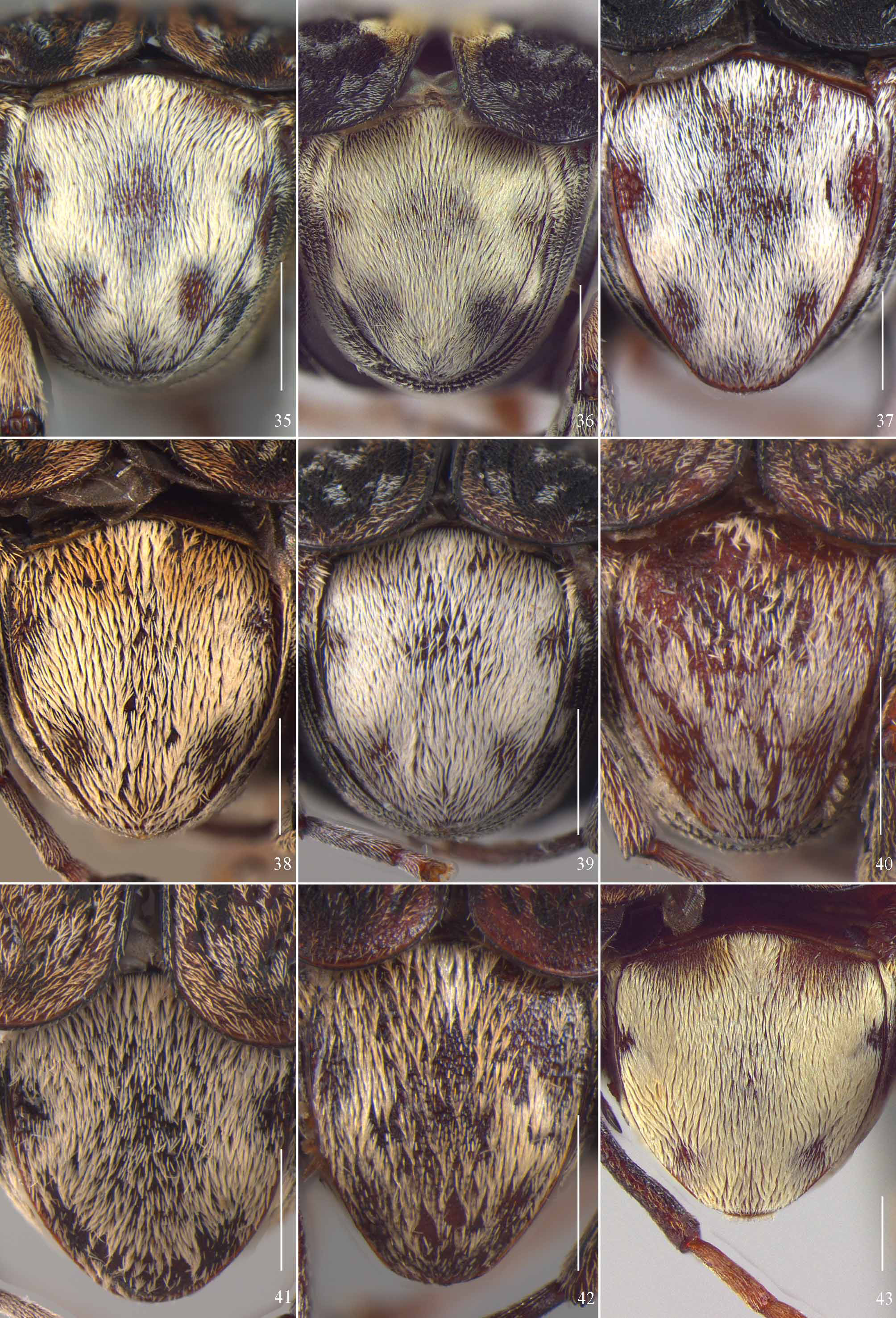
Pubescence. Pronotum reddish brown and white; sparse setae exposing the integument forming an oval, wide area from anterior to posterior region and on each lateral region one small area ( Fig. 17 View FIGURES 17 – 21 ). Elytra slightly variegated, reddish brown and black setae; interstria 3 without white setae at base and at submedian region with short sparse strip of white setae ( Fig. 17 View FIGURES 17 – 21 ). Pygidium dense, white and yellowish gray in a spotted pattern ( Fig. 42 View FIGURES 35 – 43 ). Ventral region with setae yellowish gray, brown and white ( Fig. 61 View FIGURES 61 – 65 ).
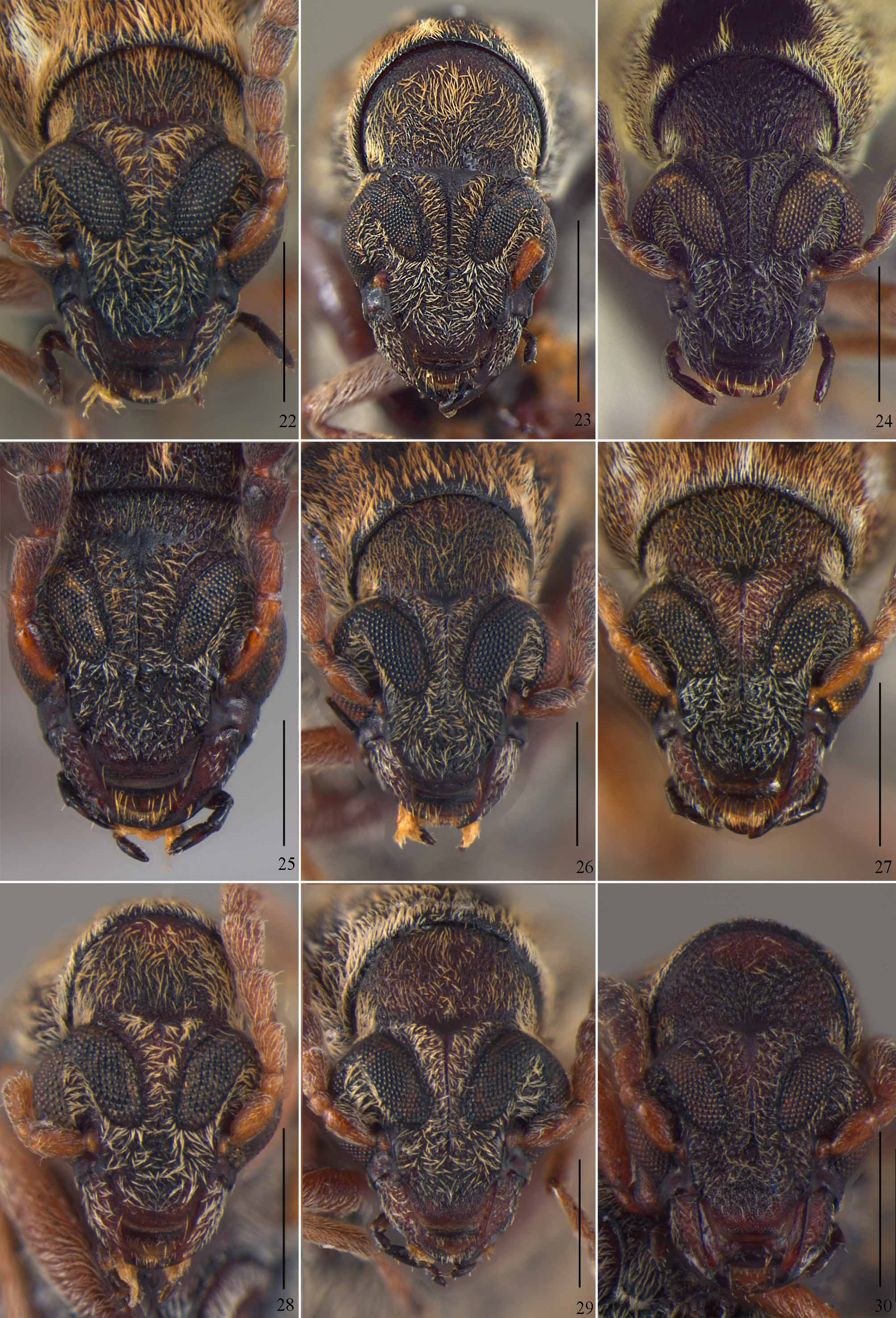
Head. Ocular sinus 0.2 mm; ocular index 4.5; length of eyes in frontal view behind sinus 0.09 mm ( Fig. 30 View FIGURES 22 – 30 ). Antenna serrate from antennomere 4-10 ( Fig. 61 View FIGURES 61 – 65 ). Frons with frontal carina ( Fig. 30 View FIGURES 22 – 30 ).
Prothorax. Pronotum with median gibbosity slightly elevated, not divided by longitudinal and transversal sulcus ( Fig. 61 View FIGURES 61 – 65 ); lateral gibbosity slightly elevated; basal lobe with depression and slightly emarginated ( Fig. 17 View FIGURES 17 – 21 ).
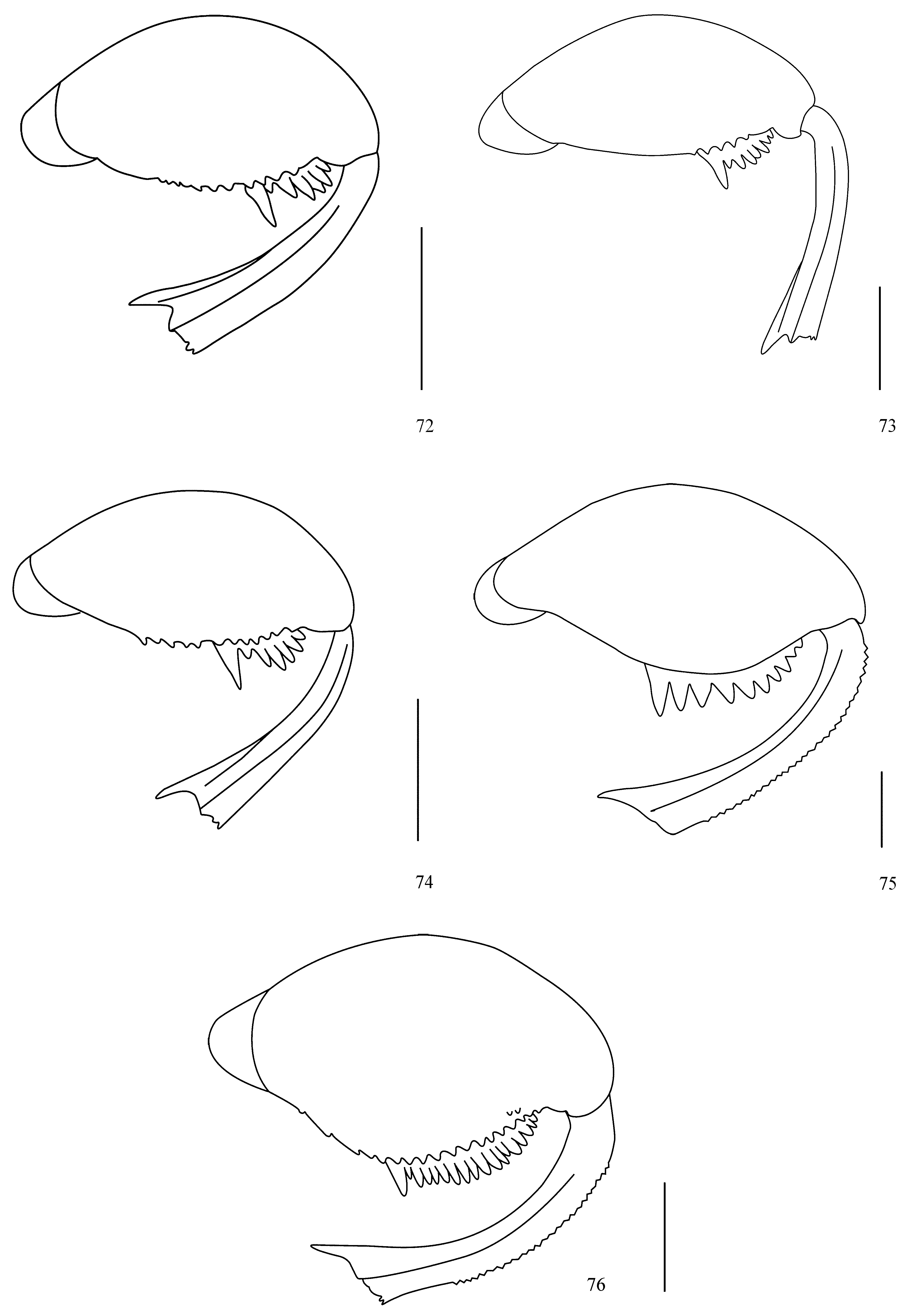
Mesothorax and metathorax. Elytra, striae with punctures moderately impressed; less conspicuous teeth at base of striae 3 and 4; tooth of stria 4 closer to base of tooth of stria 3 than to anterior margin of elytra; stria 6 conspicuously impressed. Hind femur ( Fig. 74 View FIGURES 72 – 76 ) on external ventral margin with toothed carina; without denticles above of external ventral margin; pecten with 8 teeth. Hind tibia ( Fig. 74 View FIGURES 72 – 76 ) strongly emarginated beside mucro; lateral coronal denticles present.
Abdomen. Pygidium longer than wide, oval, at median basal region with moderately impressed punctures ( Fig. 42 View FIGURES 35 – 43 ).
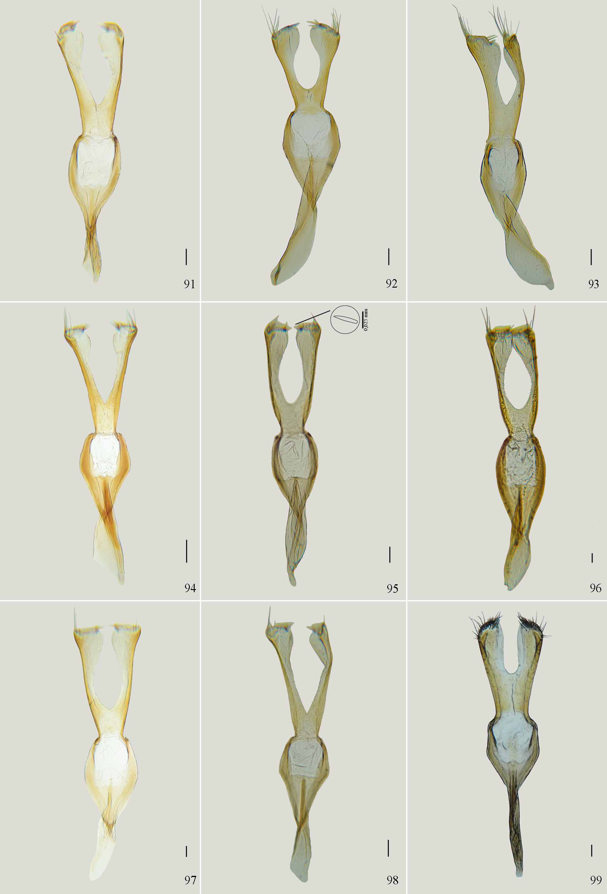
Male genitalia. Median lobe, ventral valve as long as wide, lateral margin concave on subapical region ( Fig. 86 View FIGURES 85 – 90 ), basal margin not emarginated. Internal sac, lateral apex with short tuft of setae, hinge sclerite with curved apex, long, extending through subapical region; subapical region with homogeneously distributed spicules, dense medially and in lateral forming elongated strips; median region with homogeneously distributed spicules; submedian region with squamous subtriangular- shape sclerite ( Fig. 86 View FIGURES 85 – 90 ); basal region with spicules. Tegmen ( Fig. 98 View FIGURES 91 – 99 ), lateral lobes separated by emargination about 0.85 times the length of lateral lobes; internal margin near end of emargination straight, forming a “V”; expanded at apex, about 2.5 times the smallest width on median region; without membranous projection at apex.
Note. The female is unknown.
Etymology. The specific name “ triangulatus ” refers to the squamous subtriangular-shaped sclerite of male genitalia.
Distribution. Neotropical region: Colombia.


No known copyright restrictions apply. See Agosti, D., Egloff, W., 2009. Taxonomic information exchange and copyright: the Plazi approach. BMC Research Notes 2009, 2:53 for further explanation.
|
Kingdom |
|
|
Phylum |
|
|
Class |
|
|
Order |
|
|
Family |
|
|
SubFamily |
Bruchinae |
|
Genus |