
Opistognathus solorensis Bleeker, 1853
|
publication ID |
https://doi.org/ 10.12782/specdiv.23.233 |
|
persistent identifier |
https://treatment.plazi.org/id/03FF87D6-E849-FFC6-20E2-FC4BFEF9D971 |
|
treatment provided by |
Felipe |
|
scientific name |
Opistognathus solorensis Bleeker, 1853 |
| status |
|
Opistognathus solorensis Bleeker, 1853 View in CoL
[New standard Japanese name: Hoshikage-ago-amadai] ( Figs 1–3 View Fig View Fig View Fig ; Table 1)
Opistognathus solorensis Bleeker, 1853: 81 View in CoL (Solor Island, Indonesia); Bleeker 1874: 471, fig. 3 (Solor, Amboina, and Goram Islands, Indonesia); Allen et al. 2003: 298, unnumbered fig. (underwater photograph); Allen and Erdmann 2012: 355, unnumbered fig. ( Brunei); Smith- Vaniz 2016: 284, figs 5–9 ( Palau, Papua New Guinea, Timor Leste, Indonesia, Brunei, Philippines, and Taiwan).
Opistognathus sp. 1 : Motomura and Harazaki 2017, 36, pl. 3E (Yaku-shima island, Japan).
Materials examined. 6 specimens, 48.1–75.1 mm SL, all from the Osumi Islands , Kagoshima Prefecture, southern Japan . KAUM –I. 62138, 70.5 mm SL, off Urata , Nishinoomote, Tanega-shima island, 30°49′36″N, 131°02′11″E, 10–15 m, S GoogleMaps . Tashiro , 13 June 2014; KAUM –I . 67972, 48.1 mm SL, off Isso , Yaku-shima island, 30°27′N, 130°29′E, 5–13m, S GoogleMaps . Tashiro , 25 December 2014; KAUM –I . 68000, 75.1 mm SL, off Isso , Yaku-shima island, 30°27′45″N, 130°29′40″E, 10–15 m, Y GoogleMaps . Kanade , 26 December 2014; KAUM –I . 95905, 59.2 mm SL, KAUM –I. 95906, 59.6 mm SL, KAUM –I. 95907, 60.7 mm SL, Katatomari Port , Kuroshima island, 30°49′20″N, 129°54′26″E, 25 m, D GoogleMaps . Uyeno , 22 November 2016 .
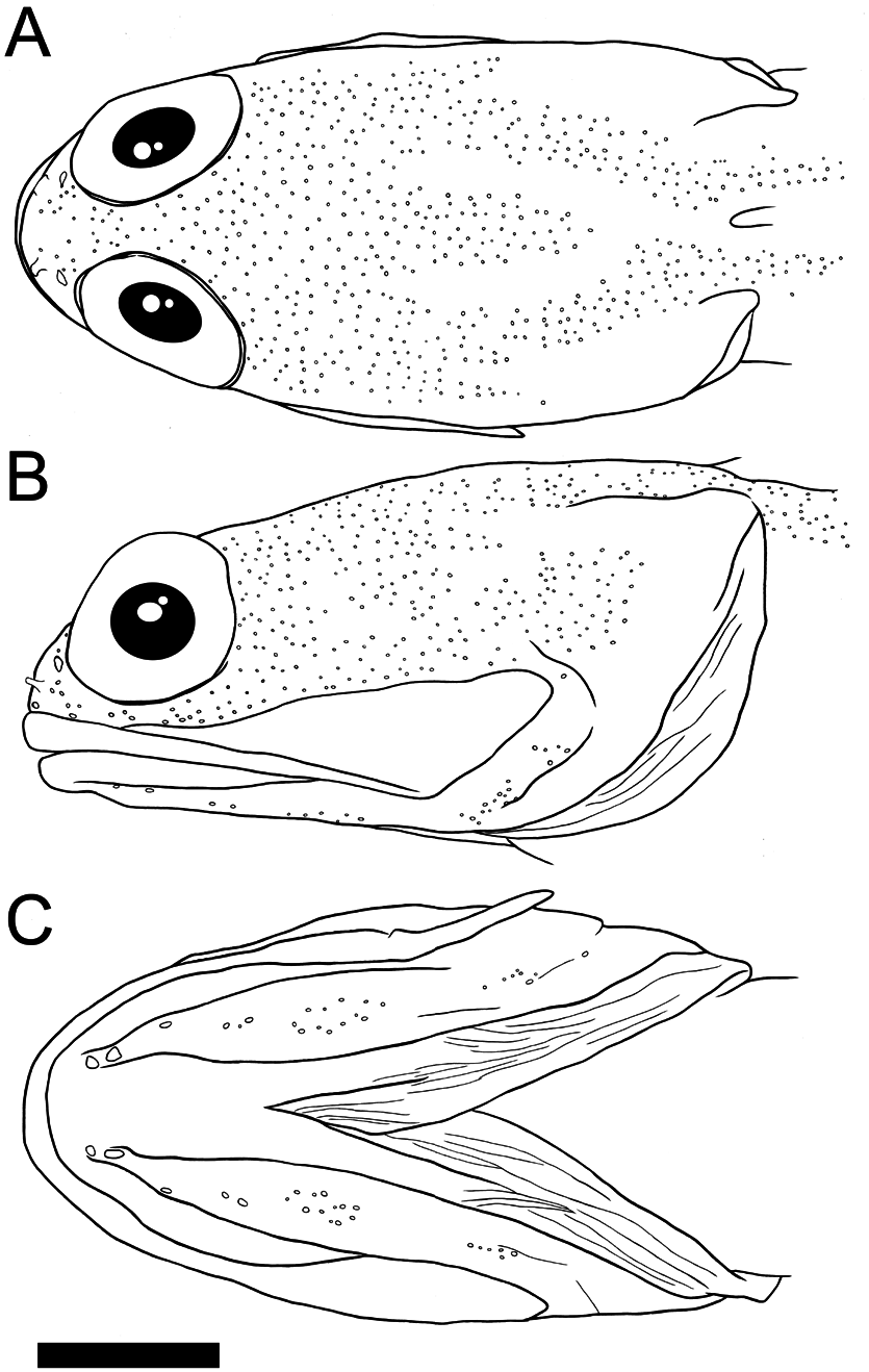
Description. Counts and measurements are given in Table 1. Cephalic sensory pores are illustrated in Fig. 2 View Fig .
Body elongate, compressed anteriorly, progressively more compressed posteriorly. Head and body scaled, except for pre-dorsal area above lateral-line, pectoral-fin base and chest. Anterior nostril a short membranous tube with a tiny tentacle on posterior rim, when depressed not reaching posterior nostril; situated about mid-way between posterior nostril and dorsal margin of upper lip (or slightly closer to posterior nostril). Posterior nostril opening elliptical. Posterior margin of preopercle indistinct, covered with skin, without free margin. Posterior upper jaw elongate, produced posteriorly as a thin flexible lamina; posterior margin of maxilla not reaching preopercle; supramaxilla elongate horizontally. Cephalic sensory pores relatively numerous ( Fig. 2 View Fig ). Mandibular pore positions 1 and 2 each with a single large pore; position 3 with a single pore, its size slightly smaller than those of anterior positions; positions 4 and 5 with 2–3 and 6–12 pores, respectively ( Fig. 2C View Fig ). Lateral-line pores arranged in two rows, with small sparsely distributed pores between. Lateral-line ending below third to fifth segmented dorsal-fin rays.
Premaxilla with an outer row of similarly-sized conical teeth; 1–6 irregular rows of smaller inner teeth anteriorly. Dentary with an outer row of conical teeth, of similar size to premaxillary teeth; 1–6 irregular inner rows of smaller conical teeth anteriorly. No teeth on vomer.
Dorsal fin moderately low anteriorly, profile relatively uniform with a very slight change in fin height at junction of spinous and segmented rays; all dorsal rays branched distally. Anal-fin origin vertically level with base of last dorsal-fin spine; all fin rays branched distally. Pelvic-fin origin anterior to vertical through base of first spine of first dorsal fin; first ray of pelvic-fin robust, not tightly bound to second ray; membrane between first and second rays incised distally; second ray longest, innermost 3 rays branched distally. Pectoral-fin base below 2nd and 3rd dorsal-fin spine bases. Caudal fin margin rounded.
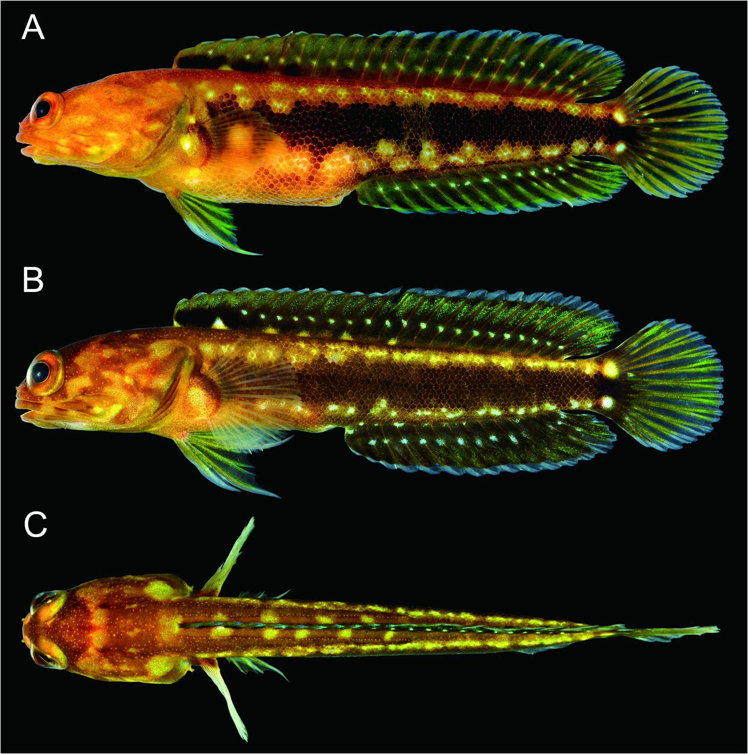
Fresh color ( Fig. 1 View Fig ). Head and body yellowish to brown; lateral and dorsal surfaces of head mottled with pale yellowwhite blotches. Lateral surface of body with broad darkbrown to blackish band extending from posterior pectoralfin base to basal part of caudal fin; bright white blotches scattered along with upper and lower sides of band. Anterior basal part of dorsal fin membrane with one or two (one of six specimens present three) blackish, marginally indistinct blotches from the 1–8th dorsal-fin spines; anterior half of dorsal fin base with 2–6 whitish blotches; basal half of fin darker, distal half greenish-yellow, except for translucent distal margin; middle of fin with several bright whitish spots in two horizontal dotted lines (distal line sometimes absent); basal line extending from 4th spine to posteriormost ray, distal line from 5th spine to posteriormost ray; spots on spinous and anterior soft-rayed portion of fin pale, indistinct. Pectoral-fin base with a pale blotch; reddish-brown stripes along lower fin margin; rays pale yellow, membrane translucent. Pelvic-fin rays greenish-yellow, membranes dusky; rays and membranes distally translucent. Basal part of anal fin darker; remainder of fin greenish-yellow, except for translucent distal margin; irregular whitish spots on anal-fin base; middle of fin with several bright whitish spots in two horizontal dotted lines; basal line extending from first to posteriormost rays; distal line pale, indistinct anteriorly. Caudal fin base blackish with paired bright whitish blotches; rays greenish-yellow, membranes dusky; rays and membranes distally translucent. Oral areas (posterior regions of upper and lower pharyngeal plates) with blackish pigmentation.
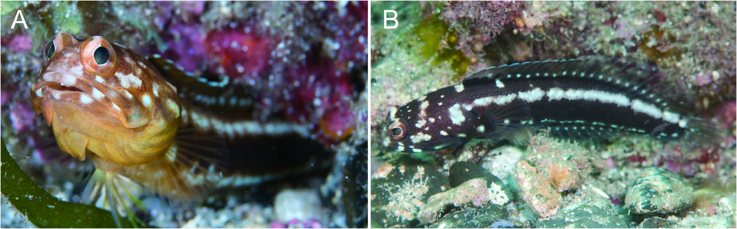
Life color ( Fig. 3 View Fig ). Body blackish to yellow. Bright white mottled blotches on upper and lower jaws, snout, eye, and lateral and dorsal surfaces of head. All banded and blotched patterns similar to those of fresh color, but brighter and more defined.
Distribution. Opistognathus solorensis occurs in the western Pacific from Palau, Papua New Guinea, Timor Leste, Indonesia, Brunei, the Philippines, Taiwan ( Bleeker 1874; Allen and Erdmann 2012; Smith-Vaniz 2016) and Japan (Osumi Islands). In the latter group, the species was collected from Tanega-shima, Yaku-shima, and Kuro-shima islands in depths from 5–25 m (this study).
Remarks. The six specimens of Opistognathus solorensis from Osumi Islands agree well with the species diagnosis given by Smith-Vaniz (2016) (except for the three blackish blotches anteriorly on the dorsal fin): dorsal-fin rays XI, 14; anal-fin rays III, 14; 64–68 longitudinal scale rows; lateral line ending below 3–4th dorsal-fin soft ray; broad areas above and below esophageal opening with blackish pigmentation; posterior part of maxilla with flexible lamina; and inner lining of flexible part of upper jaw, with two blackish stripes.
Bleeker (1874) described O. solorensis as having 1–3 blotches anteriorly on the dorsal fin (based on 8 specimens from Indonesia), but Smith-Vaniz (2016) subsequently noted only one or two blotches in the species, regarding Bleeker’s (1874) count of three as probably erroneous. However, specimen KAUM–I. 62138, 70.5 mm SL had three such blotches ( Fig. 1A View Fig ), extending the degree of intraspecific variation within the species.
Smith-Vaniz (2016) defined the Opistognathus solorensis species group as having the upper jaws with a flexible lamina posteriorly; maxilla widest before the end and weakly pointed posteriorly; supramaxilla relatively elongate; dorsalfin rays XI, 14; anal-fin rays III, 14; all dorsal and anal fin soft-rays branched distally; vertebrae 10+18; vomer without teeth; and 44–69 longitudinal scale rows. In addition to O. solorensis , the species group includes Opistognathus ensiferus Smith-Vaniz, 2016 and Opistognathus verecundus Smith- Vaniz, 2004.
Opistognathus solorensis is distinguished from O. ensiferus by having a shorter lateral line, ending below 1–4th (3rd or 4th in this study) dorsal-fin soft rays (vs. 6–7th in O. ensiferus ); inner lining of flexible part of upper jaw with two blackish stripes (vs. single stripe); and areas above and below esophageal opening with blackish pigmentation (vs. without pigmentation) ( Smith-Vaniz 2016; this study). Opistognathus solorensis is also distinguished from O. verecundus by having 58–69 longitudinal scale rows (vs. 45–55 in O. verecundus ); 27–33 total gill rakers (vs. 23–26); and 1–3 (rarely 3) blotches anteriorly on the dorsal fin (vs. blotches absent) ( Smith-Vaniz 2004, 2016; this study).
Opistognathus solorensis was originally described by Bleeker (1853) from Solor Island , Indonesia . The species was redescribed by Smith-Vaniz (2016), based on 102 specimens from the western Pacific (see “distribution”), with designation of a neotype, and a northernmost record from southern Taiwan . The present Osumi Islands specimens represent the first records of the species in Japanese waters, the single specimen from Tanega-shima island being the northernmost known . In fact, the species has already been noted (as Opistognathus sp. 1 ) in the Yaku-shima island fish species checklist ( Motomura and Harazaki 2017) .
A new standard Japanese name “Hoshikage-ago-amadai” is herein proposed for O. solorensis , based on the single specimen (KAUM–I. 62138) from Tanega-shima island, Osumi Islands. “Hoshikage” means starlight in Japanese, referring to the bright white spots and blotches on the body and fins, and “ago-amadai”, a fish of the family Opistognathidae .
| KAUM |
Kagoshima University Museum |
No known copyright restrictions apply. See Agosti, D., Egloff, W., 2009. Taxonomic information exchange and copyright: the Plazi approach. BMC Research Notes 2009, 2:53 for further explanation.
|
Kingdom |
|
|
Phylum |
|
|
Class |
|
|
Order |
|
|
Family |
|
|
Genus |
Opistognathus solorensis Bleeker, 1853
| Tashiro, Satokuni, Uyeno, Daisuke & Motomura, Hiroyuki 2018 |
Opistognathus solorensis
| Allen, G. R. & Erdmann, M. V. 2012: 355 |
| Allen, G. R. & Steene, R. & Humann, P. & DeLoach, N. 2003: 298 |
| Bleeker, P. 1874: 471 |
| Bleeker, P. 1853: 81 |