
Echinothrix diadema (Linnaeus, 1758)
|
publication ID |
https://doi.org/ 10.5281/zenodo.5401706 |
|
persistent identifier |
https://treatment.plazi.org/id/03FE87C2-070F-FFD1-FD3A-FA69FEC2A9B8 |
|
treatment provided by |
Marcus |
|
scientific name |
Echinothrix diadema (Linnaeus, 1758) |
| status |
|
Echinothrix diadema (Linnaeus, 1758) View in CoL
Specimens were collected from Malmahera ( Indonesia), Haunomu Bay, Kahe Point (Hawaii), Bougainville Is. ( Papua New Guinea), Mariana Is. ( Guam), Ignoitijala ( Maldives), Luzon ( Philippines), Dravuni Is., Taveuni Is., Yanuca Is., Suva ( Fiji), and Nouméa ( New Caledonia).
Th e tests were black with a blue-green sheen in young specimens. Th is feature was lost with age. Juveniles had very diff erent colouration to the adults. However, this was mainly associated with the spines as the test remained black.
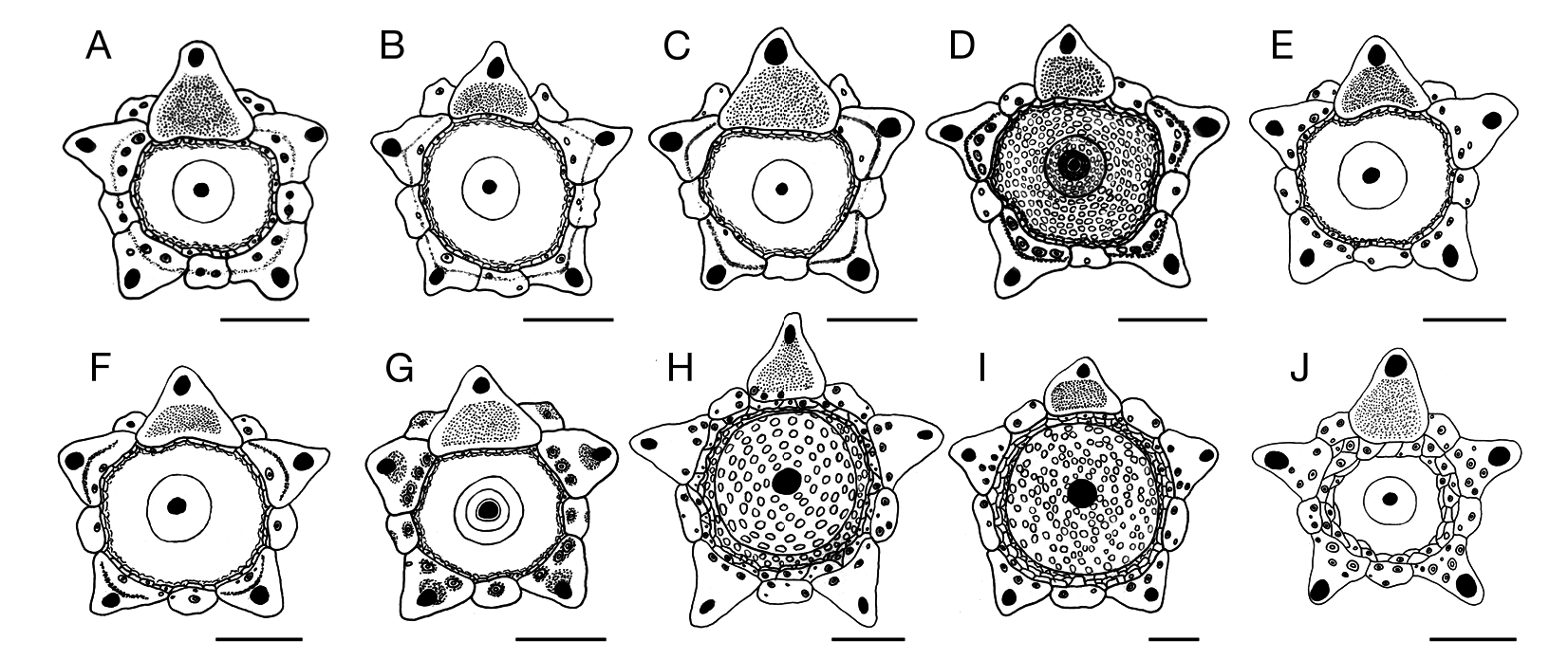
Adult specimens had mean test diameters of 92 mm (h.d.) (SD ± 6.3 mm) and 55 mm (v.d.) (SD ± 4.4 mm). Th e maximum test diameters recorded for this species in this study were 110 mm (h.d.) by 61 mm (v.d.) from 200 specimens examined. Th e tests were not as flattened aborally as seen in E. calamaris , while the ambulacra were not conspicuously raised or the interambulacra conspicuously sunken. Th e tests had a greater vertical diameter to horizontal diameter ratio than E. calamaris and thus had a more rounded appearance. Th e ambulacra measured 22-26% of the interambulacra measured at the ambitus, increasing in width towards the periproct but not increasing in width towards the peristome.
Th e apical system was monocyclic ( Fig. 1J View FIG ) and measured 20-25% (h.d.). Th erefore, it was smaller than in E. calamaris . Th e periproct measured 12- 15% (h.d.) and had a small black anal cone (not swollen as in E. calamaris ) lacking white platelets or other distinguishing features.
Th e genital plates ( Fig. 1J View FIG ) had four to eight tubercles present, not just along their inner edge but distributed all over the plate beneath the gonopores. Th e gonopores were particularly large in this species, made more noticeable by the reduced size of the apical system.
Th e peristome was black and measured 38-46% (h.d.), being somewhat smaller than in E. calamaris .
COMPARISON BETWEEN SPECIES WITHIN GENERA
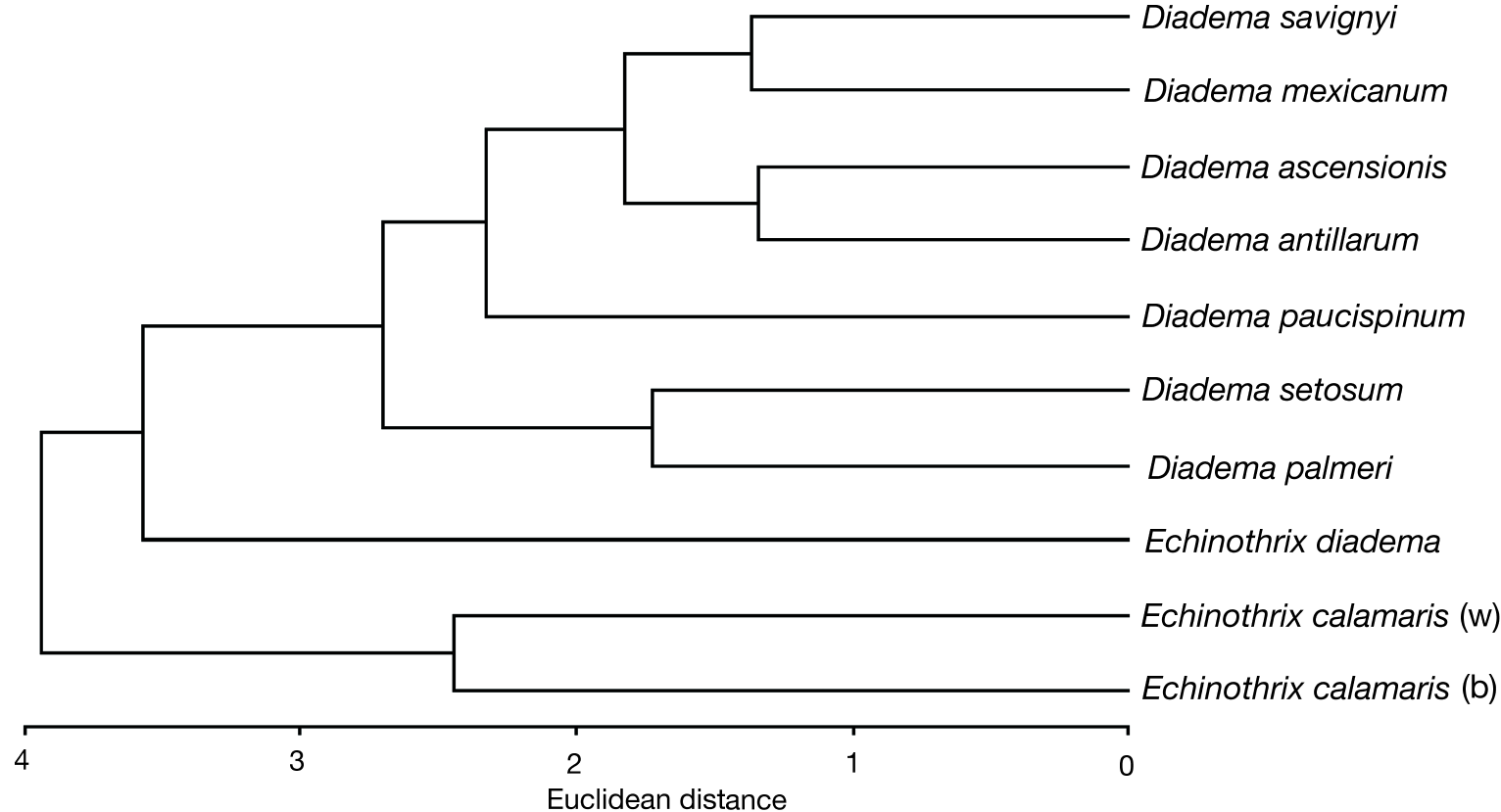
Comparisons using ordination by multi-dimensional scaling (MDS), and cluster analysis using Euclidean distances of taxonomic similarity, based on 28 test features, are illustrated in Figures 3 View FIG and 4 View FIG .
From the dendrogram ( Fig. 4 View FIG ) a distinct association can be seen between the white and brown colour morphs of E. calamaris . Although this shows a reasonable level of similarity, it is not as high as would be expected for two colour morphs of the same species (zero Euclidean distance). Th is level of similarity is considerably less than for different species within the genus Diadema . Th e MDS plot ( Fig. 3 View FIG ) substantiates this, with a large distance between the white and the brown colour morphs of E. calamaris . From this information it can be surmised that based on test structures and proportions, the brown and white colour morphs of E. calamaris are possibly separate species.
The next group incorporates all the species of the genus Diadema , as well as E. diadema . Echinothrix diadema is the first divergent species, indicating only slight association with species of Diadema . Each of the two species in a group composed of D. palmeri and D. setosum , possessed species-specific test features. This is supported by the MDS plot which indicates their disassociation from other members of the genus. The next group comprises all other species of Diadema . Diadema paucispinum is the first divergent species from this grouping, which then subdivides into two groups composed of D. mexicanum and D. savignyi , and D. antillarum and D. ascensionis . These clusterings are illustrated on the MDS plot, with D. antillarum and D. ascensionis showing the closest association.

No known copyright restrictions apply. See Agosti, D., Egloff, W., 2009. Taxonomic information exchange and copyright: the Plazi approach. BMC Research Notes 2009, 2:53 for further explanation.