
Canrightiopsis crassitesta E.M.FRIIS, G.W.GRIMM, M.M.MENDES et K.R.PEDERSEN, 2015
|
publication ID |
https://doi.org/10.37520/fi.2022.016 |
|
DOI |
https://doi.org/10.5281/zenodo.7535246 |
|
persistent identifier |
https://treatment.plazi.org/id/03FD87F2-FFF5-FFE4-FF15-F96BC3ADFC4D |
|
treatment provided by |
Felipe (2023-01-10 20:28:08, last updated 2023-01-13 21:03:26) |
|
scientific name |
Canrightiopsis crassitesta E.M.FRIIS, G.W.GRIMM, M.M.MENDES et K.R.PEDERSEN, 2015 |
| status |
|
Canrightiopsis crassitesta E.M.FRIIS, G.W.GRIMM, M.M.MENDES et K.R.PEDERSEN, 2015
Text-fig. 6a–c, g, h View Text-fig
D e s c r i p t i o n a n d r e m a r k s. Canrightiopsis crassitesta was established based on fruits, seeds and adhering pollen from the Catefica mesofossil flora ( Friis et al. 2015a). The fruits are elliptical to spherical in outline and are interpreted as berries with a single seed ( Text-fig. 6a, b View Text-fig ). They are derived from bisexual flowers and remains of a hypanthium, as well as scars from stamens, are present on the probable abaxial face of the fruit, about one third to two thirds of the distance from the base ( Text-fig. 6a, d View Text-fig ). The seeds are orthotropous, pendent and endotestal with a distinct, thick and finely crystalliferous endotesta (Textfig. 6b, c). The outer surface of endotesta is characterized by relatively large pits arranged in longitudinal rows that are also visible where the fruit wall is compressed or poorly preserved ( Text-fig. 6a View Text-fig ). The tegmen is three cell layers thick. In some specimens, remains of an endothelium are seen as slightly elongated cells, but the distinct endothelium seen in other species of Canrightiopsis has not been observed. Pollen grains attached to the fruits are similar to dispersed pollen assigned to the extinct pollen genus Clavatipollenites COUPER ( Text-fig. 6g, h View Text-fig ). Grains are 12–14 µm in equatorial diameter, monocolpate, semitectate-reticulate with a long, extended colpus with an irregular margin. The reticulum is composed of narrow, beaded muri supported by long, scattered columellae ( Text-fig. 6g, h View Text-fig ). The embryo is minute and surrounded by a nutritive tissue of thin-walled, isodiametric cells ( Text-fig. 6b, c View Text-fig ).
A f f i n i t y a n d o t h e r o c c u r r e n c e s. Analysis of the phylogenetic relationships of Canrightiopsis placed the genus in the Chloranthaceae as part of the Ascarina J.R.FORST. et G.FORST. - Sarcandra GARDNER- Chloranthus SW. clade, particularly close to Sarcandra and Chloranthus ( Friis et al. 2015a) , a result also supported by a subsequent analysis ( Doyle and Endress 2018).
Fruits and seeds of Canrightiopsis are common in Early Cretaceous mesofossil floras from Portugal. In addition to C. crassitesta , two other species have been recognized including C. intermedia and C. dinisii E.M.FRIIS, G.W.GRIMM, M.M.MENDES et K.R.PEDERSEN. Only C. crassitesta and C. intermedia are present in the Catefica mesofossil flora. C. crassitesta is distinguished from C. intermedia by its much thicker endotesta, but the two species are similar in fruit morphology and without internal details, the fossils are difficult to separate. All Canrightiopsis specimens from Catefica studied using SEM are typical C. crassitesta , while only one specimen is a distinct C. intermedia . Other specimens from Catefica for which internal features are unknown are referred to as Canrightiopsis sp. ( Friis et al. 2015a).
Fruits and seeds of Canrightiopsis are particularly common in the mesofossil flora from Famalicão, but are also reported from the Arazede, Buarcos, Chicalhão, Vale de Água and Vila Verde mesofossil floras ( Friis et al. 2015a). Currently C. crassitesta is reported only from the Catefica mesofossil flora.
Pollen grains found on fruits of Canrightiopsis crassitesta are similar in size and general morphology to those found in situ in isolated stamens and inflorescence fragments from Catefica with Clavatipollenites -type pollen ( Text-figs 10– 13 View Text-fig View Text-fig View Text-fig View Text-fig , Tab. 1 View Table 1 ), but the reticulum of the pollen associated with xy1485) through fruit in the region of the hypanthium rim showing sections through the two seeds close to the chalazal region; note endotesta (oi-end) surrounded by larger cells of exotesta (oi-o) and fruit wall (fr). Specimen, Catefica 49-S174249 (holotype, a–f). Scale bars = 300 Μm (a–c, e, f), 100 Μm (d).
Canrightiopsis crassitesta is more open and the grains are smaller.
Doyle, J. A., Endress, P. K. (2018): Phylogenetic analyses of Cretaceous fossils related to Chloranthaceae and their evolutionary implications. - The Botanical Review, 84: 156 - 202. https: // doi. org / 10.1007 / s 12229 - 018 - 9197 - 6
Friis, E. M., Grimm, G. W., Mendes, M. M., Pedersen, K. R. (2015 a): Canrightiopsis, a new Early Cretaceous fossil with Clavatipollenites - type pollen bridge the gap between extinct Canrightia and extant Chloranthaceae. - Grana, 54: 184 - 212. https: // doi. org / 10.1080 / 00173134.2015.1060750
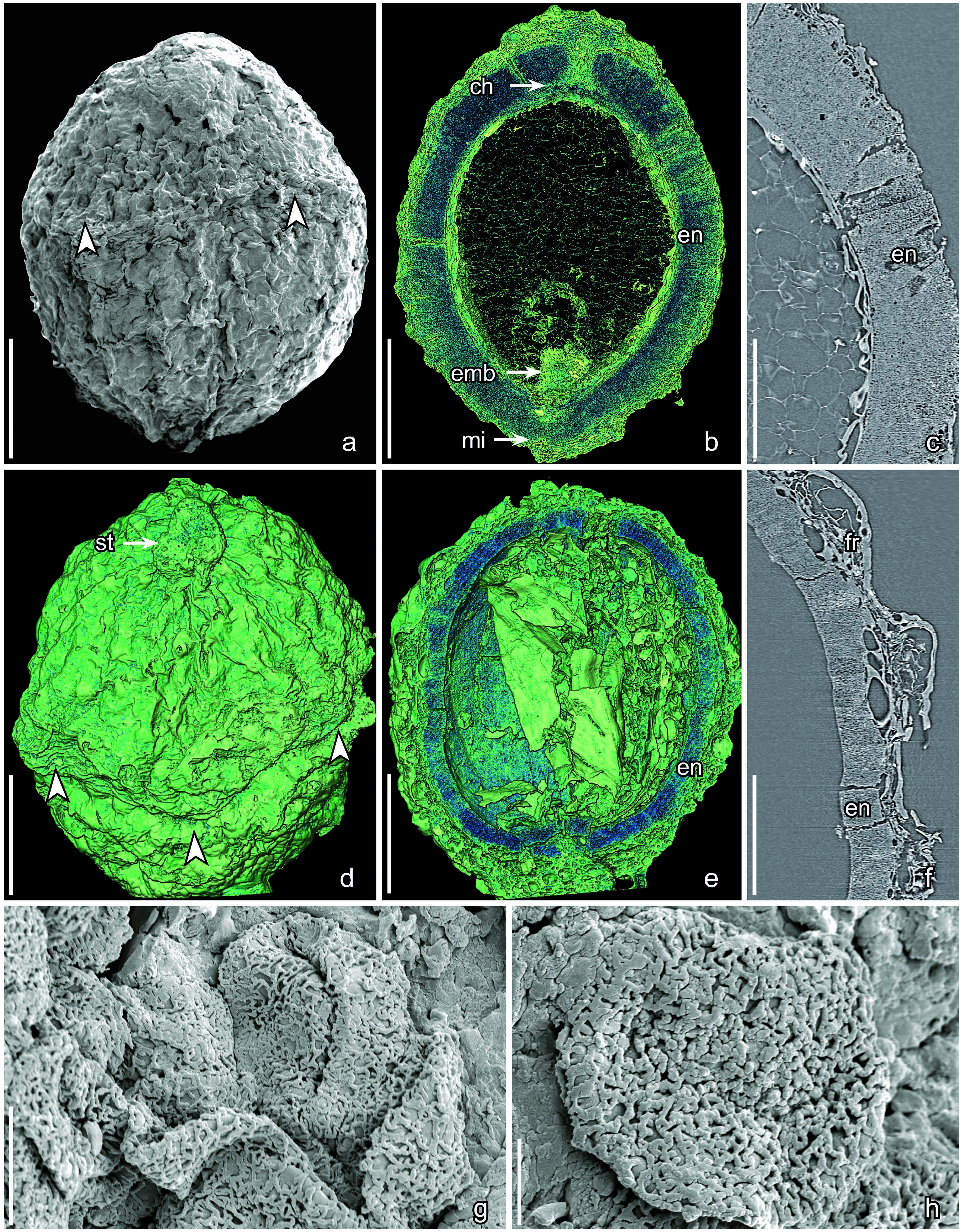
Text-fig. 6. Scanning electron microscope (SEM, a, g, h) and synchrotron radiation X-ray tomographic microscopy (SRXTM, b–f) images of fruits and pollen grains of Canrightiopsis crassitesta (a–c, g, h) and fruit of Canrightiopsis intermedia (d–f); Catefica locality, Portugal. a) Dorsal view of fruit showing rim of hypanthium (arrowheads); b) Surface rendering of longitudinal section in the median plane of fruit (cut between orthoslices yz0440-0510) showing the thin fruit wall, thick endotesta of the seed coat (en, dark blue) and the orthotropous, pendent seed with the chalaza (ch) near the fruit apex and the micropyle (mi) at the fruit base; note the tiny embryo (emb) adjacent to the micropyle at the base of the fruit; c) Longitudinal section (orthoslice xz0511) through the seed wall showing the thick, finely crystalliferous endotesta (en) surrounding the nutritive tissue of the seed; d) Surface rendering of fruit in dorsal view showing rim of the hypanthium (arrowheads) and apical stigmatic region (st); e) Surface rendering of
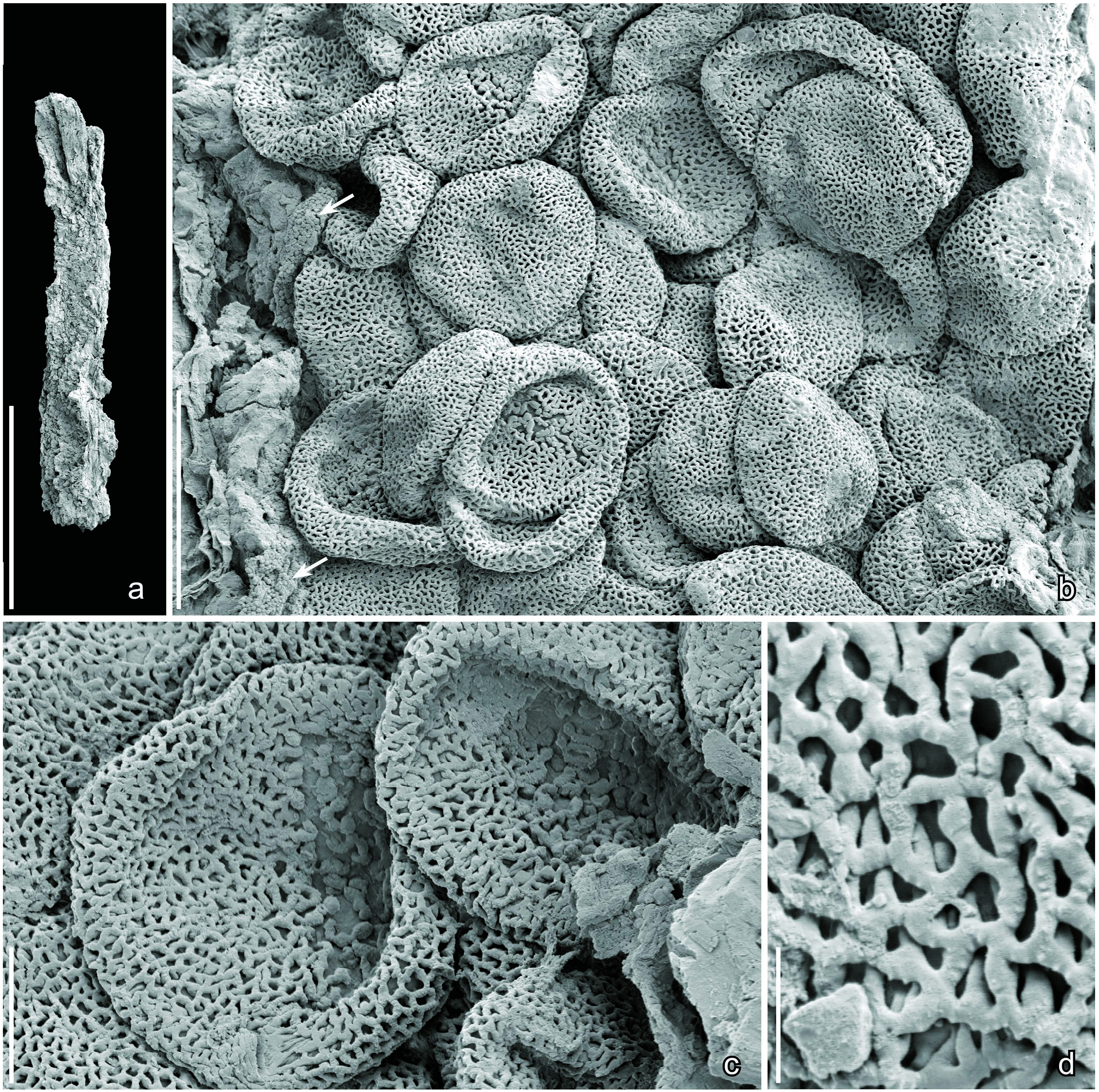
Text-fig. 10. Scanning electron microscope (SEM) images of isolated “Stamen fragment with Clavatipollenites-type pollen sp. 1”; Catefica locality, Portugal. a) Fragment of tetrasporangiate stamen with pollen in situ; b) Detail from stamen fragment showing distal and proximal surfaces of in situ pollen grains and tiny orbicules on the inner surface of the anther wall (arrows); c) Pollen grains in distal view showing short colpi with irregular margins and aperture membrane with irregular verrucae; d) Detail of pollen wall showing the semitectate-reticulate tectum and long, scattered columellae supporting the narrow muri with finely verrucate supratectal ornamentation. Specimen, Catefica 50-S170387 (a–d). Scale bars = 600 Μm (a), 20 Μm (b), 6 Μm (c), 1.5 Μm (d).
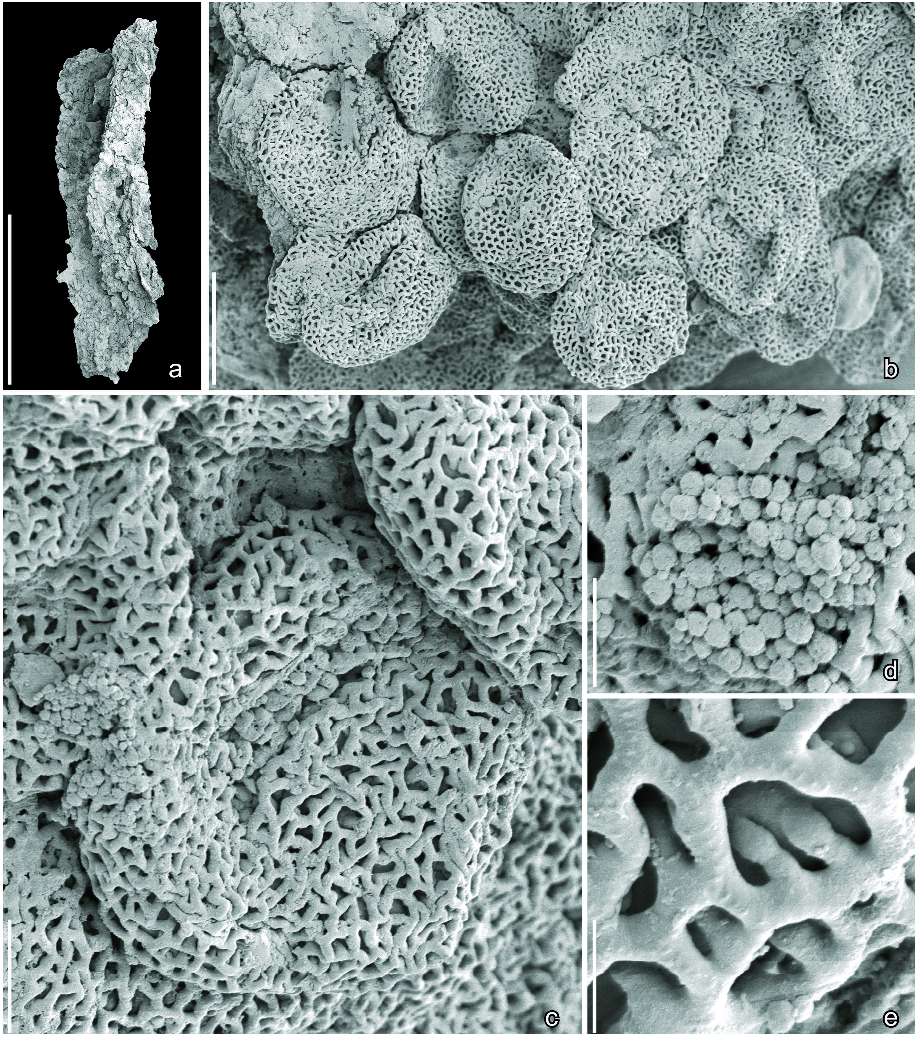
Text-fig. 11. Scanning electron microscope (SEM) images of isolated “Stamen fragment with Clavatipollenites-type pollen sp. 2”; Catefica locality, Portugal. a) Fragment of tetrasporangiate stamen with pollen in situ; b) Detail from stamen fragment showing distal and proximal surfaces of in situ pollen grains; c) Pollen grain in distal view showing short colpus with irregular margin and aperture membrane covered by irregular verrucae; d) Detail of pollen wall showing tiny spherical orbicules; e) Detail of pollen wall showing the semitectate-reticulate tectum and long, scattered columellae supporting the narrow muri with finely verrucate supratectal ornamentation. Specimen, Catefica 50-S170389 (a–e). Scale bars = 600 Μm (a), 20 Μm (b), 6 Μm (c), 3 Μm (d), 1.5 Μm (e).
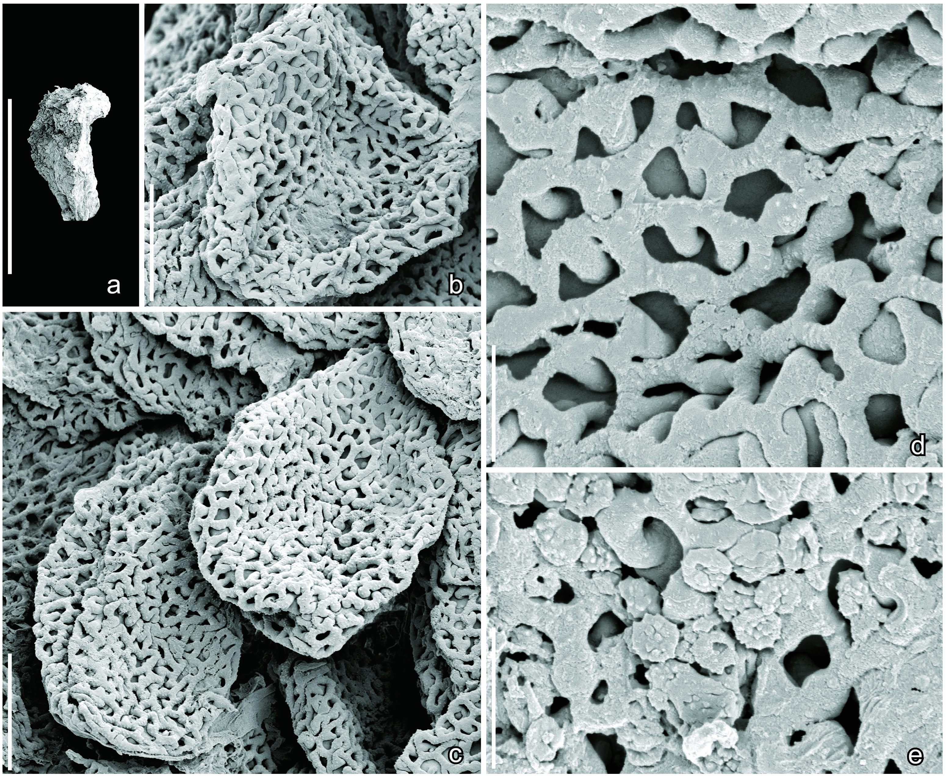
Text-fig. 12. Scanning electron microscope (SEM) images of isolated “Stamen fragment with Clavatipollenites-type pollen sp. 3”; Catefica locality, Portugal. a) Stamen fragment with pollen in situ; b, c) Pollen grains showing the semitectate-reticulate pollen wall and folds (c) indicating a possible monocolpate aperture; d) Detail of pollen wall showing the semitectate-reticulate tectum and long, scattered columellae supporting the narrow muri with finely verrucate supratectal ornamentation; e) Detail of pollen wall showing tiny, spherical, finely spiny orbicules. Specimen, Catefica 50-S170449 (a–e). Scale bars = 600 Μm (a), 6 Μm (b, c), 1.5 Μm (d, e).
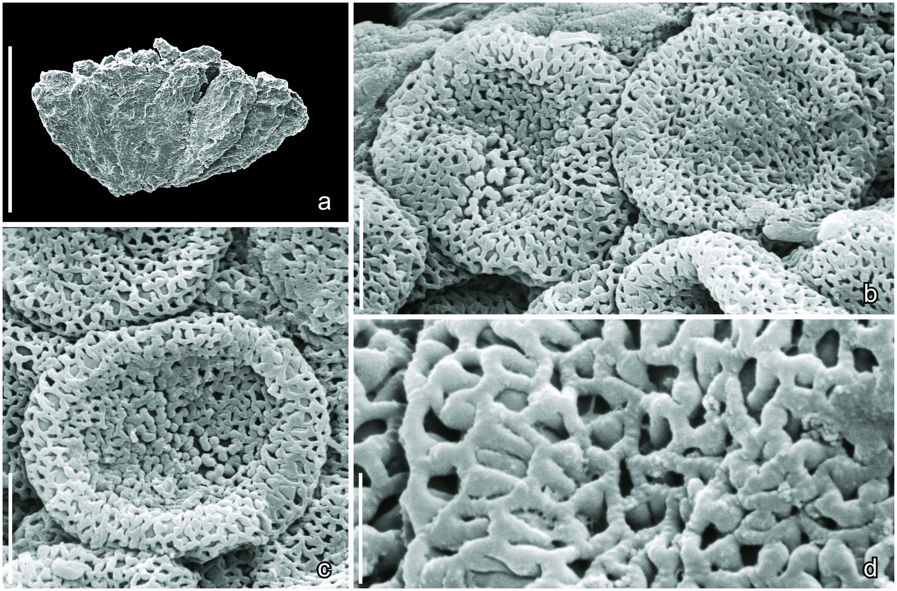
Text-fig. 13. Scanning electron microscope (SEM) images of “Staminate inflorescence fragment with Clavatipollenites-type pollen sp. 4”; Catefica locality, Portugal. a) Fragment of stamen whorl from staminate inflorescence showing several closely packed, almost sessile stamens that lack a well-developed filament; b, c) Distal and proximal views of pollen grains showing poorly defined aperture with verrucate aperture membrane; d) Detail of pollen wall showing the semitectate-reticulate tectum and long, scattered columellae supporting the narrow muri with finely verrucate supratectal ornamentation. Specimen, Catefica 49-S107782 (a–d). Scale bars = 600 Μm (a), 6 Μm (b, c), 1.5 Μm (d).
No known copyright restrictions apply. See Agosti, D., Egloff, W., 2009. Taxonomic information exchange and copyright: the Plazi approach. BMC Research Notes 2009, 2:53 for further explanation.
|
Kingdom |
|
|
Phylum |
|
|
Class |
|
|
Order |
|
|
Family |
|
|
Genus |
1 (by felipe, 2023-01-10 20:28:08)
2 (by ExternalLinkService, 2023-01-10 20:42:41)
3 (by juliana, 2023-01-13 19:26:10)
4 (by ExternalLinkService, 2023-01-13 19:37:09)
5 (by ExternalLinkService, 2023-01-13 21:03:26)
6 (by plazi, 2023-11-08 12:21:23)
7 (by ExternalLinkService, 2023-11-08 20:02:36)
8 (by juliana, 2024-10-10 23:48:11)