
Procyrnea sinica, Zhang & Song & Zhang, 2011
|
publication ID |
https://doi.org/ 10.1080/00222933.2011.622451 |
|
DOI |
https://doi.org/10.5281/zenodo.10537044 |
|
persistent identifier |
https://treatment.plazi.org/id/03FD0778-4103-FFA5-FE10-FDF7B46CF98A |
|
treatment provided by |
Felipe |
|
scientific name |
Procyrnea sinica |
| status |
sp. nov. |
Procyrnea sinica sp. nov.
( Figures 3 View Figure 3 and 4 View Figure 4 )
Type host
Other host
Type locality
Beijing, China (39 ◦ 54’ N, 116 ◦ 28’ E) GoogleMaps .
Site of infection
Under the lining of the gizzard.
Prevalence
This was 7.4% (two of 27 birds), 2–4 (3) specimens in Asio otus ; 2.6% (one of 39 birds) in Athene noctua .
Type specimens
Holotype: male (HBNU-1120); allotype: female (HBNU-1121); paratypes: two males (HBNU-1122) and two females (HBNU-1123).
Etymology
The species name refers to its geographic location ( China).
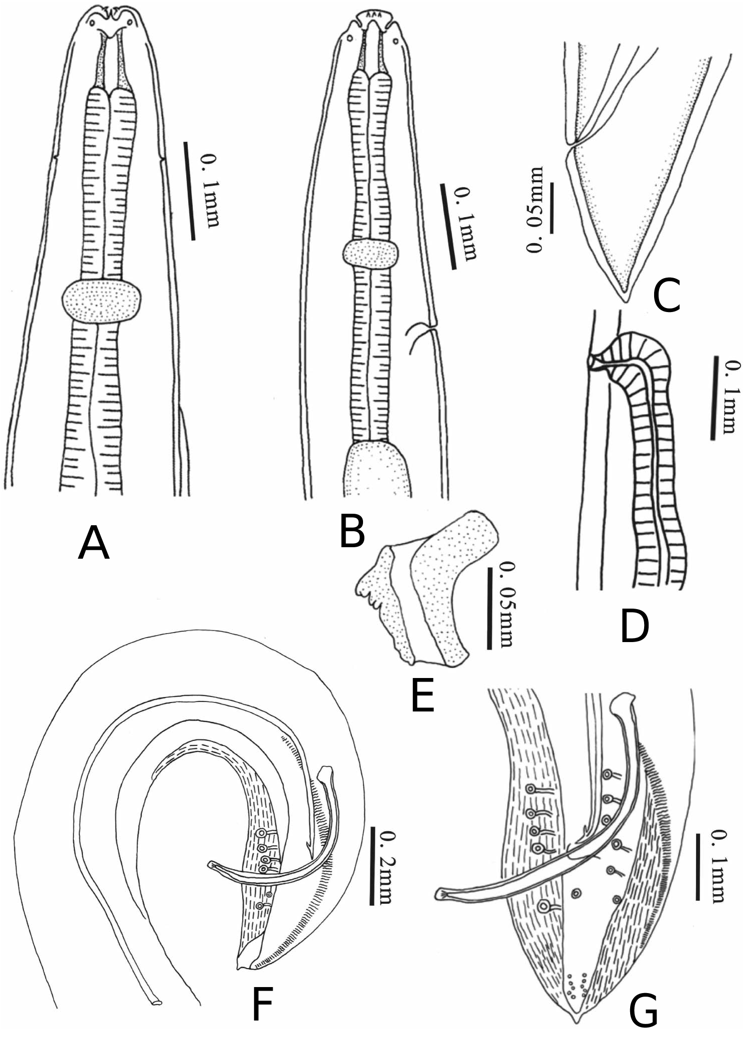
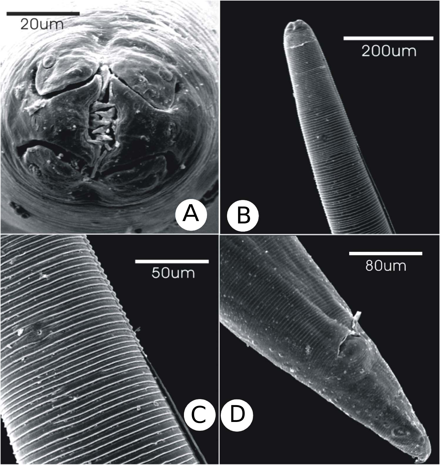
Diganosis
Body medium sized with distinct transverse striations. Labial region consisting of two pseudolabia and dorsal and ventral labia. Pseudolabia wider distally than at base, amphids located on base of pseudolabia; three small teeth on interior border of each pseudolabium. Dorsal and ventral labia each consisting of two submedian lobes, with two papillae on each, and a median internal process. Single lateral ala on left side of body lacking sinuous striations, originating just posterior to excretory pore. Buccal capsule laterally compressed. Oesophagus clearly divided into short anterior muscular part and long posterior glandular part. Muscular part 3.1–4.3% (3.8%) TBL in male and 3.6–4.5% (4.0%) TBL in female; glandular part 27.1–31.4% (29.8%) TBL in male and 22.2–32.0% (25.8%) TBL in female. Nerve ring located at middle level of muscular oesophagus. Cervical papillae anterior to nerve ring. Excretory pore posterior to nerve ring.
Male (n = 3). Body length 9.2–10.3 (9.7) mm. Maximum width 243–319 (277). Lateral ala 1.2–1.8 (1.6) mm long, 11.3–19.2% (16.3%) TBL from anterior end. Buccal capsule 31–48 (43) long and 12–19 (14) wide. Muscular oesophagus 319–392 (368) long and 29–39 (33) wide; glandular oesophagus 2.5–3.3 (2.9) mm long and 100–147 (123) wide. Nerve ring 221–245 (234) from anterior end; excretory pore 245–319 (291) from anterior end; cervical papillae 123–172 (139) from anterior end. Posterior end of body curved. Caudal alae well developed and symmetrical, 1.0–1.2 (1.1) mm long. Caudal alae with longitudinal striations on ventral surface and transverse striation on dorsal surface. Ventral surface of caudal region with prominent longitudinal ridges. Tail 196–245 (225) long, with pointed tip, with four pairs of pedunculate pre-cloacal papillae arranged symmetrically and two pairs of pedunculate post-cloacal papillae arranged asymmetrically. Five pairs of sessile papillae located near the tail tip. Spicules unequal and dissimilar. Left spicule 1.1–1.4 (1.3) mm long, with a barb on the distal end. Right spicule 343–490 (441) long, with rounded distal end. Ratio of right spicule: left spicule 1: 2.8–3.3 (1: 3.0). Gubernaculum trowel shaped, 48–51 (48) long and 39–41 (40) wide.
Female (n = 3). Body length 10.7–12.1 (11.5) mm long. Maximum width 243–320 (295). Lateral ala 2.1–2.5 (2.3) mm long, 19.8–20.8% (20.2%) TBL from anterior end. Buccal capsule 36–39 (37) long and 10–14 (12) wide. Muscular oesophagus 417–534 (464) long and 25–39 (31) wide; glandular oesophagus 2.70–3.43 (2.98) mm long and 123–175 (143) wide. Nerve ring 221–340 (269) from anterior end. Excretory pore 319–388 (342) from anterior end. Cervical papillae 157–172 (164) from anterior end. Vulva located behind middle of body, 4.2–5.0 (4.7) mm from posterior end. Tail pointed, 147–196 (168) long. Eggs ellipsoid, thick shelled, embryonated, 34–39 (35) long, 17–19 (18) wide.
Remarks
Procyrnea sinica sp. nov. resembles Procyrnea fotedari ( Gupta and Kumar, 1980) , Procyrnea monoptera (Gendre, 1922) , Procyrnea rauschi ( Gupta and Kumar, 1980) , Procyrnea suraiyae ( Ali, 1961) , Procyrnea tulostoma (Hemprich and Ehrenberg, 1866) , Procyrnea unilateralis (Molin, 1860) , Procyrnea urophasiana (Wehr, 1931) and Procyrnea vinodi ( Gupta and Kumar, 1980) in possessing a single lateral ala. However, it can be easily distinguished from all these related species except P. monoptera in the single lateral ala beginning posterior to the excretory pore instead of from the lip region, and in the distal end of the left spicule with a barb versus the distal end of left spicule with a simple pointed tip. The new species is very similar to P. monoptera in having a barb on distal end of the left spicule, but it differs from the latter in the single left ala beginning posterior to excretory pore rather than beginning anterior to the nerve ring, in having a shorter lateral ala (1.2–1.8 mm long in P. sinica vs 2.58 mm long in P. monoptera ), and in the length of the left spicule (1.1–1.4 mm long in P. sinica vs 1.58–1.68 mm long in P. monoptera ). Finally, P. monoptera males possess a single pre-cloacal sessile papilla, which P. sinica males lack.
No known copyright restrictions apply. See Agosti, D., Egloff, W., 2009. Taxonomic information exchange and copyright: the Plazi approach. BMC Research Notes 2009, 2:53 for further explanation.
|
Kingdom |
|
|
Phylum |
|
|
Class |
|
|
Order |
|
|
Family |
|
|
Genus |
Procyrnea sinica
| Zhang, Shuqian, Song, Jie & Zhang, Luping 2011 |
Procyrnea sinica
| Zhang & Song & Zhang 2011 |
P. sinica
| Zhang & Song & Zhang 2011 |
P. sinica
| Zhang & Song & Zhang 2011 |
P. sinica
| Zhang & Song & Zhang 2011 |