
Tyrannomolpus, Nadein & Leschen, 2017
|
publication ID |
https://doi.org/ 10.11646/zootaxa.4294.2.9 |
|
publication LSID |
lsid:zoobank.org:pub:7EAE9AD8-F840-40DB-843C-D1DD80568E76 |
|
DOI |
https://doi.org/10.5281/zenodo.6032326 |
|
persistent identifier |
https://treatment.plazi.org/id/F2AD6850-5644-4AAE-8ABC-86522E2266D6 |
|
taxon LSID |
lsid:zoobank.org:act:F2AD6850-5644-4AAE-8ABC-86522E2266D6 |
|
treatment provided by |
Plazi |
|
scientific name |
Tyrannomolpus |
| status |
gen. nov. |
Tyrannomolpus gen. nov.
Figs 1–31 View FIGURES 1, 2 View FIGURES 3 – 12 View FIGURES 13 – 25 View FIGURES 26 – 31 .
Type species: Tyrannomolpus rex spec. nov.
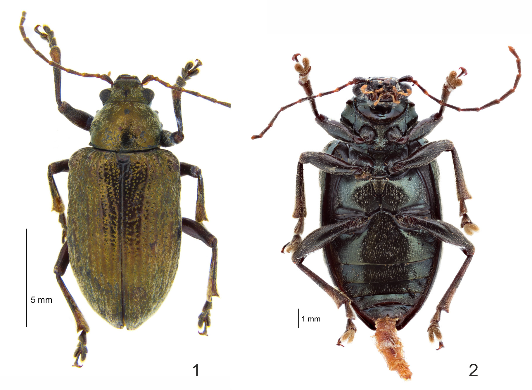
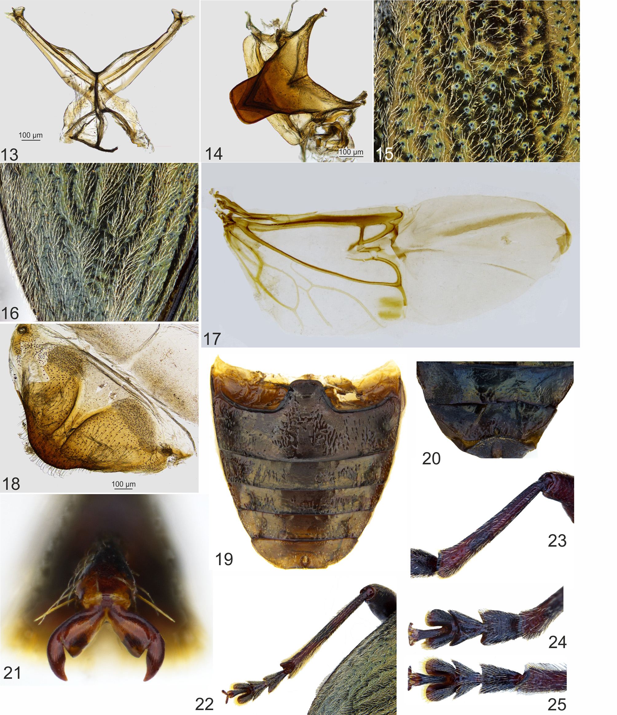
Diagnosis. Colour dark bronzy-greenish with metallic luster ( Figs 1, 2 View FIGURES 1, 2 ). Vertex of head and dorsum setose ( Fig. 3 View FIGURES 3 – 12 ). Head with midcranial suture ( Fig. 3 View FIGURES 3 – 12 ). Pronotum ( Fig. 9 View FIGURES 3 – 12 ) widest at middle, converging anteriorly, lateral margin distinctly undulate; base without a row of dense punctures. Hypomera weakly convex medially. Prosternum anteriorly deflexed with leading edge hidden in ventral view; medially concave with distinct carinae confluent with sides of prosternal process, sexually dimorphic with male having a transverse anterior fovea, prosternal process with well-developed lateral coxal braces, apex expanded behind coxae with sides parallel-sided and depressed apically ( Figs 10, 12 View FIGURES 3 – 12 ). Procoxal cavities externally closed ( Figs 2 View FIGURES 1, 2 , 10 View FIGURES 3 – 12 ). Elytra ( Fig. 1 View FIGURES 1, 2 ) moderately convex, humeral calli present, elytral punctures arranged in 10 partially confused rows with weak carinae. Hind wings fully developed ( Fig. 17 View FIGURES 13 – 25 ). Dorsal shaft of meso- and metatibiae ( Fig. 22 View FIGURES 13 – 25 ) without groove along entire flank, excavation confined to apex and lacking dorsal carinae, tarsal claws appendiculate ( Fig. 21 View FIGURES 13 – 25 ). Pygidium ( Fig. 18 View FIGURES 13 – 25 ) medially with deep groove. Aedeagus ( Figs 29–31 View FIGURES 26 – 31 ) with tegmenal strut subequal in length to penis, basal hood and main body of penis subequal in length.
Description. Body ( Figs 1, 2 View FIGURES 1, 2 ) elongate robust and broad, moderately convex; cuticle glabrous and dorsally setose ( Figs 1 View FIGURES 1, 2 , 9 View FIGURES 3 – 12 , 15, 16 View FIGURES 13 – 25 ).
Head ( Fig. 3 View FIGURES 3 – 12 ) with vertex wide, flat, with midcranial suture, covered with long and dense setae. Frons long, trapezoidal, flat, limited by thin grooves, anterior margin of frons emarginate with pair of short and acute processes, small portions of frons above antennal insertions convex; genae short, not longer than diameter of antennal insertion. Eyes ( Fig. 4 View FIGURES 3 – 12 ) large, elongate-elliptic, convex. Exposed part of labrum distinctly transverse, entire labrum weakly longer than wide ( Fig. 5 View FIGURES 3 – 12 ), anterior margin concave, anterolaterally with three long setae, labral plate with pair of pores; tormae shorter than labral plate. Mandibles large, strongly projecting. Maxillae ( Fig. View FIGURES 3 – 12
6) large, lacinia broad, shorter than galea, the latter about twice longer than wide, apically with a bunch of dense setae, maxillary palp long, 4-segmented, palpomere 1 much shorter than following, palpomere 2 longer than following, about 3 times longer than wide, palpomere 3 and 4 equal in length, about twice longer than wide, last palpomere somewhat fusiform with truncated apex. Labium ( Fig. 7 View FIGURES 3 – 12 ) small, submentum and mentum short, transverse, labial palpi long, 3-segmented, palpomere 1 shortest, about as long as wide, palpomere 2 the longest, about 3 times longer than wide, palpomere 3 about 2.5 times longer than wide, conical with obtuse apex. Antennal insertions placed at the distance of diameter of insertions from margin of eye and anterior part of frons. Antennae ( Fig. 8 View FIGURES 3 – 12 ) long, filiform, 11-segmented, extending to middle of the body; scapus thickened and slightly curved, about twice longer than wide, antennomere 2 small, globose, antennomeres 3–10 long and thin, at least three times longer than wide or longer, last antennomere shorter than previous, hardly three times longer than wide.
Prothorax ( Fig. 9 View FIGURES 3 – 12 ) with pronotum transverse (1.20–1.42 times wider than long), convex, widest at middle, converging anteriorly; anterior margin straight in middle, lateral margins with two short angular projections, undulate, posterior margin convex, rounded; hind margin with a distinct bead, anterior angles protruding, with obtuse apex, posterior angles weakly protruding, acute, each angle bear setiferous pore; pronotal surface densely punctate, punctures small, bearing long setae, densest at sides, sparser at disc; base without a row of dense punctures. Prosternum ( Figs 2 View FIGURES 1, 2 , 10, 12 View FIGURES 3 – 12 ) short in front of procoxae, much shorter than wide; anteriorly deflexed with leading edge hidden in ventral view; medially concave with distinct carinae confluent with the sides of the prosternal process, sexually dimorphic with male having a transverse anterior fovea, prosternal process with welldeveloped lateral coxal braces, apex expanded behind coxae with sides parallel-sided and depressed apically ( Figs 10, 12 View FIGURES 3 – 12 ) hypomera weakly convex medially, hypomeral suture distinct; prosternal process comparatively narrow, between coxae about twice narrower than diameter of coxa, strongly expanded behind coxae laterally and contacting the hypomeral process; procoxal cavities externally closed ( Figs 2 View FIGURES 1, 2 , 10 View FIGURES 3 – 12 ); anterior margin straight.
Scutellary shield ( Fig. 14 View FIGURES 13 – 25 ) transverse and obtuse. Mesoventrite partly hidden by prosternum, T-shaped, flat, intercoxal process ( Fig. 11 View FIGURES 3 – 12 ) long and relatively narrow, longer than wide, apical half of process expanded, rectangular and elevated, apex straight; mesanepisternum triangular, nearly medially transversely impressed; mesepimeron narrow and long. Metaventrite long, as long as abdominal ventrite 1, convex, intercoxal metaventral process rather short, wide with nearly straight apex, posterior margin medially angularly and deeply emarginate and impressed; metanepimeron narrow, flat. Metendosternite ( Fig. 13 View FIGURES 13 – 25 ) with short and broad stalk, arms long, proximal half about twice wider than distal.
Elytra ( Fig. 1 View FIGURES 1, 2 ) elongate (3.29–3.76 x longer than pronotal length) and broad (1.52–1.62 x the greatest elytral width), moderately convex, base 1.45 times wider than the combined based of the pronotum, lateral sides vertical; humeral calli large and projecting; elytral surface punctate ( Fig. 15 View FIGURES 13 – 25 ), punctures larger than those on pronotum, arranged in 10 long rows, punctures in rows somewhat confused and irregular, sutural row short, not reaching middle of elytra, of multiply, dense, confused punctures; intervals between rows convex, finely punctate, surface covered with dense setae ( Fig. 16 View FIGURES 13 – 25 ). Epipleura rather narrow and feebly concave, widest at base and visible in lateral view. Hind wings fully developed ( Fig. 17 View FIGURES 13 – 25 ).
Abdominal ( Fig. 19 View FIGURES 13 – 25 ) ventrite I moderately convex, intercoxal process long and wide, its apex triangularlyrounded; ventrites II–IV short, equal in length, much shorter than ventrite I, last ventrite nearly as long as previous or slightly longer, its margin rounded with straight apex, in male ( Fig. 20 View FIGURES 13 – 25 ) and evenly arcuate in female, subapically with a medial deep spherical setose fovea; pygidium (tergum VII) wide and relatively short, medially with wide and deep groove ( Fig. 18 View FIGURES 13 – 25 ), not reaching to apex.
Legs ( Figs 1, 2 View FIGURES 1, 2 , 22, 23 View FIGURES 13 – 25 ) large and somewhat thin, femora lacking teeth, tibiae ( Figs 22, 23 View FIGURES 13 – 25 ) thin, gradually widened towards apex, dorsal shaft of tibiae with two thin longitudinal ridges, without groove along entire flank, tibial spur indistinct, apex of middle and hind tibiae weakly expanded in two short and thin blades, with excavation confined to apex and lacking dorsal carinae; tarsomere ( Figs 22, 24, 25 View FIGURES 13 – 25 ) I somewhat longer than following, triangular, its apex hardly narrower than apex of following, tarsomere II triangular with wide apex, tarsomere III widest, long, deeply bilobed, last tarsomere as long as first, weakly projecting from third, tarsal claws appendiculate ( Fig. 21 View FIGURES 13 – 25 ); tarsi of males ( Fig. 24 View FIGURES 13 – 25 ) slightly wider and broader than those of female ( Fig. 25 View FIGURES 13 – 25 ).
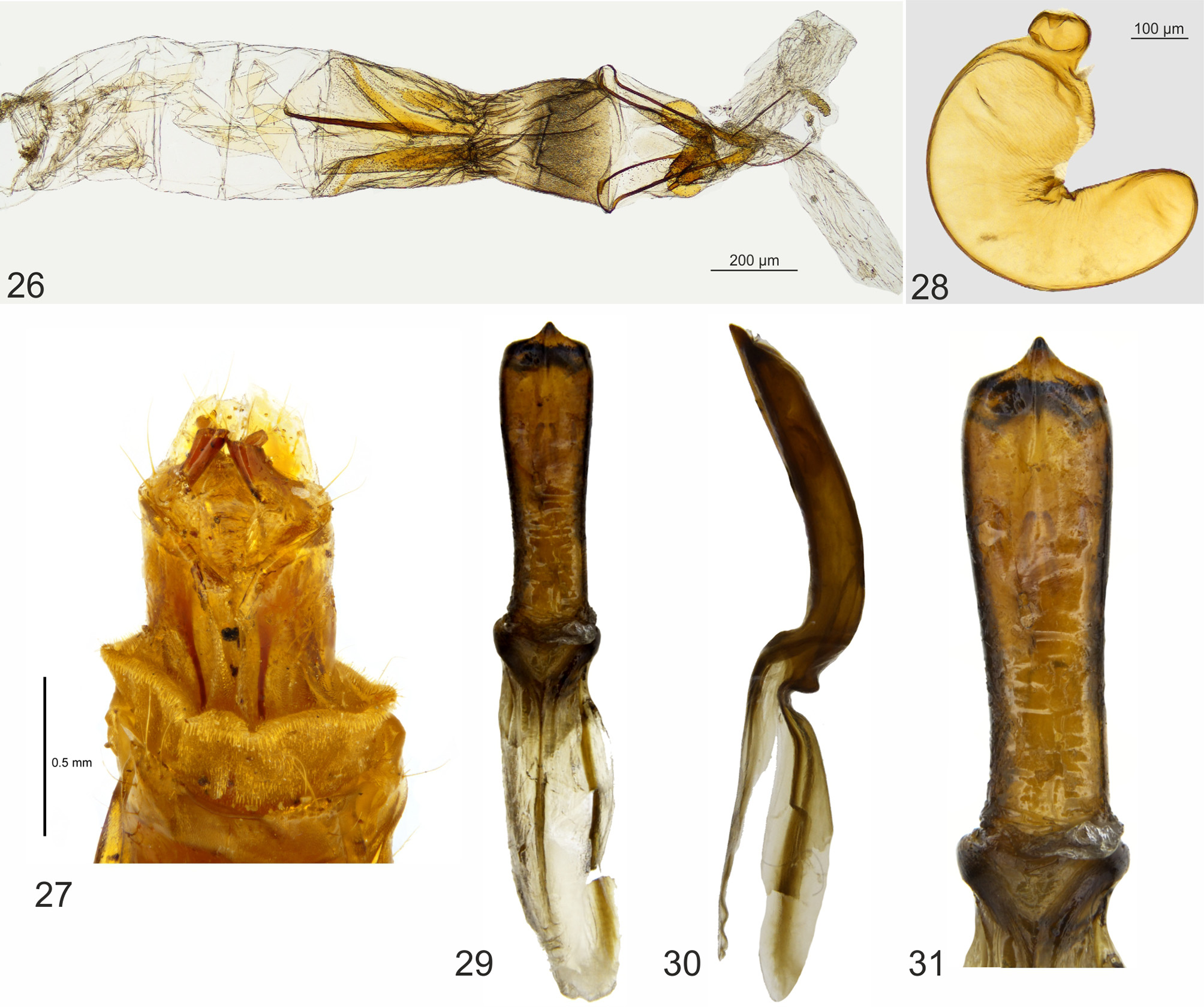
Aedeagus ( Figs 29–31 View FIGURES 26 – 31 ) with tegmenal strut (tegmen) subequal in length to penis, basal hood and main body of penis subequal in length; overall shape long and broad, weakly depressed dorsoventrally, slightly curved in lateral view, basal hood weakly sclerotized.
Ovipositor ( Figs 26, 27 View FIGURES 26 – 31 ) long; gonocoxites laterally compressed and somewhat crescent shaped, about 2 times longer than wide and gradually expanded apically with short subapical gonostyli about the length of coxite. Spermatheca ( Fig. 28 View FIGURES 26 – 31 ) c-shaped.
Etymology. The genus name combines the dinosaur genus name Tyrannosaurus with the suffix “molpus, referring to the relatively large size of the new genus to other chrysomelids endemic to New Zealand. Gender masculine.
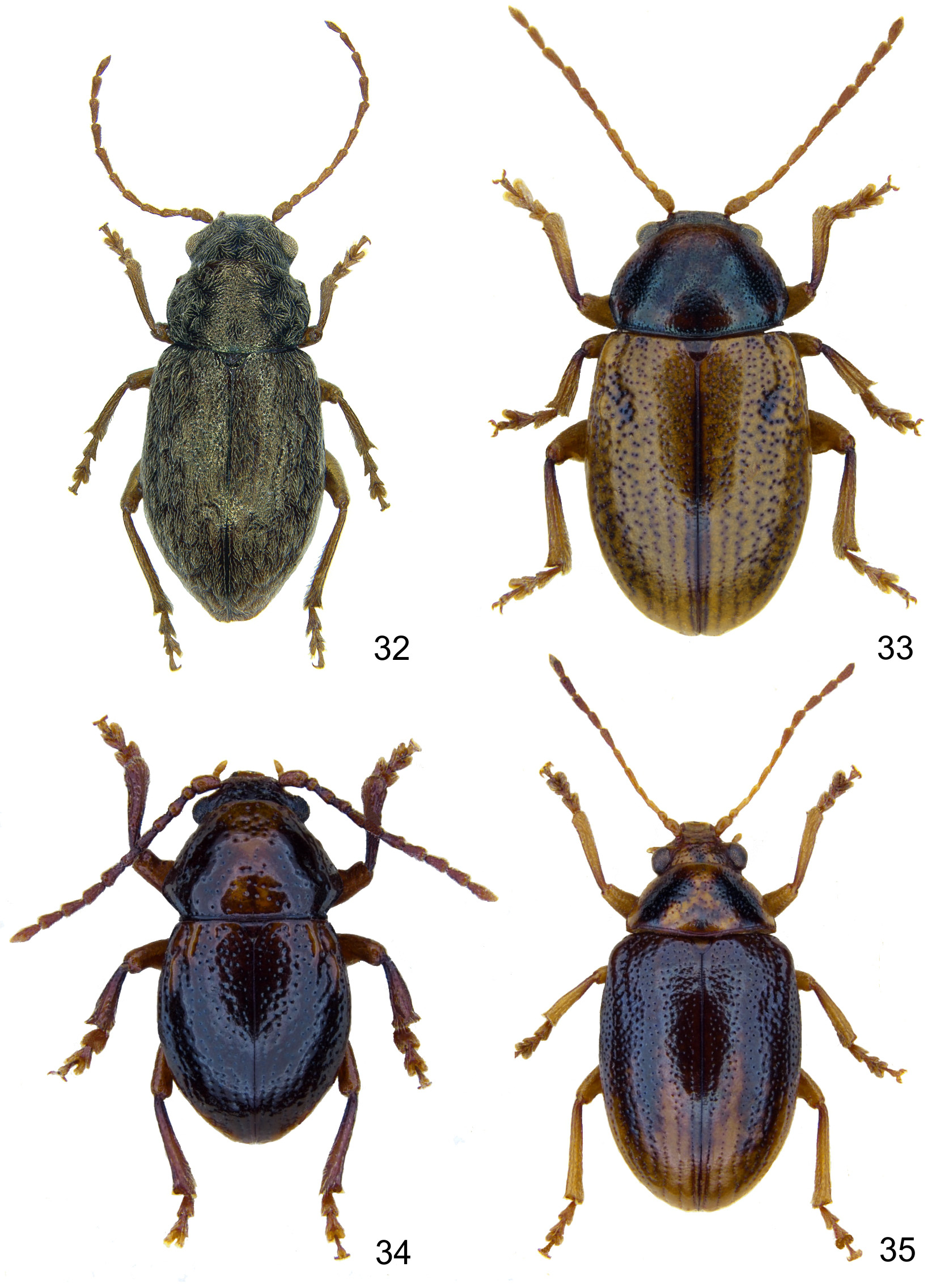
Comments. The attribution of the new genus to the tribe Eumolpini is based on the following combination of characters: simple apices of meso- and metatibiae, carinate and irregularly curved sides of prothorax, appendiculate tarsal claws, pygidium with a longitudinal medial groove, aedeagus with a constriction between a median lobe and a basal hood (after Chen 1940; Selman 1965; Moseyko & Kirejtshuk 2013). The eumolpines are in need in revision from most regions in the Pacific and Australia ( Reid 2017), and Tyrannomolpus can be distinguished from most Australian ( Chapuis 1874), New Caledonian (e.g., Gómez-Zurita & Cardoso 2014; Jolivet et al. 2007, 2009; Jolivet & Verma 2010; Samuelson 2010) and New Zealand eumolpine genera ( Figs 32–35 View FIGURES 32 – 35 ) by the prothorax that is narrower than the elytral base. This character is also present in at least one species of Taophila Heller from New Caledonia, but lacks the setose vestiture present in Tyrannomolpus . Morphologically, Tyrannomolpus is closest to Pilacolaspis ( Fig. 32 View FIGURES 32 – 35 ) from the mainland of New Zealand from which it differs by the much larger size (10.3 mm or more versus up to about 5.9 mm in Pilacolaspis ) and undulate outline of the pronotum (simple in Pilacolaspis ) as well as the longer eyes (length/width ratio is about 2, in Pilacolaspis 1.2), metaventrite as long as abdominal ventrite I (shorter in Pilacolaspis ) and the vertex of head with a distinct midcranial suture.
The most notable feature of Tyrannomolpus is its large body size, which might be an example of island gigantism well known to vertebrates (e.g., Jaffe et al. 2011) and occurs among New Zealand invertebrates, including weevils ( Buckley et al. 2015; Kuschel & Worthy 1996; Leschen & Rhode 2002). But the exact sister relationships of Tyrannomolpus among Melanesian Rift eumolpines is unknown, though it is most similar to Pilacolaspis based on similar elytral punctuation and pubescence (especially in the apical half). In a biogeographic analysis of the region, Ung et al. (2017) propose a sister relationship of the Three Kings with New Zealand, but also that Australia is the sister to the Melanesian Rift with New Caledonia and the Loyalty Islands at the base. We may predict that Tyrannomolpus may indeed be related to the New Zealand Pilacolaspis , but at present, phylogenetic relationships among eumolpines are incomplete (e.g., Papadopoulou et al. 2013), though strides in the taxonomic and phylogenetic relationships of the New Caledonia fauna have been made recently (e.g., Gómez- Zurita 2011 and references above) while discussions about its origin are mostly verbal ( Jolivet et al. 2008a, b) and require a complete phylogenetic backbone to understand the evolution of the group.
Tyrannomolpus is known by only by a few specimens, and the single specimen with plant host data indicate it was collected at night on Meryta sinclairii , referred to as Puka, a tree that is endemic to the Three Kings Islands and now planted commonly in gardens on the mainland. Despite further collecting to the islands, including a short visit by the second author in 2008 to Great Island with the intention to re-collect this and other rare endemic species (see Buckley & Leschen 2013), there are no other specimens we know in existence. Additional surveys are needed to determine whether the species is still thriving on the islands.

No known copyright restrictions apply. See Agosti, D., Egloff, W., 2009. Taxonomic information exchange and copyright: the Plazi approach. BMC Research Notes 2009, 2:53 for further explanation.
|
Kingdom |
|
|
Phylum |
|
|
Class |
|
|
Order |
|
|
Family |