
Diasemopsis comoroensis Carr & Földvári, 2006
|
publication ID |
https://doi.org/ 10.5281/zenodo.172483 |
|
DOI |
https://doi.org/10.5281/zenodo.6259139 |
|
persistent identifier |
https://treatment.plazi.org/id/03FB8784-CA4E-FF97-4673-FACA8441FEE5 |
|
treatment provided by |
Plazi |
|
scientific name |
Diasemopsis comoroensis Carr & Földvári |
| status |
sp. nov. |
Diasemopsis comoroensis Carr & Földvári View in CoL , new species
( Figs 1–9 View FIGURES 1 – 2 View FIGURES 3 – 8 View FIGURES 9 & 10 , 11, 13–16 View FIGURES 13 – 16 )
Description
Type material: Holotype, male (Natural History Museum, London). Paratypes 4 males, 5 females (Natural History Museum, London), 5 males, 5 females (Zoologische Staatssammlung, München), 5 males, 5 females (Hungarian Natural History Museum, Budapest), 2 males, 2 females (Centre National de Documentation et de Recherche Scientifique, Moroni, Comoro Islands). All type specimens (dried, double mounted, excellent condition) taken from a laboratory culture housed at University College, London in May 2005. Parent specimens collected at Comoro Islands, Mohéli, creek uphill from Hoani on way toward Chalet St. Antoine, leg. M. Kotrba 21.iv.2002.
Head. Completely black (including eye stalks) and covered with minute pale hairs. Facial teeth smaller than half of the width of the eye stalks in the middle. Outer vertical bristles at least as long as width of the eye stalk in the middle. Inner vertical bristles short, but distinct, as long as 1/3rd of the width of the eye stalk in the middle.
Thorax. Uniformly grayish pollinose, except the surface of the meron. Metapleural spine dark brown to black, at most the tip can be yellow. The spine is curved upwards (dorsally) in anterior view. Scutellar spines 2.5–3 times as long as the scutellum.
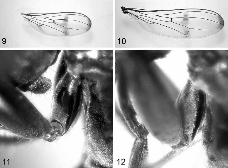
Wing Fig. 9 View FIGURES 9 & 10 . Completely hyaline, except for three infuscated brownish bands. The apical band is as broad as 1/20th of the wing length and is restricted to the space between M and R2+3 veins (not reaching these veins). The central band is darkest around R4+5 and it is situated between R2+3 and Cu slightly extended towards the anterior cross vein (R–M). The proximal band is more an infuscated brownish spot below the cell cup.
Legs Fig. 11. Front coxae are shiny posteriorly and also with a lateral shiny spot at proximal 1/3rd of the coxae. Legs are yellow–brown, brown in general; tarsi 3–5 on the front leg are paler and yellow. Front femora incrassate (length/width approximately 4), bearing on their ventral side two longitudinal rows of 3–5 prominent bristles each and between these two rows of 18–22 much shorter peglike tubercles each.
Preabdomen. Subshining black except for the following silver pollinose areas: an uninterrupted band along posterior margin of tergite 1, lateral triangles at posterior margin of tergite 2, distal half of tergite 3, and subsequent tergites.
Postabdomen (male) Figs 13–16 View FIGURES 13 – 16 . In ventral view ( Fig. 13 View FIGURES 13 – 16 ) the connection of the hypandrium to the aedeagal apodeme is clearly visible. The membranous tip of the hypandrium is divided into two lobes anteriorly. There are two thick hairs on the medial inner surface of the hypandrium, the bilobed surstyli have numerous short, distinct hairs and the gonopods bear minute hairs as well ( Fig. 14 View FIGURES 13 – 16 ). In lateral view the aedeagal apodeme is curved (more that that of D. meigenii ) and not broadening on anterior half. The ligament connecting to the hypandrium joins in the middle of the aedeagal apodeme ( Fig. 15 View FIGURES 13 – 16 ). The epandrium and cerci have long, dispersed hairs along their surface. Hairs on the hypandrium are more restricted to the ventral part and are shorter than those of D. meigenii . The distal half of the paramere is broadening towards the tip ( Fig. 16 View FIGURES 13 – 16 ).
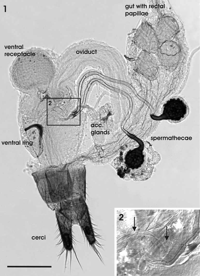
Postabdomen (female) Fig. 1 View FIGURES 1 – 2 . The internal female genital organs of D. comoroensis are most similar to those of D. meigenii as described and depicted by Kotrba (1995).
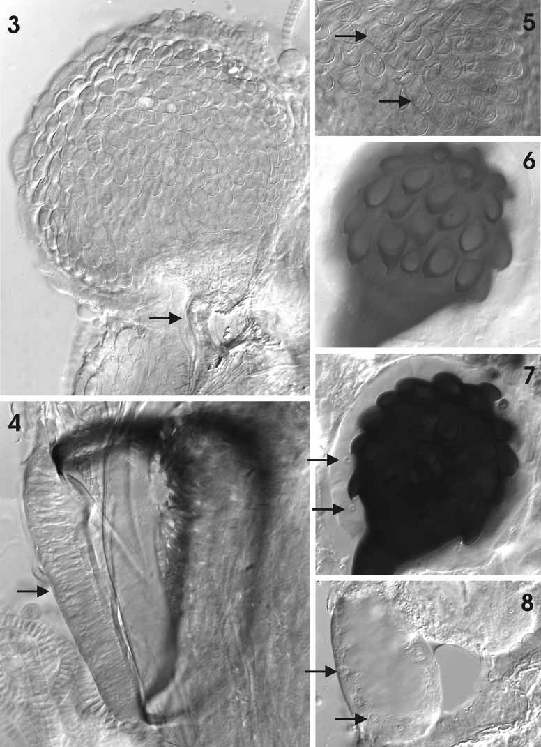
The tubular vagina is surrounded by a thick layer of muscles particularly in its anterior region. It is anteriorly connected to the common oviduct, which descends from the paired ovaries and lateral oviducts. The posterior end of the vagina is attached to the vulva behind sternum 8. From the ventral anterior portion of the vagina emanates the voluminous roundish ventral receptacle, which is composed of more than 300 tubular chambers, each with a diameter of about 15–20 μm ( Fig. 3 View FIGURES 3 – 8 ). In mated females these chambers house individual tightly coiled spermatozoa ( Fig. 5 View FIGURES 3 – 8 ). The ventral receptacle is connected with the lumen of the vagina via a narrow, tubular duct. A dense structure dorsal of the entrance may be part of a valve mechanism. Opposite the entrance of the ventral receptacle a pouchlike dorsal evagination of the vaginal lumen receives the spermathecal ducts and, ventrolateral of that, the ducts of the accessory glands ( Fig. 2 View FIGURES 1 – 2 ). Posterior to this a ringshaped ventral sclerite is embedded in the ventral wall of the vagina. Part of the vaginal musculature inserts on this ringshaped sclerite, thus sparing a cushion of specialized epithelium within its centre ( Fig. 4 View FIGURES 3 – 8 ). Two spherical spermathecae are present, one with a diameter of about 100 μm, the other slightly larger. Their strongly sclerotized, dark brown capsules are ornamented with short hollow denticles ( Fig. 6 View FIGURES 3 – 8 ). Each of these denticles is connected to the end apparatus of an epithelial gland cell ( Fig. 7 View FIGURES 3 – 8 ). The base of the spermathecae is not telescoped but drawn out into a tubular portion which merges smoothly with the apical ends of the long spermathecal ducts. The bases of the spermathecal ducts are slightly sclerotized as well next to their opening into the vagina. Like the spermathecae, the membranous ovoid reservoirs of the accessory glands are surrounded by epithelial gland cells with cuticular end apparatuses ( Fig. 8 View FIGURES 3 – 8 ). The delicate ducts of the accessory glands are only about 1/3rd as long as those of the spermathecae.
A description of the entire reproductive system of another stalk eyed fly, T. whitei (Curran) (detailed under its former name Cyrtodiopsis whitei ), was given by Kotrba (1993) including further details as well as physiological and functional aspects.
Etymology: The name refers to the type locality, Comoro Islands. Distribution: Mohéli, Comoro Islands

No known copyright restrictions apply. See Agosti, D., Egloff, W., 2009. Taxonomic information exchange and copyright: the Plazi approach. BMC Research Notes 2009, 2:53 for further explanation.
|
Kingdom |
|
|
Phylum |
|
|
Class |
|
|
Order |
|
|
Family |
|
|
Genus |