
Diasemopsis meigenii ( Westwood, 1837 )
|
publication ID |
https://doi.org/10.5281/zenodo.172483 |
|
DOI |
https://doi.org/10.5281/zenodo.6259143 |
|
persistent identifier |
https://treatment.plazi.org/id/03FB8784-CA47-FF96-4673-FE0384D1FB3F |
|
treatment provided by |
Plazi |
|
scientific name |
Diasemopsis meigenii ( Westwood, 1837 ) |
| status |
|
Diasemopsis meigenii ( Westwood, 1837) View in CoL
( Figs 10 View FIGURES 9 & 10 , 12, 1720 View FIGURES 17 – 20 )
syn. Diopsis breviseta Bezzi, 1908: 167 , Type locality: Ethiopia ( Eritrea)
Séguy (1955) established the new genus Chaetodiopsis for D. meigenii ( Westwood, 1837, originally in Diopsis ). However, Chaetodiopsis was treated as a junior synonym of Diasemopsis by the Catalogue of the Diptera of the Afrotropical Region ( Crosskey 1980) as well as by Meier & Baker (2002), based on sound molecular and morphological analysis. Chaetodiopsis is embedded deeply within Diasemopsis , i.e. its recognition would render Diasemopsis polyphyletic. Here we follow the revised classification suggested by Meier & Baker (2002).
Material studied: 5 males, 5 females taken from a laboratory culture housed at University College, London in May 2005. This culture was derived from a culture in the laboratory of G. S. Wilkinson, University of Maryland at College Park, which was originally founded from flies caught near Pietermaritzburg, South Africa in December 1994 by M. Kotrba. The dried, doublemounted specimens are deposited in the Hungarian Natural History Museum, Budapest.
Other material: 3 males, 3 females, Lourenço marques, Mozambique, Diopsis meigeni Westw. Det Lindner. Cameroun: 2 males, 3 females, Yaounde Obili, 20–2–1963, coll. L. Segers: 1 male, same data, 3–1–1963 (all Zoologische Staatssammlung, München); 1 female, Nkolbisson, Dept. NyongSanaga, IX. [19]68, L.G. Segers leg., van Schuytbroek det. 19 [no year given], Chaetodiopsis meigeni (Royal Museum for Central Africa). Republic of Congo: 1 male, Mayambé: Kiniati, 7–VI–1911, R. Mayné; 1male Ikengé, IX– 1912, R. Mayné, 5 males, Congo da Lemba, R. Mayné; dates: V–1912, V–1912, I– II– 1913, III–1913, IV–1913; 1 male, Lemfu, P. Vandereijst; 1 male, Mandungu, 25–XI–1912, R. Mayné (all Royal Museum for Central Africa).
Head. Completely covered with minute pale hairs, generally black, but the eye stalks are brown. Facial teeth 1.5 times longer than width of eye stalks in the middle. Outer vertical bristles at least as long as width of the eye stalk in the middle. Inner vertical bristles minute and weak, shorter than 1/3rd of width of the eye stalk in the middle.
Thorax. Uniformly grayish pollinose, except the surface of the meron. Metapleural spine yellow or orange yellow and straight in anterior view. Scutellar spines 2.5–3 times as long as the scutellum.
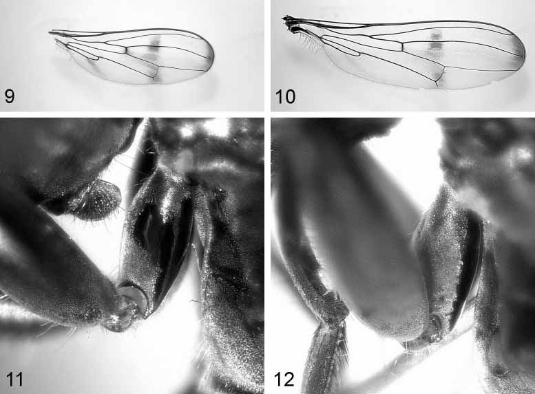
Wing Fig. 10 View FIGURES 9 & 10 . Completely hyaline, except for three infuscated brownish bands. The apical band is as broad as 1/14th of the wing length and is restricted to the space between M and R2+3 veins (the band reaches these veins). The central band is darkest around R4+5 and it is situated between R2+3 and Cu slightly extended towards the anterior cross vein (R–M). The proximal band is more an infuscated brownish spot below the cell cup.
Legs Fig. 12. Front coxae are only shiny on the posterior surface. Legs are yellowbrown in general, tibiae and first two tarsi of the front leg are black, tarsi 3–5 on the front leg are whitish yellow (clearly contrasting the other tarsi). Front femora incrassate (length/ width approximately 4), bearing on their ventral side two longitudinal rows of 2–4 prominent bristles each and between these two rows of 24–28 much shorter peglike tubercles each.
Preabdomen. Subshining black except for the following silver pollinose areas: an uninterrupted band along posterior margin of tergite 1, lateral triangles at posterior margin of tergite 2, distal half of tergite 3, and subsequent tergites.
Postabdomen (male) Figs 17–20 View FIGURES 17 – 20 . The hypandrium is connected to the aedeagal apodeme and the membranous tip of the hypandrium is continuous, not divided into two lobes anteriorly ( Fig. 17 View FIGURES 17 – 20 ). There are 3(–4) thick hairs on the medial surface of the hypandrium, the bilobed surstyli have numerous short, distinct hairs and the gonopods bear minute hairs ( Fig. 18 View FIGURES 17 – 20 ). In lateral view the aedeagal apodeme is more straight (that of D. comoroensis is curved) and also broadening towards tip. The ligament connecting to the hypandrium joins at basal 1/3rd of the aedeagal apodeme ( Fig. 20 View FIGURES 17 – 20 ). The epandrium and cerci have long, dispersed hairs along their surface. Hairs on the hypandrium are reaching to the lateral part and are longer than those of D. comoroensis . The distal half of the paramere is slightly narrowing towards the tip ( Fig. 19 View FIGURES 17 – 20 ).
Distribution: Widespread in Afrotropical regions.

No known copyright restrictions apply. See Agosti, D., Egloff, W., 2009. Taxonomic information exchange and copyright: the Plazi approach. BMC Research Notes 2009, 2:53 for further explanation.
|
Kingdom |
|
|
Phylum |
|
|
Class |
|
|
Order |
|
|
Family |
|
|
Genus |