
Russula luteolamellata C. L. Hou, H. Zhou & G. Q. Cheng, 2022
|
publication ID |
https://doi.org/ 10.11646/phytotaxa.556.2.3 |
|
DOI |
https://doi.org/10.5281/zenodo.6974813 |
|
persistent identifier |
https://treatment.plazi.org/id/03F987D9-FFB5-FF96-FF09-FD8DFF3D4DAD |
|
treatment provided by |
Plazi |
|
scientific name |
Russula luteolamellata C. L. Hou, H. Zhou & G. Q. Cheng |
| status |
sp. nov. |
Russula luteolamellata C. L. Hou, H. Zhou & G. Q. Cheng View in CoL sp. nov. ( Figs. 3 View FIGURE 3 , 4 View FIGURE 4 , 5 View FIGURE 5 )
MycoBank: MB 843987
Diagnosis:— Russula luteolamellata is diagnosed by a yellowish to pale orange pileus, white to yellowish lamellae with a few rust-colored spots on the margins, a low density of warts on the basidiospores, and pileocystidia that turn gray in SV. Morphologically, R. luteolamellata is easily confused with R. laevis but that species has an ochre cream to light brown very recurved pileus.
Holotype:— CHINA, Beijing, Yanqing District, Baihebao Village, 40°38’13.5” N, 116°9’43.1” E, elev. 628 m, 4 August , 2018, coll. C. L. Hou, J. Q. Li and H. Zhou ( BJTC 0534 About BJTC ). GoogleMaps
Etymology:—‘ luteolamellata ’ refers to the yellowish lamellae.
Description:— Basidiomata small to medium-sized. Pileus 30–94 mm in diam., hemispherical when young, applanate with a depressed center when mature;margin smooth,recurved slightly,no striae, cuticle peeling approximately to 1/2 of the pileus radius; yellowish (#ffffed) to pale orange (#fff2cd). Lamellae white (#ffffff) to yellowish (#ffffed), a few rust-colored (#b7410e) spots on the edges, adnate to adnexed, approximately 9–12 per cm near the pileus margin, not forked, lamellulae absent. Stipe 20–61 × 9–22 mm, white (#ffffff), sometimes light yellowish brown (#ffffba) at base, cylindrical, smooth, brittle, initially stuffed and compact, then spongy to slightly fistulose. Context 5–10 mm thick at halfway of the pileus radius, white (#ffffff), unchanging when bruised, odor not obvious, taste mild. Spore print not observed.
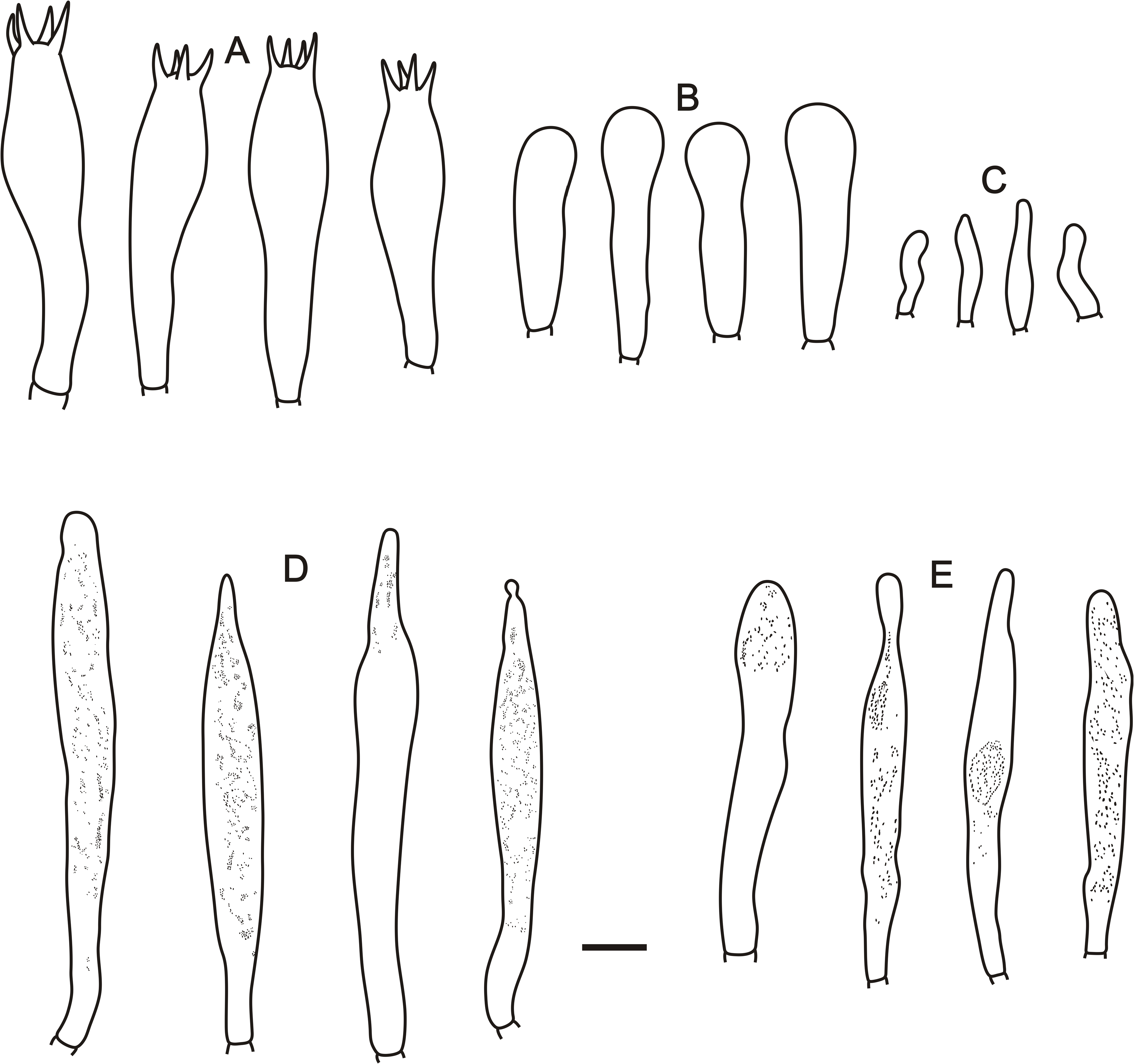
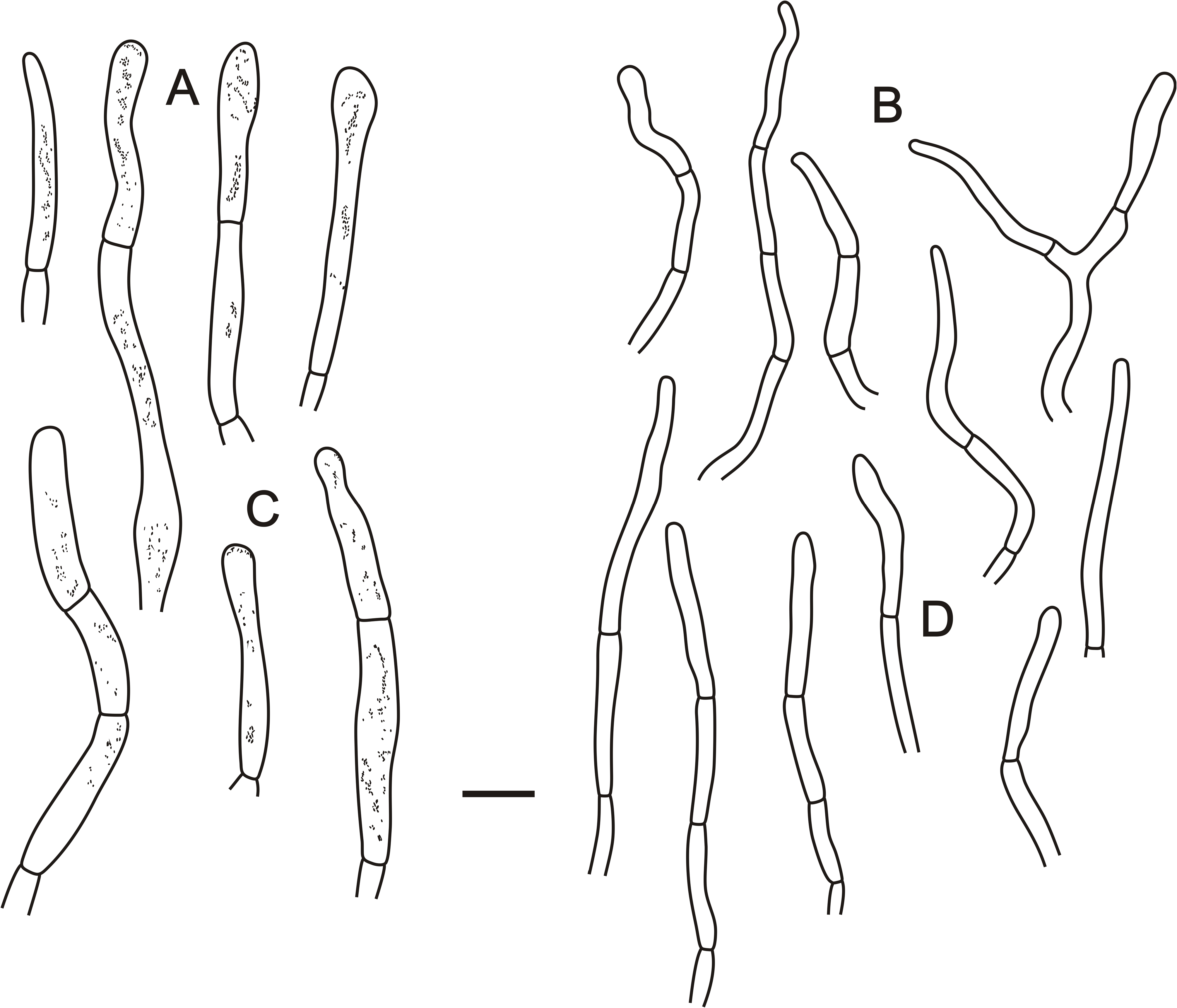
Basidiospores (8.6–)9.0–9.7–10.4(–11.8) × (7.4–)8.0–8.6–9.2(–10.3) μm, [Q = (1.00–)1.05–1.13–1.21(–1.35)], subglobose to broadly ellipsoid, ornamentation moderately large, moderately distant to dense [5–7 in a 3 μm diameter circle] amyloid warts, 0.4–1.2 μm high, forming a reticulated network, occasionally fused in pairs, triplets or short chains [0–2 in the circle], frequently connected by short or long, fine line connections [(1–) 2–4 in the circle], suprahilar spot large, amyloid. Basidia (60.5–)66.1–72.6–79.2(–83.7) × (14.8–)15.5–16.8–18.0(–20.1) μm, 2-, 4-spored, thinwalled, with guttate or granular contents; basidioles clavate or subcylindrical, ca. 7–14 μm wide. Hymenial cystidia on lamellae sides dispersed to moderately numerous, (80.4–)93.7–105.6–117.5(–127.2) × (–9.8)10.4–11.7–13.0(–14.2) μm, subcylindrical, clavate or subfusiform, apically mainly obtuse, few moniliform, sometimes with 4–10 μm long appendage, with heteromorphous-crystalline, occasionally banded contents, becoming yellowish brown (#ffff00) in sulfovanillin. Hymenial cystidia on lamellae edges (63.9–)69.8–75.3–80.9(–84.2) × (6.0–)7.8–9.5–11.2(–13.1) μm, clavate, occasionally cylindrical or subfusiform, sometimes with 2–8 μm long appendage, with crystalline or granulose. Marginal cells (11–)16.6–21.2–26.8(–30) × (3–)4.06–5.2–6.28(–7) μm, subcylindrical, often flexuous. Pileipellis orthochromatic in cresyl blue, sharply delimited from the underlying context, 190–240 μm thick, twolayered. Suprapellis 90–140 μm thick, not gelatinized, composed of erect, repent or ascending and near the surface loose hyphal terminations, inflated at base and attenuated towards terminal cells; Subpellis 100–150 μm thick, less gelatinized, composed of interwoven hyphae of 2–9 μm wide. Hyphal terminations near the pileus margin not branched, occasionally flexuous, thin-walled; terminal cells (24.2–)22.2–32.7–43.3(–51.6) × (2.9–)3.1–3.8–4.5(–5.1) μm, subcylindrical to cylindrical, sometimes apically attenuated, subterminal cells often short or infated, 4–6 μm wide, not forked. Hyphal terminations near the pileus center like those near the pileus margin, terminal cells (20.4–)25.3–32.3–39.4(–49.6) × (3.5–)3.6–4.3–5.0(–5.5) μm, mostly subcylindrical. Pileocystidia near the pileus margin (22.4–)24.2–46.7–69.0(–108.1) × (4.1–)4.7–6.4–8.1(–9.7) μm, thin-walled, cylindrical, clavate or narrowly fusiform, sometimes with 3–5 μm long appendage, contents with heteromorphous granulose or banded, becoming gray (#808080) in sulfovanillin. Pileocystidia near the pileus center (24.3–)26.2–44.4–58.6(–84.4) × (4.3–)5.0–6.5–7.9(–8.6) μm, thin-walled, subcylindrical, clavate or narrowly fusiform, apically mainly obtuse, sometimes with 2–4 μm long appendage, contents with heteromorphous-granulose. Cystidioid hyphae with heteromorphous-granulose contents in subpellis and context, oleiferous hyphae with yellow refringent contents, frequent in the subpellis. Clamp connections absent.
Habitat and distribution:—Scattered in a coniferous forest of Pinus tabuliformis Carrière in a warm temperate region of North China (Beijing).
Additional specimens examined: CHINA. Beijing, Yanqing District, Baihebao Village , 40°38’13.5” N, 116°9’43.1” E, elev. 1210 m, 4 August 2018, coll. C. L. Hou , J. Q. Li and H. Zhou ( BJTC T2201 ) GoogleMaps . CHINA. Beijing, Yanqing District, Baihebao Village , 40°38’43.6”, 116°9’13” E, elev. 628 m, 4 August 2018, coll. C.L. Hou, J.Q. Li and H. Zhou ( BJTC T2202 ) .

| C |
University of Copenhagen |
| L |
Nationaal Herbarium Nederland, Leiden University branch |
| J |
University of the Witwatersrand |
| Q |
Universidad Central |
| H |
University of Helsinki |
| BJTC |
Capital Normal University |
No known copyright restrictions apply. See Agosti, D., Egloff, W., 2009. Taxonomic information exchange and copyright: the Plazi approach. BMC Research Notes 2009, 2:53 for further explanation.
|
Kingdom |
|
|
Phylum |
|
|
Class |
|
|
Order |
|
|
Family |
|
|
Genus |