
Mymarothecioides xinguensis Soares and Domingues, 2019
|
publication ID |
https://doi.org/10.11646/zootaxa.4700.2.3 |
|
publication LSID |
lsid:zoobank.org:pub:C10998AB-822A-4B17-8FE1-596DC44C7D6A |
|
DOI |
https://doi.org/10.5281/zenodo.5614570 |
|
persistent identifier |
https://treatment.plazi.org/id/EE0E7DDE-5148-44C5-A496-D0DC0D75F398 |
|
taxon LSID |
lsid:zoobank.org:act:EE0E7DDE-5148-44C5-A496-D0DC0D75F398 |
|
treatment provided by |
Plazi |
|
scientific name |
Mymarothecioides xinguensis Soares and Domingues |
| status |
sp. nov. |
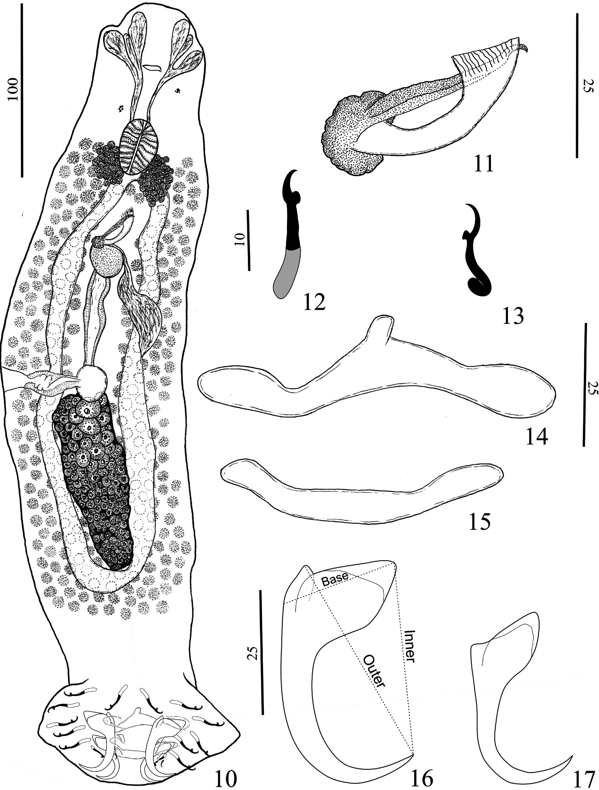
Mymarothecioides xinguensis Soares and Domingues n. sp.
( Figs. 10–17 View FIGURES 10–17 ; 43 View FIGURES 42–45 )
Type host: Hydrolycus armatus (Jardine & Schomburgk) .
Site: Gills.
Type locality: Volta Grande , Xingu River, municipality of Altamira, Pará State, Brazil ( 03°21’15,7’’S; 52°11’47,5’’W), collected on June 13, 2015 GoogleMaps .
Prevalence: 100% of three hosts examined.
Mean intensity: 10 parasites per infected host.
Specimens deposited: Holotype, MPEG nº 181; 7 paratypes, MPEG n° 182–188.
Etymology: The specific name is derived from the type locality, the Xingu River, Pará State, Brazil.
Zoobank Life Science Identifier: (LSID) for Mymarothecioides xinguensis n. sp. is urn:lsid:zoobank.org:act:
Comparative measurements: see Table 3 View TABLE 3 .
Description (based on eight specimens, three mounted in Hoyer, five mounted in Gomori’s trichrome): Body fusiform, total length excluding haptor 396 (315–450; n= 4), total width at level of germarium 129 (122–142; n=4) ( Fig. 10 View FIGURES 10–17 ). Cephalic margin tapered; moderately developed terminal lobes; three bilateral pairs of head organs, with rod-shaped secretion; cephalic glands unicellular, posterolateral to the pharynx. Accessory chromatic granules present in cephalic area. Pharynx ovate, 29 (24–33; n= 3) long, 22 (19–24; n=3) wide. Testis pyriform, 83 (71–103; n=4) long, 45 (41–47; n=4) wide. Prostatic reservoir saccate, posterior to MCO. MCO, 33 (30–36; n=5) long, with small grooves at edges of distal opening; base of MCO with sclerotized margin ( Figs. 11 View FIGURES 10–17 , 43 View FIGURES 42–45 ). Accessory piece comprising straight rod, with hook-shaped distal portion. Germarium fusiform, 75 (71–79; n=3) long, 24 (n=3) wide. Eggs, Mehlis’ glands, ootype not observed. Vaginal aperture marginal (dextral), opening at level of vitelline commissure; vaginal vestibule heavily muscular; vaginal canal sigmoid. Seminal receptacle subspherical. Uterus delicate. Vitelline follicles dense. Peduncle broad; haptor pentagonal, 95 (85–100; n=4) long, 119 (100–137; n=4) wide. Anchors similar. Ventral anchor, outer 43 (39–47; n=5) long, inner 42 (36–47; n=5) long; base 24 (22–25; n=5); superficial root triangular, with delicate sclerotization on distal extremity; deep root poorly developed; slightly curved shaft and point; point extending to level of tip of superficial root ( Fig. 16 View FIGURES 10–17 ). Dorsal anchor, outer 32 (27–38; n=5) long, inner 34 (30–39; n=5) long; base 24 (22–25; n=5); superficial root triangular, with delicate sclerotization on distal portion; deep root poorly developed; slightly curved shaft and point, point extending just past level of tip of superficial root ( Fig. 17 View FIGURES 10–17 ). Ventral bar, 63 (55–71; n=6) long, 12 (11–14; n=5) wide, slightly curved rod, with anteromedial projection, enlarged ends ( Fig. 14 View FIGURES 10–17 ). Dorsal bar, 51 (44–61; n=5) long, 6 (5–8; n=5) wide, broadly V-shaped rod with rounded ends ( Fig. 15 View FIGURES 10–17 ). Hooks similar in shape, hook pairs 1–4, 6 –7 26 (20–33; n=19) long, with upright thumb, rounded, short shaft, slightly curved, shank divided into two units; filamentous hook loop not observed ( Fig. 12 View FIGURES 10–17 ); hook pair 5, 17 (15–20; n=4) long, shank with proximal dilatation comprising approximately 1/3 of the shank length; filamentous hook loop not observed ( Fig. 13 View FIGURES 10–17 ).
Remarks: Features that distinguish the new species from most of its congeners include a hook-shaped accessory piece without the subterminal branch at the distal portion, a dextromarginal vaginal pore, and ventral and dorsal anchors with triangular superficial roots.

| MPEG |
Museu Paraense Emilio Goeldi |
No known copyright restrictions apply. See Agosti, D., Egloff, W., 2009. Taxonomic information exchange and copyright: the Plazi approach. BMC Research Notes 2009, 2:53 for further explanation.
|
Kingdom |
|
|
Phylum |
|
|
Class |
|
|
Order |
|
|
Family |
|
|
Genus |