
Syspira, Simon, 1895
|
publication ID |
https://doi.org/ 10.11646/zootaxa.4894.3.7 |
|
publication LSID |
lsid:zoobank.org:pub:37DC47AA-45C7-4729-A28F-5539FD26F21B |
|
DOI |
https://doi.org/10.5281/zenodo.4331414 |
|
persistent identifier |
https://treatment.plazi.org/id/03F9306A-9729-FFCE-FF1F-F9E40D9CFB62 |
|
treatment provided by |
Plazi |
|
scientific name |
Syspira |
| status |
|
Key for Hispaniolan Syspira View in CoL View at ENA species
1. Male............................................................................................... 2
- Female............................................................................................. 6
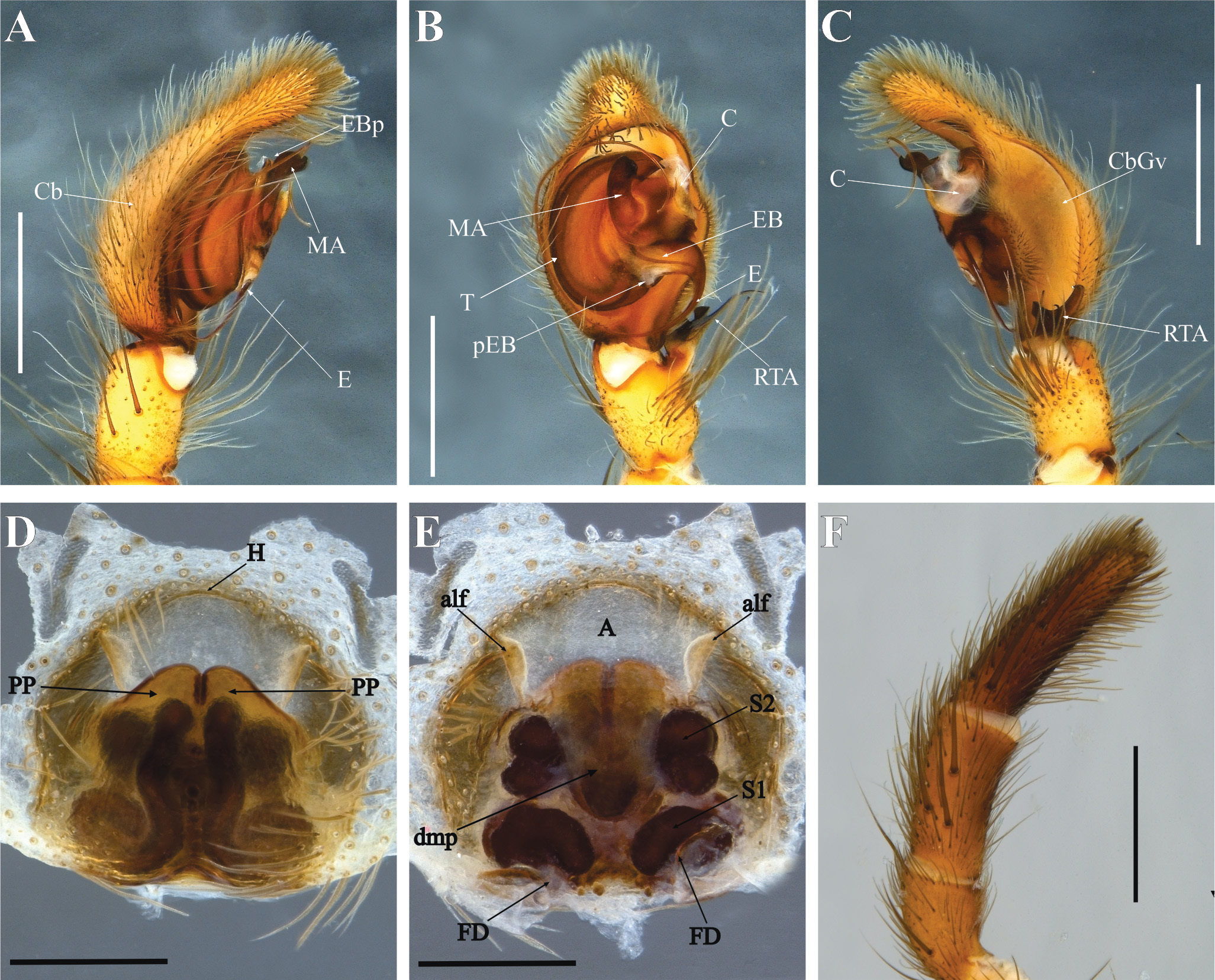
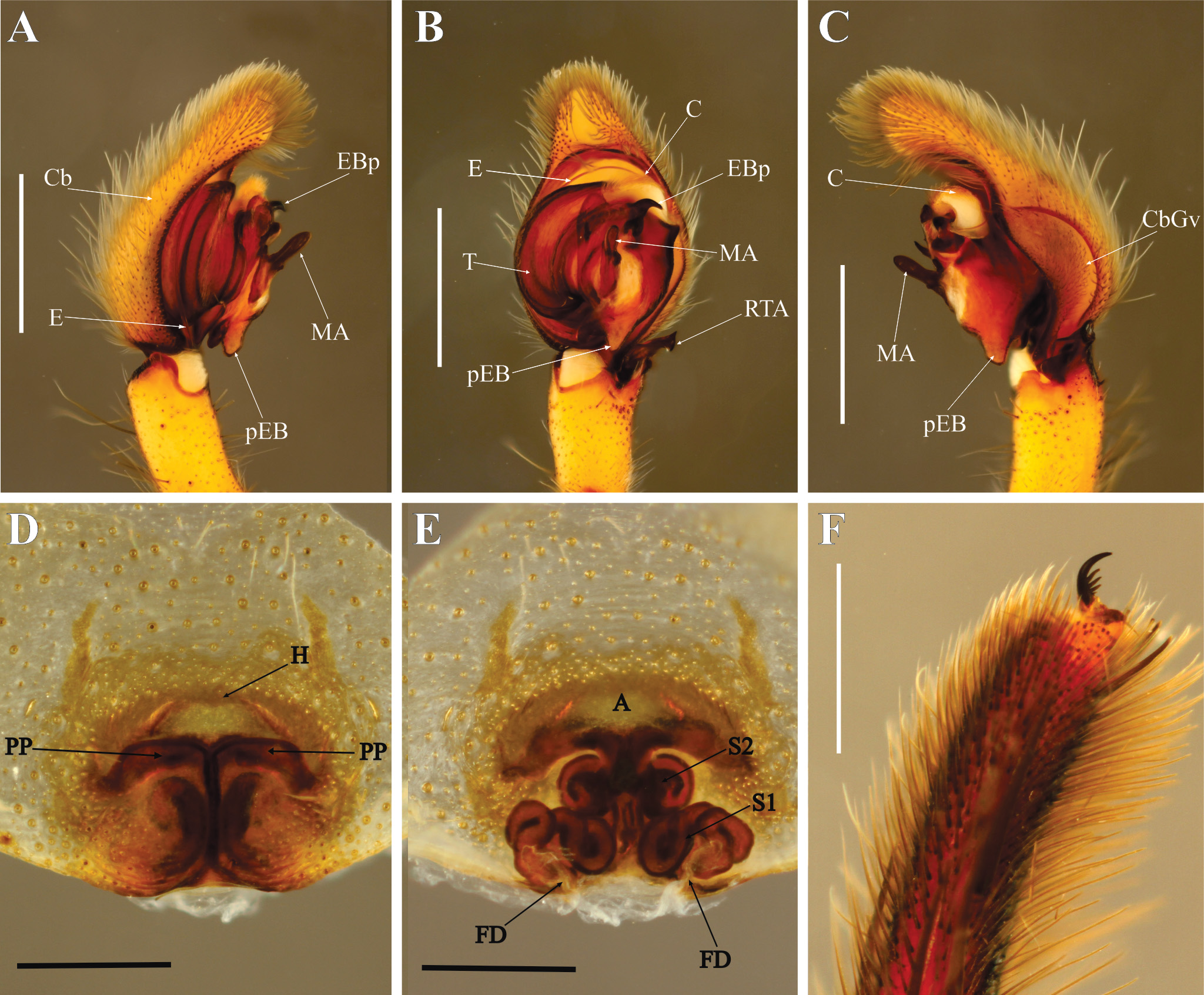
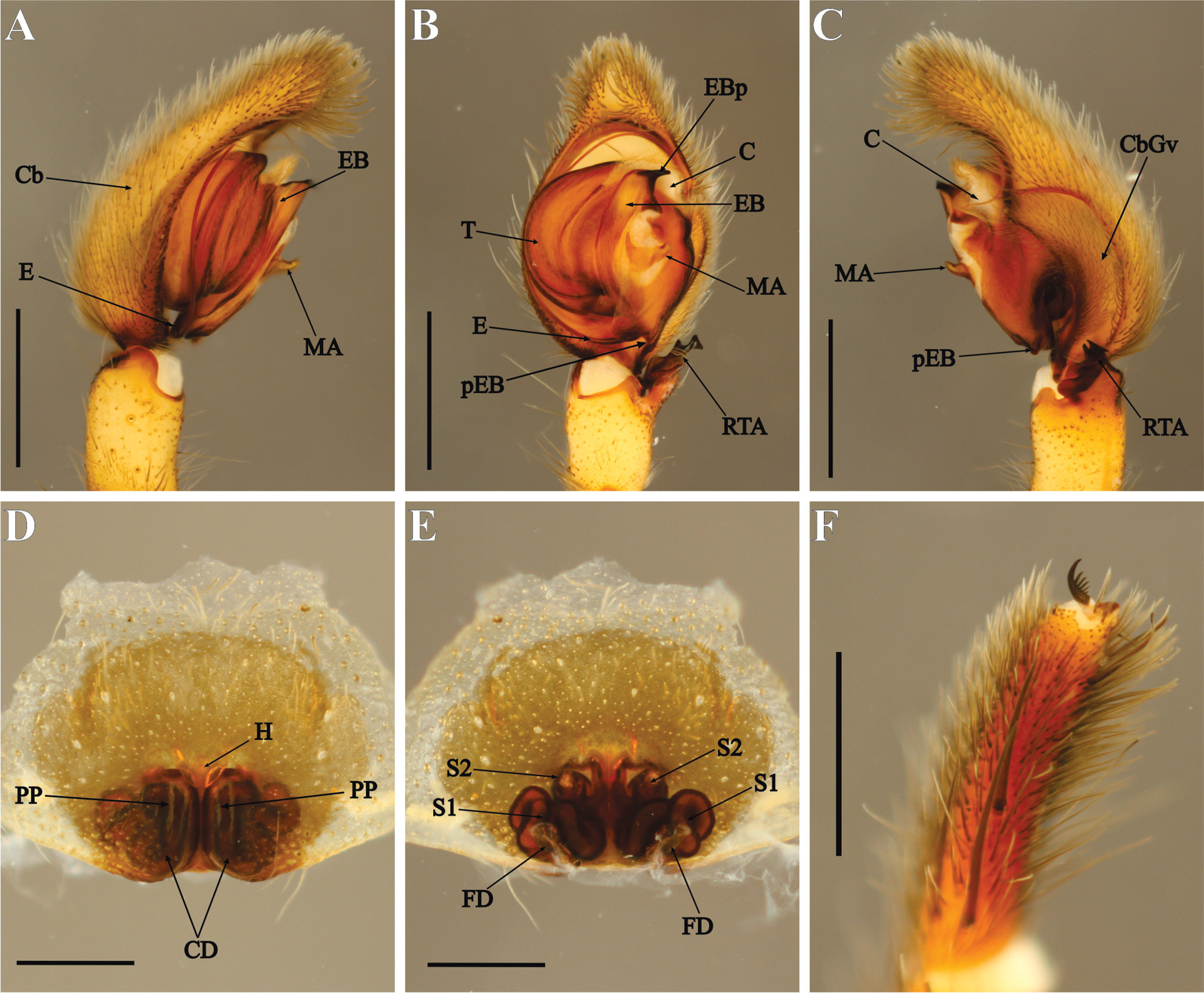
2. Cymbial groove not reaching the ventral distal margin of cymbium ( Figs 1C View FIGURE 1 , 2C View FIGURE 2 ); RTA enlarged and curved ( Figs 1B View FIGURE 1 , 2B View FIGURE 2 ); apical projection of embolar base small and not curved and with lighter sclerotization ( Figs 1A View FIGURE 1 , 2A View FIGURE 2 ); process on embolar base inconspicuous ( Figs 1A View FIGURE 1 , 2B View FIGURE 2 ) (the cimitarra View in CoL group).......................................................... 3
- Cymbial groove distally closed, reaching the ventral margin of cymbium ( Figs 4C View FIGURE 4 , 5C View FIGURE 5 , 6C View FIGURE 6 ); RTA short and truncated ( Figs 4B View FIGURE 4 , 5B View FIGURE 5 , 6B View FIGURE 6 ); bigger and curved embolar base apical projection with heavy sclerotization ( Figs 4B View FIGURE 4 , 5B View FIGURE 5 , 6B View FIGURE 6 ); process on embolar base developed ( Figs 4A View FIGURE 4 , 5A View FIGURE 5 , 6A View FIGURE 6 ) (the agujas View in CoL group)........................................................ 4
3. Median apophysis with finger-shaped tip ( Fig. 1B View FIGURE 1 ; Brescovit et al. 2018: fig. 2C); RTA elongated and curved distally, with bifurcated projections unequal in size and with the longer projection striated at tip ( Fig. 1C View FIGURE 1 , Brescovit et al. 2018: fig. 2F)............................................................................................. S. cimitarra View in CoL
- Median apophysis with bifurcated tip ( Fig. 2 A View FIGURE 2 , Brescovit et al. 2018: fig. 4C); RTA shorter, sickle-shaped, with bifurcated projections almost equal in size ( Fig. 2B – C View FIGURE 2 , Brescovit et al. 2018: fig. 4F)................................. S. jimmyi View in CoL
4. Median apophysis enlarged and heavily sclerotized ( Fig. 4B, C View FIGURE 4 ); apical projection on embolar base bowed, claw-shaped ( Fig. 4A, B View FIGURE 4 )........................................................................................ S. agujas View in CoL
- Median apophysis small ( Figs 5B – C View FIGURE 5 , 6B – C View FIGURE 6 ); apical projection of embolar base not claw-shaped ( Figs 5A – B View FIGURE 5 , 6A – B View FIGURE 6 )...... 5
5. Embolar base process fairly developed ( Fig. 5A, C View FIGURE 5 ); spatula-shaped median apophysis in ventral view ( Fig. 5B View FIGURE 5 ); apical projection of embolar base slightly curved in ventral view ( Fig. 5B View FIGURE 5 )........................................ S. medialuna View in CoL
- Embolar base process small ( Fig. 6A – C View FIGURE 6 ); very small finger-shaped median apophysis ( Fig. 6A, C View FIGURE 6 ); apical projection of embolar base strongly curved in ventral view ( Fig. 6B View FIGURE 6 ).................................................. S. armasi View in CoL sp. n.
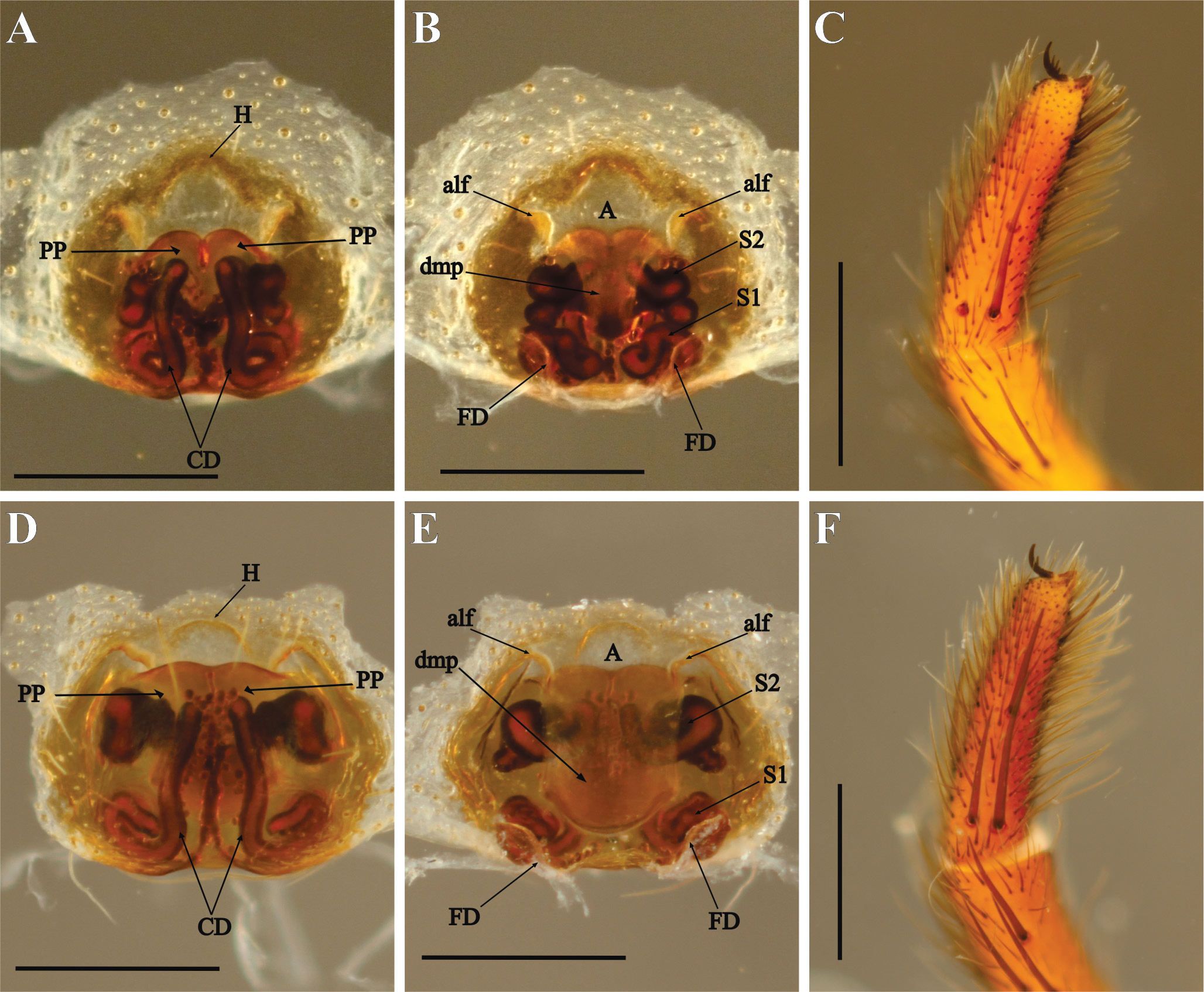
6. Epigynal posterior plates (PP) medially fused at the inner borders; epigynal median plate (dmp) projecting dorsally from the atrium ( Figs 1D – E View FIGURE 1 , 2D – E View FIGURE 2 , 3A – B, D – E View FIGURE 3 ) (the cimitarra View in CoL group)................................................. 7
- Epigynal posterior plates (PP) not fused, touching (or not) the inner borders and lacking the epigynal dorsal median plate (dmp) ( Figs 4D – E View FIGURE 4 , 5D – E View FIGURE 5 , 6D – E View FIGURE 6 , 7A – B View FIGURE 7 , 8D View FIGURE 8 ) (the agujas View in CoL group).................................................... 10
7. Anterior edges of epigynal posterior plates (PP) slightly straight and close together, posterior edges widely separated from each other ( Fig. 3D – E View FIGURE 3 )....................................................................... S. barbacoa View in CoL sp. n.
- Anterior edges of epigynal posterior plates (PP) rounded and elevated, and posterior edges fused ( Figs 1D – E View FIGURE 1 , 2D – E View FIGURE 2 , 3A – B View FIGURE 3 ). ................................................................................................... 8
8. Dorsal median plate (dmp) narrow, 2.5 times longer than wide, atrial hood (H) sclerotized ( Fig. 3A, B View FIGURE 3 )..... S. alayoni View in CoL sp. n.
- Dorsal median plate (dmp) wide, less than 1.5 times longer than wide, atrial hood (H) weakly sclerotized or inconspicuous ( Figs 1D View FIGURE 1 , 2D View FIGURE 2 )............................................................................................. 9
9. Dorsal median plate (dmp) truncated at the distal margin ( Fig. 1E View FIGURE 1 ), epigynal posterior plates (PP) wider, exceeding in half the atrial lateral dorsal folds ( Fig. 1D View FIGURE 1 )............................................................... S. cimitarra View in CoL
- Dorsal median plate (dmp) rounded at the distal margin, epigynal posterior plates (PP) narrower, barely exceeding the atrial lateral dorsal folds ( Fig. 2D – E View FIGURE 2 )................................................................... S. jimmyi View in CoL
10. Anterior edges of epigynal posterior plates (PP) wide and straight ( Fig. 4D View FIGURE 4 )................................. S. agujas View in CoL
- Anterior edges of epigynal posterior plates (PP) relatively narrow and rounded ( Figs 5D View FIGURE 5 , 6D View FIGURE 6 , 7A View FIGURE 7 , 8D View FIGURE 8 )................. 11
11. Atrial hood (H) dorsally projected from the atrium ( Figs 6D View FIGURE 6 , 7D – E View FIGURE 7 )............................................ 12
- Atrial hood (H) not projected, remaining as a fold ( Figs 5D View FIGURE 5 , 8D View FIGURE 8 )............................................... 13
12. Atrial hood (H) large and weakly sclerotized, completely covering anterior edges of epigynal posterior plates ( Fig. 6D View FIGURE 6 )............................................................................................ S. armasi View in CoL sp. n.
- Atrial hood (H) small and heavily sclerotized, covering only part of anterior edges of epigynal posterior plates ( Fig. 7D – E View FIGURE 7 )........................................................................................ S. bryantae View in CoL sp. n.
13. Epigynal posterior plates (PP) large, with quite high anterior edges, twice the size seen in other species ( Fig. 5D View FIGURE 5 )................................................................................................... S. medialuna View in CoL
- Epigynal posterior plates (PP) smaller, with anterior edges similar in size to the other species ( Fig. 8D View FIGURE 8 ).................................................................................................. S. monticola View in CoL comb. nov.


No known copyright restrictions apply. See Agosti, D., Egloff, W., 2009. Taxonomic information exchange and copyright: the Plazi approach. BMC Research Notes 2009, 2:53 for further explanation.