
Diognetus intonsus Distant, 1904
|
publication ID |
https://doi.org/10.37520/aemnp.2023.001 |
|
publication LSID |
lsid:zoobank.org:pub:3F2C90B1-6EA1-4B38-A218-C314D09F6E00 |
|
persistent identifier |
https://treatment.plazi.org/id/03F587DF-FFEA-E110-0DCD-6D27FD44FC04 |
|
treatment provided by |
Felipe |
|
scientific name |
Diognetus intonsus Distant, 1904 |
| status |
|
Diognetus intonsus Distant, 1904 View in CoL View at ENA
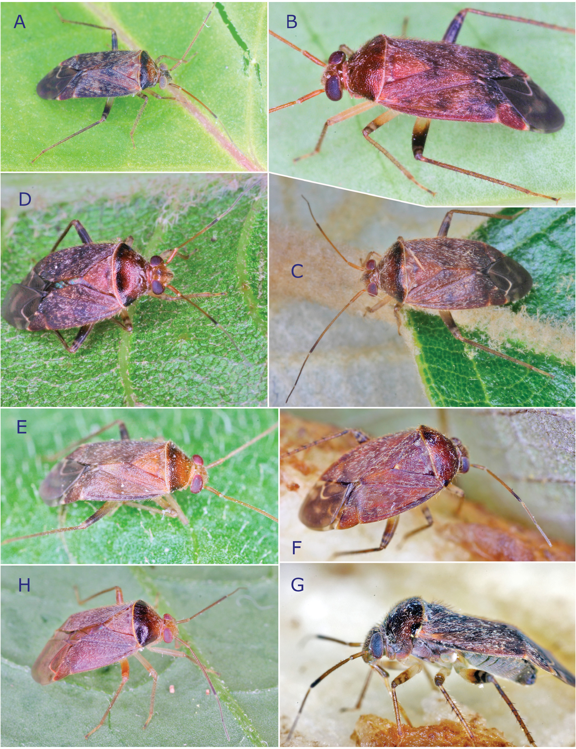
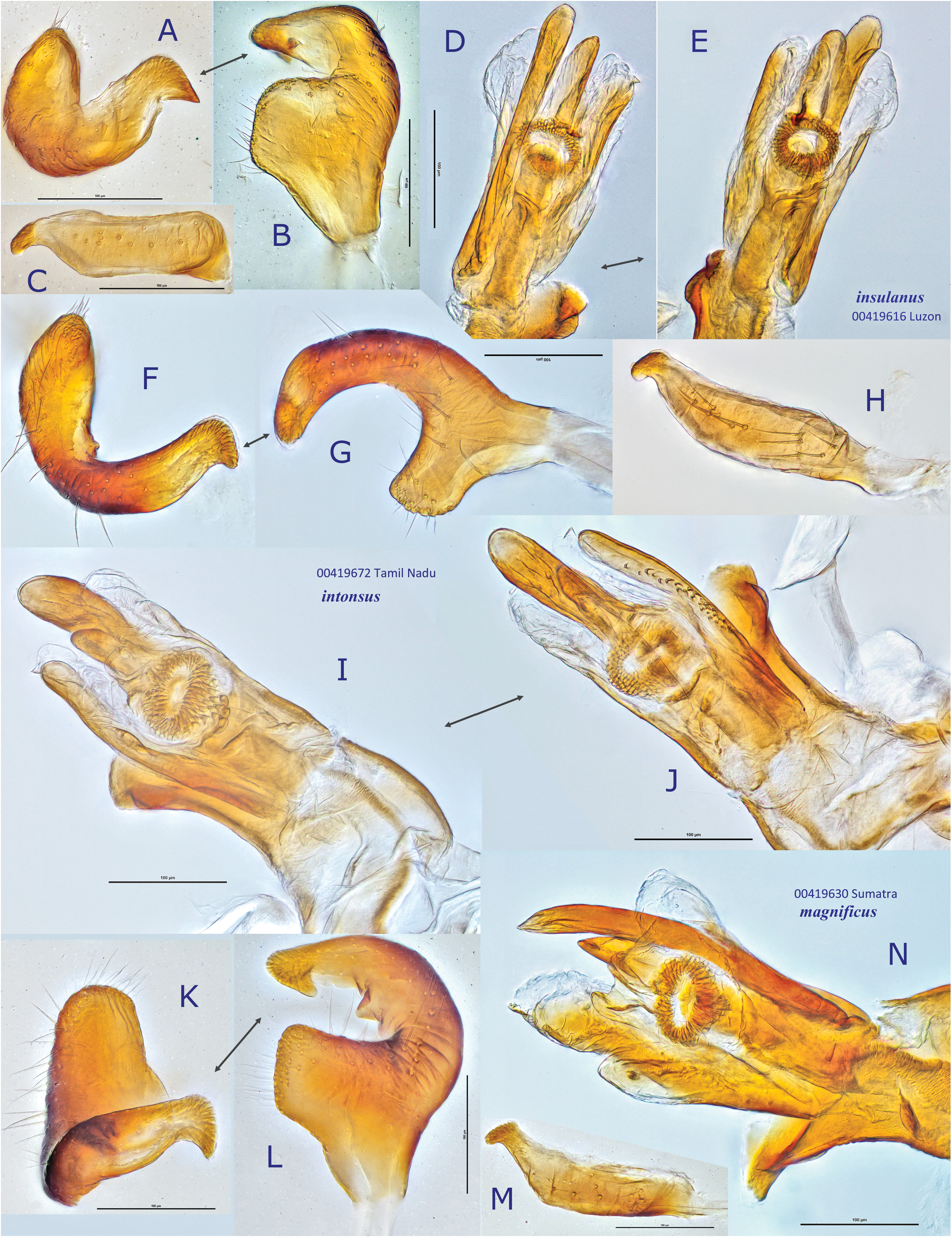
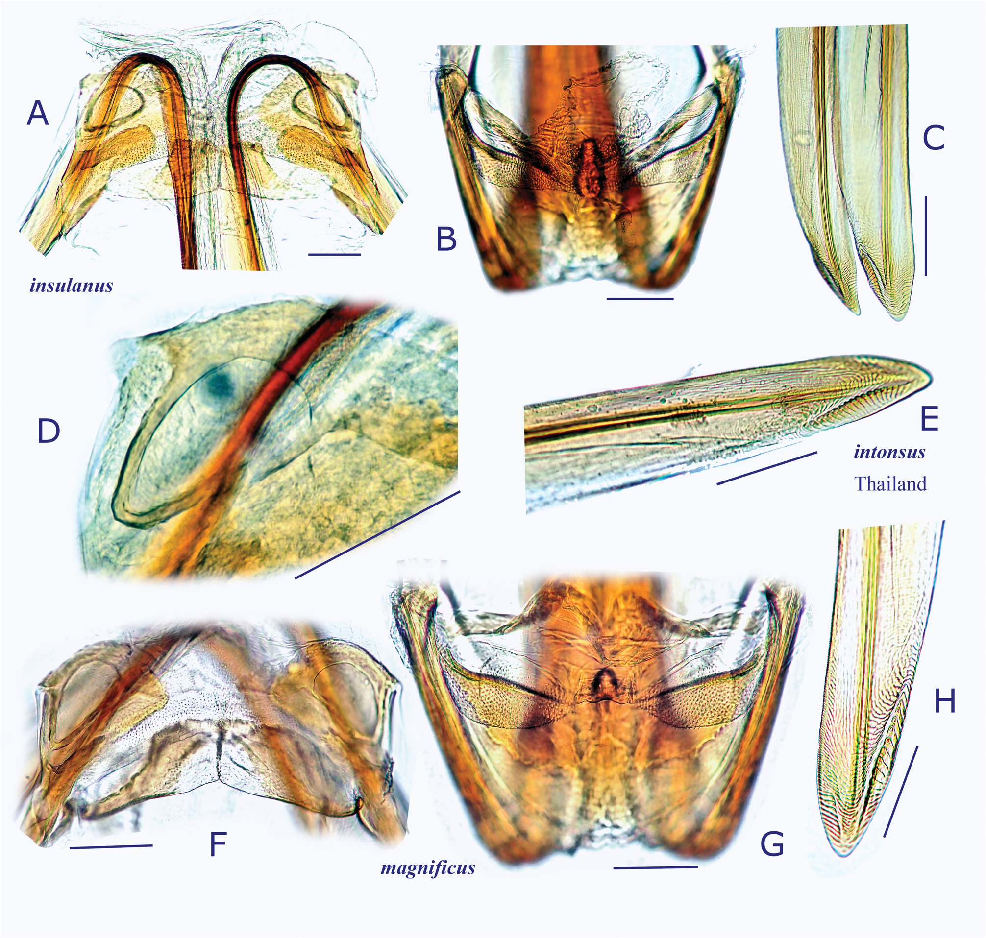
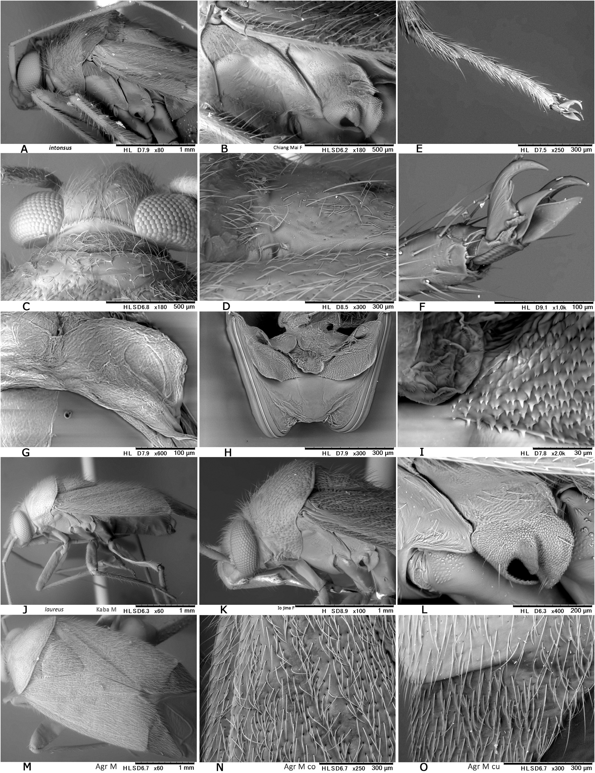
( Figs 2G View Fig , 9C− D View Fig , 14C− F View Fig , 15F− J View Fig , 16D− E View Fig , 30A− I View Fig )
Diognetus intonsus Distant, 1904b: 432 View in CoL (as new species).
Diognetus intonsus: SCHUH (1995) View in CoL : 760 (catalog), SCHUH (2002 –2013) (online catalog); CH ḖROT et al. (2017): 93–94 (diagnosis, DV, MG, FG); SAHA et al. (2020): 279 (faunal list).
Diophantus literatus Distant, 1909: 510–511 View in CoL (as new species).
Diophantus literatus: SCHUH (1995) View in CoL : 760 (catalog), SCHUH (2002 –2013) (online catalog); CH ḖROT et al. (2017): 94 (synonymy).
Diplotrichiella rufescens Poppius, 1915 a: 66 View in CoL (as new species). New junior subjective synonym.
Diplotrichiella rufescens: SCHUH (1995) View in CoL : 761 (catalog), SCHUH (2002 – 2013) (online catalog).
Type material examined. Diognetus intonsus : LECTOTYPE: ♀ ( BMNH), SRI LANKA: Ceylon , Maskelyia, Green (no further data, BMNH).
Diognetus literatus : LECTOTYPE: ♀ ( BMNH), SRI LANKA: Ceylon , Ohiya, Green (no further data, damaged specimen, BMNH).
Diplotrichiella rufescens : HOLOTYPE: ♀ ( MZHF), INDIA: TAMIL NADU: Trichinopoly, 1898, Coll. Nouhalier (vertically compressed and partly damaged condition).
Additional material examined. INDIA: TAMIL NADU: Madras,Anamalai Hills, Cinchona, 10.28228, 76.97999, 1,067 m ( 3,500 ft), Apr 1959, P. S. Nathan, 1♀ ( AMNH _ PBI 00419635) ( CNC); same locality, May 1965; P. S. Nathan, 3 JJ 2 ♀♀ (00419670–00419674) ( CNC). THAILAND: CHIANG MAI: Doi Pui, 18.826460, 98.892088, UV lighting, 23–24 Oct 1981, S. Sakurai, 3 ♀♀ ( DOAT, TYCN).
Redescription. Body oval, moderate in size, 4.8–5.3 mm. COLORATION: Dorsum castaneous, usually tinged with red ( Figs 14C− D View Fig ), usually with mottled pattern ( Fig. 14C View Fig ). Antennae yellowish brown; apical 1/6−1/5 of segment II, segment III (except for pale extreme base) and segment IV brown. Labium pale brown, partly tinged with red. Pronotum reddish-brown, relatively shining, with posterior 1/3−1/2 darkened and posterior margin pale; anterior mesal part of scutellum sometimes narrowly infuscate; pleura fuscous; scent efferent system creamy yellow. Hemelytron castaneous to reddish-brown, without mottled pattern; border between apex of corium and cuneal base narrowly pale; membrane pale smoky brown, with irregular pale, semitransparent maculae and pale veins. Coxae and legs yellowish brown; apical 1/3−1/2 of metafemur darkened, with two (subapical and apical) pale rings ( Fig. 14F View Fig ). Ventral surface of abdomen pale brown, with more or less reddish lateral margins. SURFACE AND VESTITURE: As in generic diagnosis; dorsum with densely distributed, silvery, reclining setae; base of mesepisternum with long, erect setae ( Fig. 30B View Fig ); hemelytron rather matte. STRUCTURE: Vertex relatively wide ( Fig. 30C View Fig ). Labium reaching apex of metacoxa ( Fig. 13F View Fig 13 ). Scutellum weakly swollen, shallowly and sparsely punctate, less rugose.
Metathoracic scent efferent system as in Fig 30B View Fig . Metatarsomere II as long as III ( Fig. 30E View Fig ); pretarsal structure as in Fig. 30F View Fig ; parempodia slightly shorter than claw. MALE GENITALIA ( Figs 15F− J View Fig ): Left paramere generally broad, with small, median process and flattened apex ( Fig. 15F View Fig ). Vesica with short, narrowed MS, medialy spinulate LS and tiny TP ( Figs 15I–J View Fig ). FEMALE GENITALIA ( Figs 9C–D View Fig , 16D− E View Fig , 30G− I View Fig ): Sclerotized ring with thickened anterior rim, posterolateral angle somewhat angulate ( Fig. 16D View Fig ); posterior wall ( Figs 30H− I View Fig ) with wide, rounded interramal lobe; dorsal structure lacking spinulate processes ( Fig. 30I View Fig ).
Measurements. See Table 1.
Differential diagnosis. Recognized and distinguished from other congeners by the following combination of characters: body moderate-sized, ovoid; dorsum with relatively dense, reclining, silvery setae; scutellum less rugose, with sparsely distributed, fine punctures; left paramere generally stout, with a small median process on sensory lobe; posterior wall with rather wide interramal lobe; and dorsal structure lacking spinulate processes.
Biology. Unknown; most of available specimens were collected by UV lighting method at forest zones in the Oriental Region.
Distribution. India ( Tamil Nadu), Sri Lanka, Thailand ( Chiang Mai); record from Papua New Guinea by CH ḖROT et al. (2017) most probably represents an undescribed species.


No known copyright restrictions apply. See Agosti, D., Egloff, W., 2009. Taxonomic information exchange and copyright: the Plazi approach. BMC Research Notes 2009, 2:53 for further explanation.
|
Kingdom |
|
|
Phylum |
|
|
Class |
|
|
Order |
|
|
Family |
|
|
Genus |
Diognetus intonsus Distant, 1904
| Yasunaga, Tomohide, Schwartz, Michael D. & Chérot, Frédéric 2023 |
Diognetus intonsus : SCHUH (1995)
| SAHA P. C. & KUSHWAHA S. & HASSAN M. E. & PANT H. 2020: 279 |
| SCHUH R. T. 1995: 760 |
Diophantus literatus :
| SCHUH R. T. 1995: 760 |
Diplotrichiella rufescens : SCHUH (1995)
| SCHUH R. T. 1995: 761 |
Diophantus literatus
| DISTANT W. L. 1909: 511 |
Diognetus intonsus
| DISTANT W. L. 1904: 432 |