
Naxia atlantica, Poore, Gary C. B., 2014
|
publication ID |
https://doi.org/ 10.11646/zootaxa.3861.1.5 |
|
publication LSID |
lsid:zoobank.org:pub:BBDD80D8-59DA-40BD-B50E-431A8648C0BC |
|
DOI |
https://doi.org/10.5281/zenodo.6140756 |
|
persistent identifier |
https://treatment.plazi.org/id/03F50D68-FFC3-5E7F-FF61-F981FBABF86E |
|
treatment provided by |
Plazi |
|
scientific name |
Naxia atlantica |
| status |
sp. nov. |
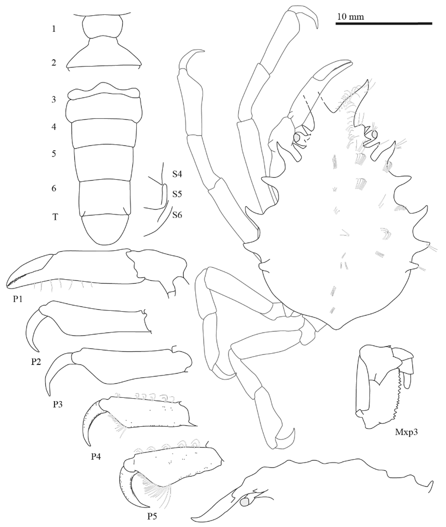
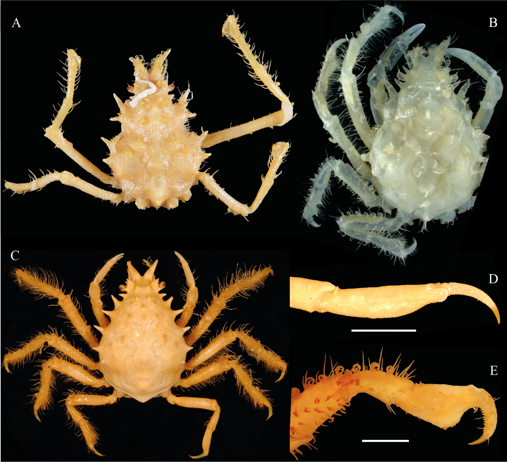
Naxia atlantica View in CoL n. sp.
( Figs 1 View FIGURE 1 , 2B View FIGURE 2. A – C )
Type Material. Holotype. Off the coast of São Paulo, Brazil, REVIZEE SUL, stn 6670, 11 Nov. 1998, 503 m: male cl 23.2 mm, cw 13.0 mm ( MZUSP 32370).
Comparative material. Naxia aries (H. Milne Edwards, 1834) : Australia, Bass Strait, 40°06.2'S, 148°25.0'E, coarse shell, 22 m: 1 male cl 31 mm ( MZUSP 32366).
Naxia aurita (Latreille, 1825) : Australia, Victoria, Port Phillip, off Swan Island, dredged in less than 1.8 m: 1 female cl 47 mm ( USNM 76543). Victoria: 1 male cl 45 mm ( MZUSP 32369).
Naxia spinosa (Hess, 1865) : Australia, Victoria, Hamers Haven: male cl 36 mm ( MZUSP 32367).
Naxia tumida (Dana, 1851) : Australia, New South Wales, Little Bay near Port Jackson, among seaweed in rock pools between tide marks: 1 male cl 30 mm ( USNM 64728). Victoria, Port Phillip Heads: 1 male cl 26 mm and 1 ovigerous female cl 17 mm ( MZUSP 32368).
Diagnosis. Length:width ratios of pereopods 2–5 propodi 3.5, 3.1, 2.8 and 2.4 respectively. Anterolateral spine of the basal antennal article with an accessory spinule. Prehepatic spine well separated from supraorbital eave.
Description. Carapace pyriform, postrostral carapace length 1.5 times width. Rostral spines (measured obliquely) 0.2 times as long as cl, separated basally by right-angled curve, unarmed, cylindrical, outwardly directed, ending in upturned acute spine, densely covered dorsally with hooked setae. Orbital margin with supraorbital eave produced anteriorly into rounded boss; eave armed posteriorly with strong, sharp spine separated from anterior boss by U-shaped notch. Hepatic region with 2 spines, anterior spine outwardly directed, large, sharp; posterior spine laterally directed, shorter than anterior spine; with small subhepatic spine. Dorsal surface with regions weakly defined by shallow grooves, with widely-spaced obsolete tubercles, bosses, each with few simple or hooked setae: 2 frontal bosses; protogastric regions with 3 tubercles each; 5 mesogastric; 4 metagastric; 2 urogastric; 1 low cardiac; 1 medial intestinal, 1 erect intestinal spine on mid-posterior margin of carapace. Branchial regions inflated laterally, greatest width 1.3 times width at branchial-hepatic juncture; epibranchial with 3 tubercles each; 3 mesobranchial stout, sharp spines, first largest, anteriorly directed, second smaller, third smallest, both laterally directed; metabranchial region naked.
Epistome concave, smooth; anterolateral margin of buccal cavity carinate, paired. Antennular articles unarmed; first short; second cylindrical; third gently compressed laterally, distally broadened. Antennal articles 2+3 ventrally flat, with prominent triangular distolateral spine (with microscopic subdistal lateral tooth), short ventral spine at base of article 3; remaining articles cylindrical. Eyestalk with 3 club setae, 3 longer stiff setae on small tubercle on disto-anterior margin. Cornea unpigmented; ommatidia very small, clearly recognizable. Maxilliped 3 ischium 2.4 times as long as wide, with 20 blunt teeth along mesial margin, strongly excavated distolaterally to accommodate merus, distal margin with small, incurved spine; merus with strong mesial triangular projection, exceeding mesial margin of ischium, with semicircular distolateral lobe, small distal spine.
Male sterno-abdominal cavity limited laterally by small, spherical tubercle at level of thoracic sternite 7, large boss at level of sternite 8. Complementary parts of abdominal locking system present, functional; thoracic sternal button adjacent to sternal suture 5/6; abdominal socket well excavated.
Cheliped smooth; palm 10 times as long as greatest depth, tapering; fingers 0.2 times upper length of palm, each with about 15 small teeth along margins. Pereopod 2 as long as cl, others decreasing in length posteriorly to 0.9 cl (pereopod 5); merus, carpus of all walking legs cylindrical. Propodi of pereopods 4, 5 about 0.8 length of pereopods 2, 3; all propodi with strong, flat distal rounded lobe on flexor margin, more pronounced on posterior legs; 2–5 pereopod propodi length:width ratios 3.5, 3.1, 2.8, 2.4 respectively; oblique distal palms of propodi with dense, multiple rows of stout acute setae; dactyli curved, with small thorns along flexor face.
Male abdomen with 6 somites, telson; somites 1–2, 4, 6 with median tubercle; widest at somite 3, tapering to semicircular telson.
Gonopod 1 strongly convergent at midline of thoracic sternum, gently curved outwards distally. Gonopod 2 straight, short (one fifth length of gonopod 1).
Etymology. The epithet atlantica, Latin feminine adjective, refers to the Atlantic Ocean where the species was collected.
Distribution. Southwestern Atlantic; known only from the type locality off São Paulo, Brazil, at a depth of 503 m.
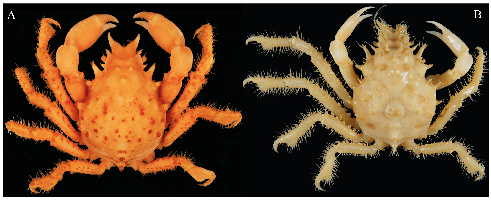
Remarks. Naxia atlantica n. sp. superficially resembles N. aries , N. spinosa and N. tumida in having pereopods 2–5 propodi strongly flattened laterally and ventrally expanded, a character that separates them from N. aurita , in which the propodi of the walking legs are only slightly expanded ventrally (cf. Figs 1 View FIGURE 1 , 2 View FIGURE 2. A – C D). Naxia atlantica n. sp. and N. tumida are unique in the genus in having the anterolateral spine of the basal antennal article with an accessory spinule. The new species can nevertheless be readily distinguished from N. tumida by a prehepatic spine that is well separated from the supraorbital eave (prehepatic spine fused to the postorbital spine or separated from it by a narrow notch in N. tumida ) (cf. Figs 1 View FIGURE 1 , 2B View FIGURE 2. A – C , 3A View FIGURE 3. A – B ). Naxia atlantica n. sp. can also be quickly distinguished from N. aries by the propodi of the walking legs being more expanded ventrally than in N. aries (cf. Figs 1 View FIGURE 1 , 2A View FIGURE 2. A – C ) and from N. aries and N. spinosa in having a rather straight rostrum that is outwardly directed and distinctly upturned at its tip (rostrum strongly curved outwards and slightly turned downward at the tip in N. aries and straight and directed outward and downward in N. spinosa ) (cf. Figs 2A, B View FIGURE 2. A – C , 3B View FIGURE 3. A – B ).
No known copyright restrictions apply. See Agosti, D., Egloff, W., 2009. Taxonomic information exchange and copyright: the Plazi approach. BMC Research Notes 2009, 2:53 for further explanation.
|
Kingdom |
|
|
Phylum |
|
|
Class |
|
|
Order |
|
|
InfraOrder |
Brachyura |
|
Family |
|
|
Genus |