
Pseudopoda mingshengi Yang & Zhang, 2022
|
publication ID |
https://doi.org/ 10.11646/zootaxa.5188.4.3 |
|
publication LSID |
lsid:zoobank.org:pub:B448106B-CCDF-4C94-8103-F38A192AB233 |
|
DOI |
https://doi.org/10.5281/zenodo.7105157 |
|
persistent identifier |
https://treatment.plazi.org/id/03F4AD1D-D566-FFAF-39B0-24F74518FE22 |
|
treatment provided by |
Plazi |
|
scientific name |
Pseudopoda mingshengi Yang & Zhang |
| status |
sp. nov. |
Pseudopoda mingshengi Yang & Zhang sp. nov.
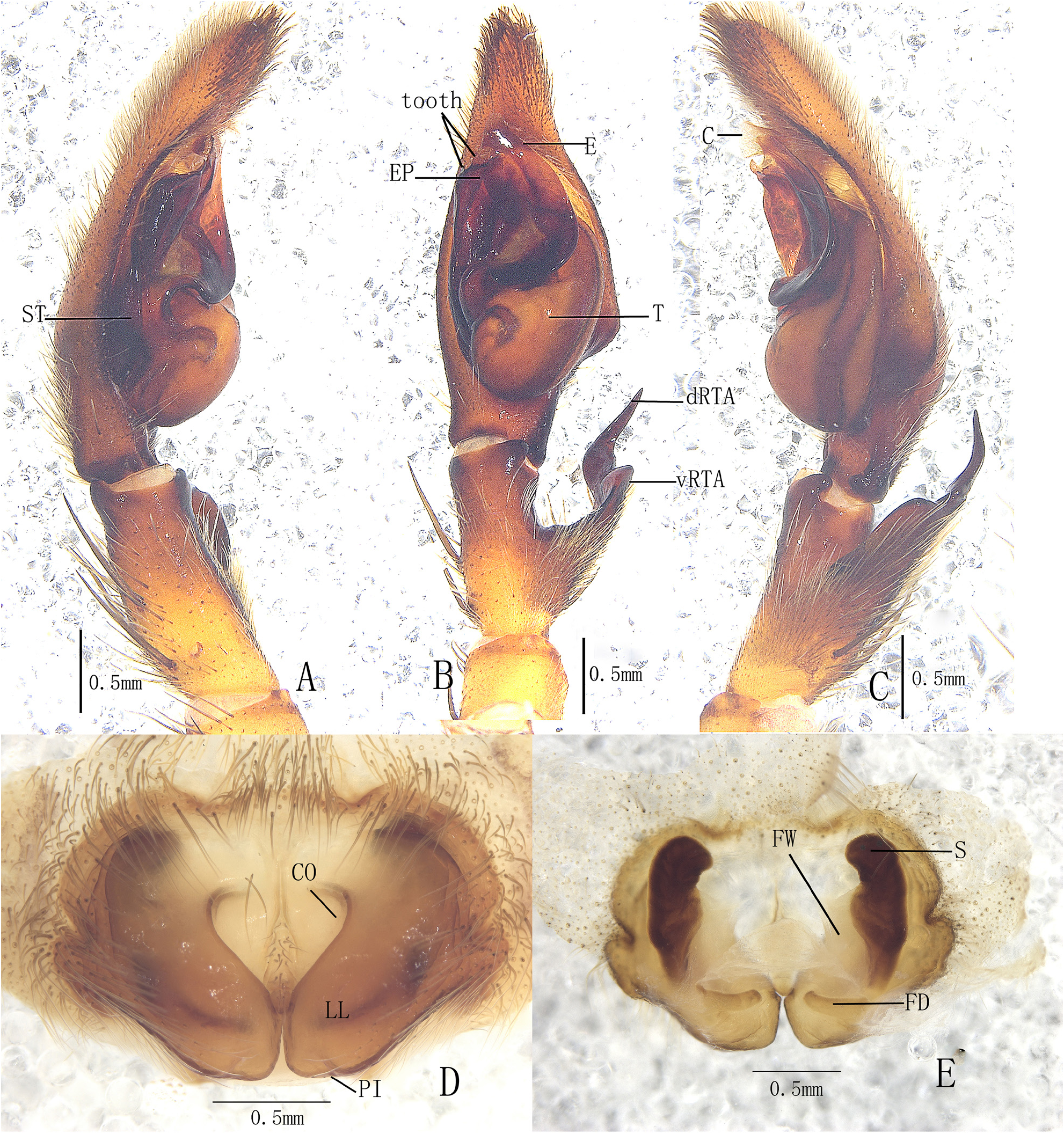
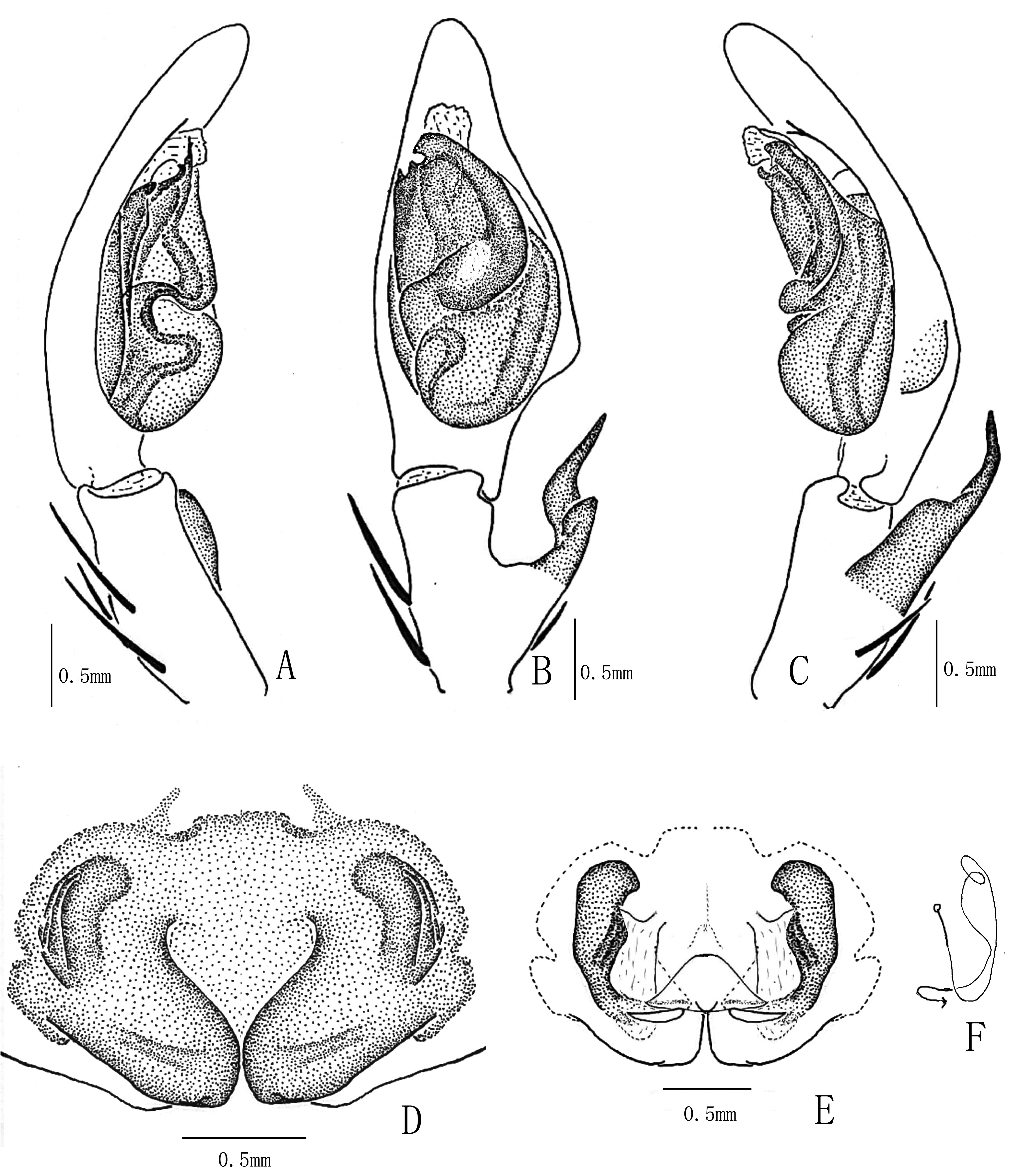
( Figs 1–3 View FIGURES 1 View FIGURES 2 View FIGURES 3 )
Type material. Holotype: ♂, CHINA : Yunnan Province: Mengjiao Township (23°16.38′N, 99°11.21′E, 1769 m), Cangyuan County, 23 May 2017, Z.Z. Yang & C.G. Li ( MHBU) GoogleMaps Paratypes: 4♀, with same data as for holotype (1♀ in MHBU, 3♀ in DUIER) GoogleMaps .
Etymology. The specific name is dedicated to the late Prof. Dr Ming-Sheng Zhu for his efforts on arachnological studies in China; nomen in genitive case.
Diagnosis. Males of this new species resemble those of P. contraria Jäger and Vedel, 2007 (see Jäger & Vedel 2007: 31, figs 114–116) in the embolus with a huge prolateral projection, and a broad apical tip of the embolic projection. It can be distinguished from the latter by the following combination of characters: 1. Apical part of embolus very broad ( Figs 2B–C View FIGURES 2 , 3B–C View FIGURES 3 ; thinner in P. contraria ); 2. Embolic projection trapezoid-shaped in ventral view ( Figs 2B View FIGURES 2 , 3B View FIGURES 3 ; square-shaped in P. contraria ); 3. Top of embolic projection with teeth ( Figs 2B View FIGURES 2 , 3B View FIGURES 3 ; absent in P. contraria ). Females of this new species resemble those of P. pingu Jäger, 2015 (see Jäger 2015: 336, figs 21–23) by the lateral windings projecting anteriorly above the median septum and the longitudinally sclerotised internal duct system. It can be distinguished from the latter by the following combination of characters: 1. lateral lobes touching each other ( Figs 2D View FIGURES 2 , 3D View FIGURES 3 ; distinctly separated in P. pingu ); 2. the first winding of internal duct system obviously wide ( Figs 2E View FIGURES 2 , 3E View FIGURES 3 ; moderately wide in P. pingu ).
Description. Male (holotype): Total length 10.83: prosoma 5.61 long, 5.24 wide; opisthosoma 5.24 long, 3.42 wide. AER slightly recurved, PER straight. Eyes diameters and interdistances: AME 0.21, ALE 0.45, PME 0.37, PLE 0.42, AME–AME 0.27, AME–ALE 0.08, PME–PME 0.29, PME–PLE 0.46. MOA 1.03 long, anterior width 0.64, posterior width 1.03. Clypeus height 0.26. Chelicerae brown, with 3 promarginal teeth and 4 retromarginal teeth, and some denticles between them. Sternum with dark setae. Legs yellowish brown, with small spots and slightly larger spine patches, metatarsus of leg with dense scopula. Leg measurements: I 23.47 (6.75, 2.80, 5.34, 6.24, 2.34), II 26.04 (7.61, 2.79, 6.62, 6.57, 2.45), III 20.12 (6.49, 2.40, 5.22, 4.36, 1.65), IV 21.18 (6.43, 2.15, 6.01, 4.52, 2.07). Leg formula: 2143. Leg spination: palp 131, 101, 2121; femur I–III 323, IV 321; patella I–IV 101; tibia I–IV 3223; metatarsus I–II 2112, III–IV 2222.
Palp as in diagnosis ( Figs 2A–C View FIGURES 2 , 3A–C View FIGURES 3 ). Cymbium slender; RTA arising proximally to medially from tibia, dRTA long and distally sharp, vRTA indistinct; spermophor running submarginally at retromargin of tegulum, turning into an S-shaped duct at promargin of tegulum to the base of embolus; embolus arising from tegulum at 9 o’clock position, very broadened in its middle part; embolic projection flat and very large, pointing prolaterally, embolic projection distally with two triangular teeth; conductor arising from tegulum at 12 o’clock position, slender, bent ventrad.
Coloration in ethanol: Carapace yellowish brown, bearing some small spots. Cervical groove and radial furrow obvious. Longitudinal fovea dark brown. Ocular area darker. Each eye surrounded with circular black patch. Labium, gnathocoxae and sternum yellowish brown, the posterior part of labium dark brown. Legs yellowish brown, with small spots and slightly larger spine patches, metatarsus darker. Dorsal opisthosoma gray black dorsally, with white lateral patterns and a big dark triangular median pattern, the posterior part whitish; venter gray black ( Figs 1A–B View FIGURES 1 ).
Female: Total length 13.65–13.71 (n=4). One paratype: Total length 13.71; prosoma 6.25 long, 5.78 wide; opisthosoma 7.47 long, 5.00 wide. Eyes diameters and interdistances: AME 0.30, ALE 0.44, PME 0.33, PLE 0.42, AME–AME 0.27, AME–ALE 0.11, PME–PME 0.38, PME–PLE 0.54. MOA 1.11 long, anterior width 0.77, posterior width 1.07. Clypeus height 0.50. Leg measurements: I 21.81 (6.27, 2.93, 5.98, 5.11, 1.52), II 23.63 (6.56, 3.43, 6.13, 5.51, 2.00), III 18.74 (5.47, 2.68, 4.57, 4.06, 1.96), IV 21.21 (6.48, 2.52, 5.22, 4.41, 2.58). Leg formula: 2143. Leg spination: palp 131, 101, 2121, 2112; femur I–II 323, III 332, IV 331; patella I–IV 001; tibia I–IV 3223; metatarsus I–II 2112, III–IV 2222.
Epigyne as in diagnosis ( Figs 2D–E View FIGURES 2 , 3D–E View FIGURES 3 ). Epigynal field wider than long, with slightly trilobate anterior margin, anterior bands short, indistinct; lateral lobes longer than wide, slightly converged on the central axis; anterior margins of lateral lobes heart - shaped; loops of internal duct system bending laterally, the posterior parts hidden behind lateral lobes; the anterior margin of longitudinally sclerotised parts beyond copulatory opening.
Coloration in ethanol: Color and markings of carapace as in male. Dorsal opisthosoma dark brown dorsally, with some irregular patches laterally and a distinct white transversal patch in the posterior part; venter brown, with small and irregular patches ( Figs 1C–D View FIGURES 1 ).
Distribution. China (Yunnan).
Note. The species P. mingshengi cannot be allocated to any of the nine known species groups according to morphological features. Males share the broad prolateral embolic projection with several species, e.g., P. daxing Zhao and Li, 2018 in Jiang et al. 2018, P. digitata Jäger and Vedel, 2007 and P. contraria , but females of this new species are completely different.

No known copyright restrictions apply. See Agosti, D., Egloff, W., 2009. Taxonomic information exchange and copyright: the Plazi approach. BMC Research Notes 2009, 2:53 for further explanation.
|
Kingdom |
|
|
Phylum |
|
|
Class |
|
|
Order |
|
|
Family |
|
|
Genus |