
Evansula arenicola, NICHOLLS, 1939
|
publication ID |
https://doi.org/10.1111/j.1096-3642.2006.00227.x |
|
persistent identifier |
https://treatment.plazi.org/id/03F487A0-FFBD-FFBC-FCAB-FEEAFAE4931D |
|
treatment provided by |
Felipe |
|
scientific name |
Evansula arenicola |
| status |
|
EVANSULA ARENICOLA NICHOLLS, 1939
Synonyms: E. incerta ( Scott, 1892) sensu Wilson (1932) .
Original description: Nicholls (1939: 299–302, figs 23, 24).
Additional description: Wilson (1932) (as E. incerta ).
Type locality: Washings from coarse sand, taken by grab at 8 m depth, at Baie de Mille Vaches on the north shore of the St. Lawrence River, Quebec ( Canada) .
Material examined: NHM, reg. nos 1940.5.1.73–78: syntypes, 8 ♀♀ and 2 ♂♂ in alcohol; 1 ♀ (on nine slides) and 1 ♂ (on six slides) dissected; leg. A. G. Nicholls, 19 August 1937.
Redescription
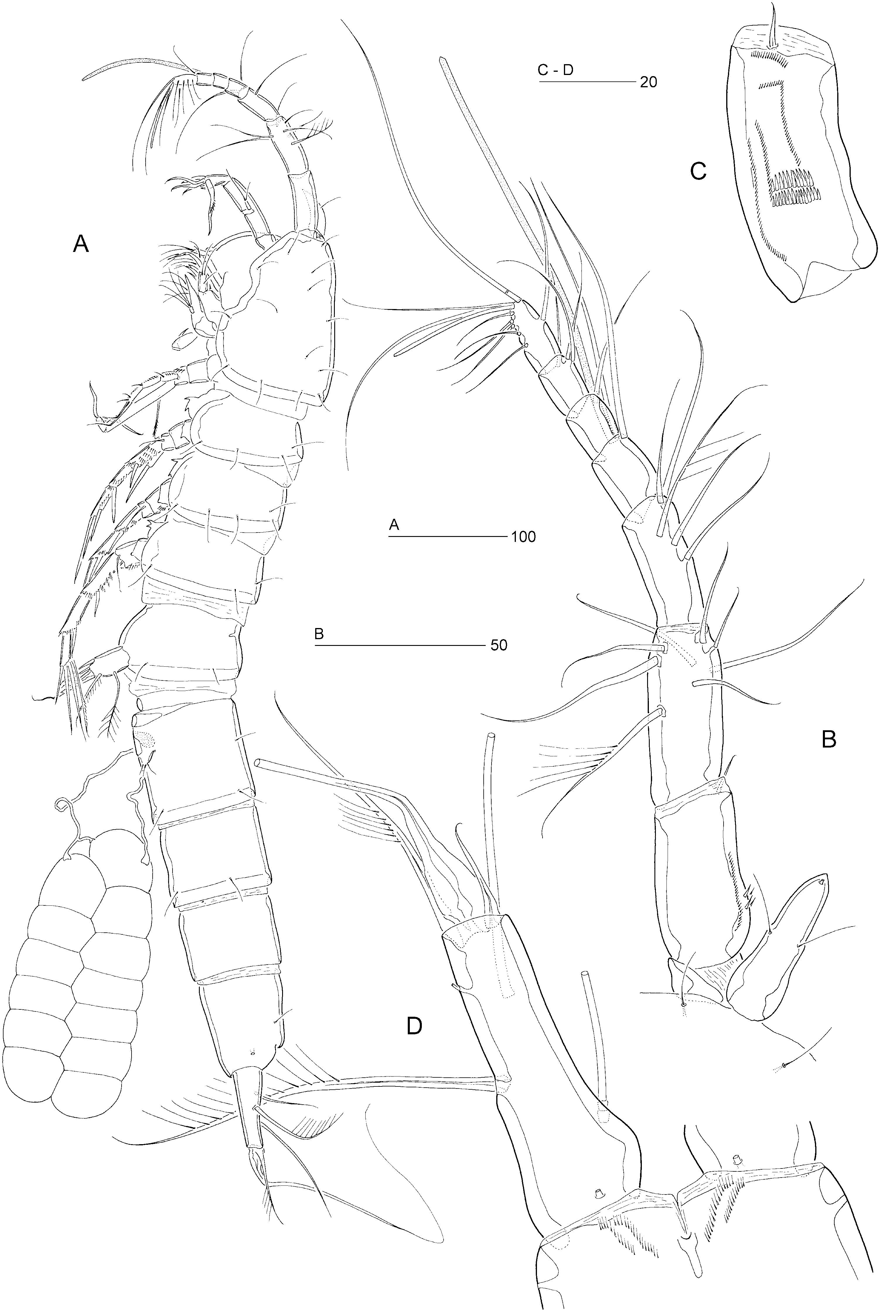
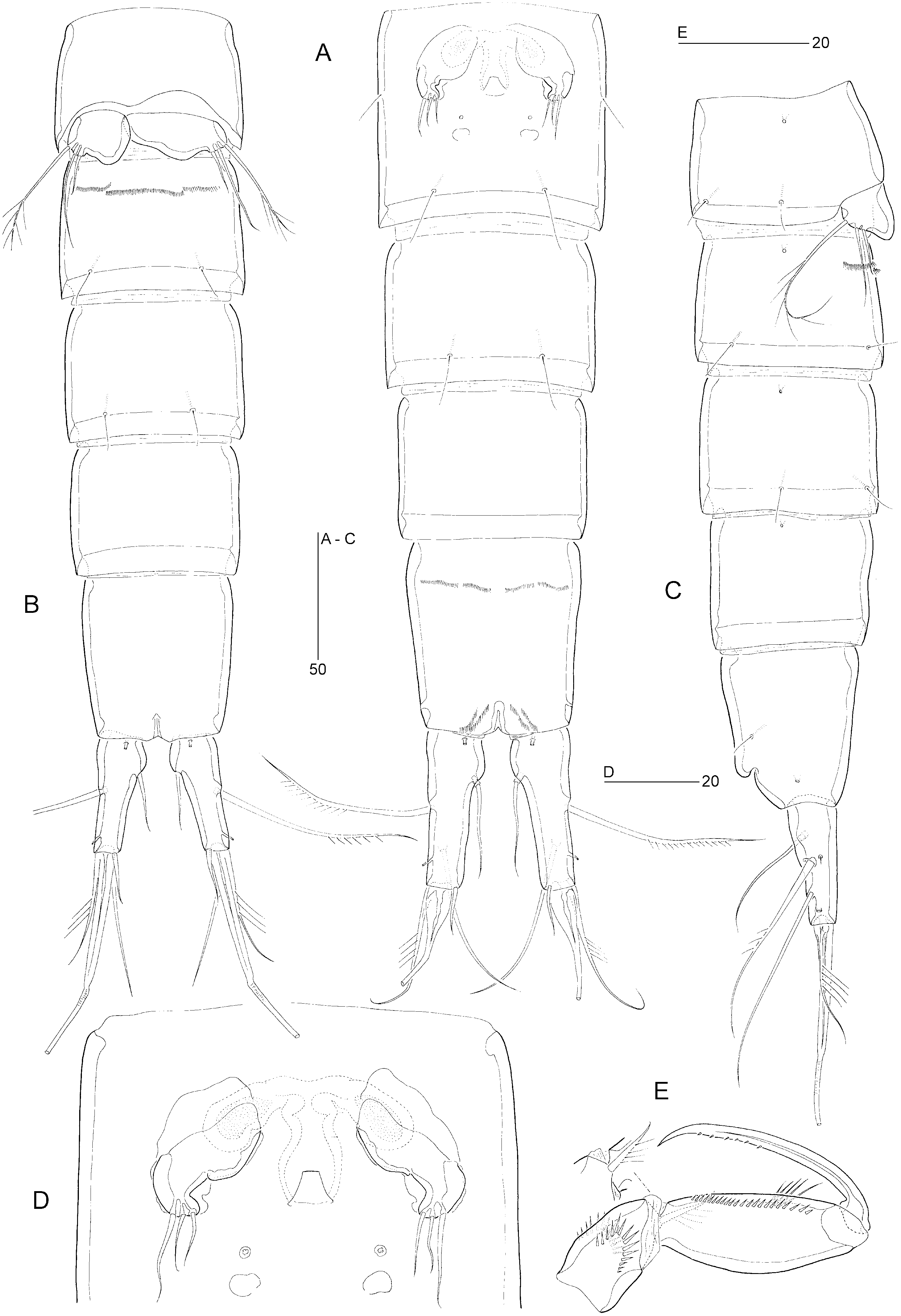
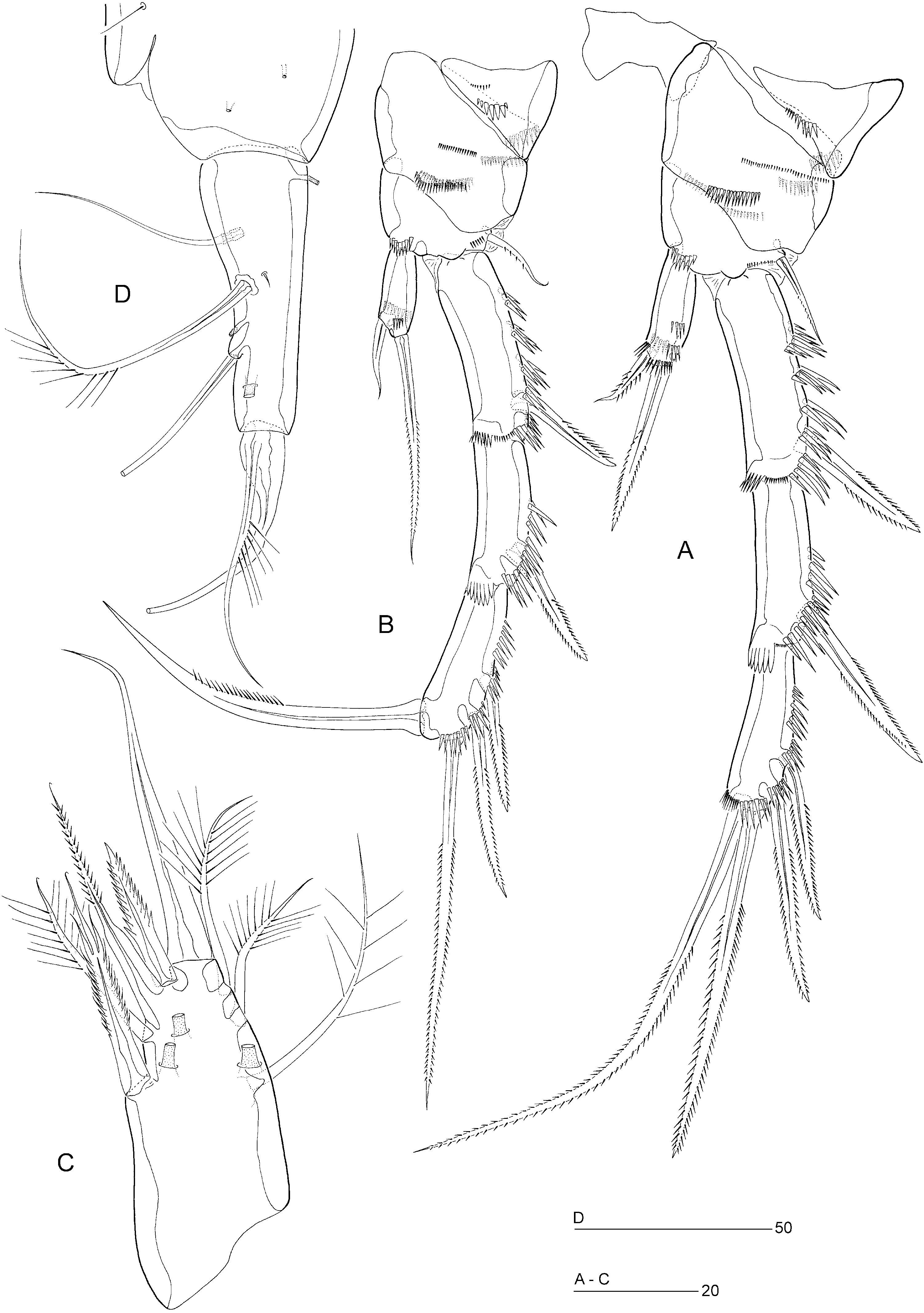
Female: Total body length: 710–750 µm ( N = 5; mean = 730 µm). Body slender, cylindrical ( Fig. 13A View Figure 13 ), semitransparent, yellowish; no distinct separation between prosome and urosome. Genital double-somite completely fused ( Figs 13A View Figure 13 , 14A View Figure 14 ); original segmentation marked dorsally by paired anterior and posterior sensillae ( Fig. 13A View Figure 13 ) and ventral chitinous patches ( Fig. 14A View Figure 14 ). Anal somite only slightly longer than wide (84 × 76 µm), with two pairs of secretory pores laterally ( Fig. 16D View Figure 16 ); ventral surface with four rows of tiny spinules near anterior margin ( Fig. 14A View Figure 14 ); posterior margin with two short spinular rows on either side of ventral midline ( Fig. 13D View Figure 13 ). Anal operculum weakly developed, unarmed ( Fig. 17E View Figure 17 ).
Caudal rami slightly divergent, cylindrical except for proximal third, which is swollen both dorsally and medially ( Figs 13D View Figure 13 , 16D View Figure 16 , 17E View Figure 17 ), length (measured along outer margin) approximately 2.8 times the proximal width; dorsal surface without chitinous spur or raised spinular row; with seven setae, seta VII in proximal third, setae I–II at approximately half ramus length and setae III–VI in distal third ( Fig. 17E View Figure 17 ); seta I diminutive; seta II long and pinnate; seta III long and bare; seta IV relatively long, unipinnate at approximately halfway its length, longer than swollen part of V; seta V long, with distinct flexure zone between short, proximal bulbous part and long, distal flagellate part, fused at base with seta IV ( Figs 13D View Figure 13 , 17E View Figure 17 ); seta VI vestigial; seta VII tri-articulate at base and located along proximal inner margin; with three tube-pores, one dorsally, one ventrally, and one laterally ( Figs 13D View Figure 13 , 16D View Figure 16 , 17E View Figure 17 ).
Rostrum elongate ( Fig. 13B View Figure 13 ), only slightly shorter than first antennulary segment; proximal third with inflated lateral margins, distinctly tapering distally; with two long sensillae; median pore positioned dorsally near apex of rostrum.
Antennule seven-segmented ( Fig. 13B View Figure 13 ). Segment 1 approximately as long as segment 2, with small sclerite around proximal posterior margin, anterior surface with pattern of spinular rows, as illustrated in Figure 13C View Figure 13 ; segment 2 without secretory pore; segment 4 with distal cylindrical process bearing large aesthetasc (115 µm). Armature formula: 1-[1], 2-[8 + 1 pinnate], 3-[5], 4-[1 + (1 + ae)], 5-[1], 6-[3], 7-[7 + acrothek]. Apical acrothek consisting of two long setae and one slender aesthetasc (40 µm).
Antenna, mandible, maxillule, and maxilla as in E. incerta .
Maxilliped ( Fig. 14E View Figure 14 ) well developed, subchelate, directed inwards. Syncoxa well developed, with one pinnate seta and two spinular rows. Basis elongate, with three to four long spinules on anterior surface and a spinular row along posterior inner margin. Endopod represented by strong, curved, sparsely pinnate claw.
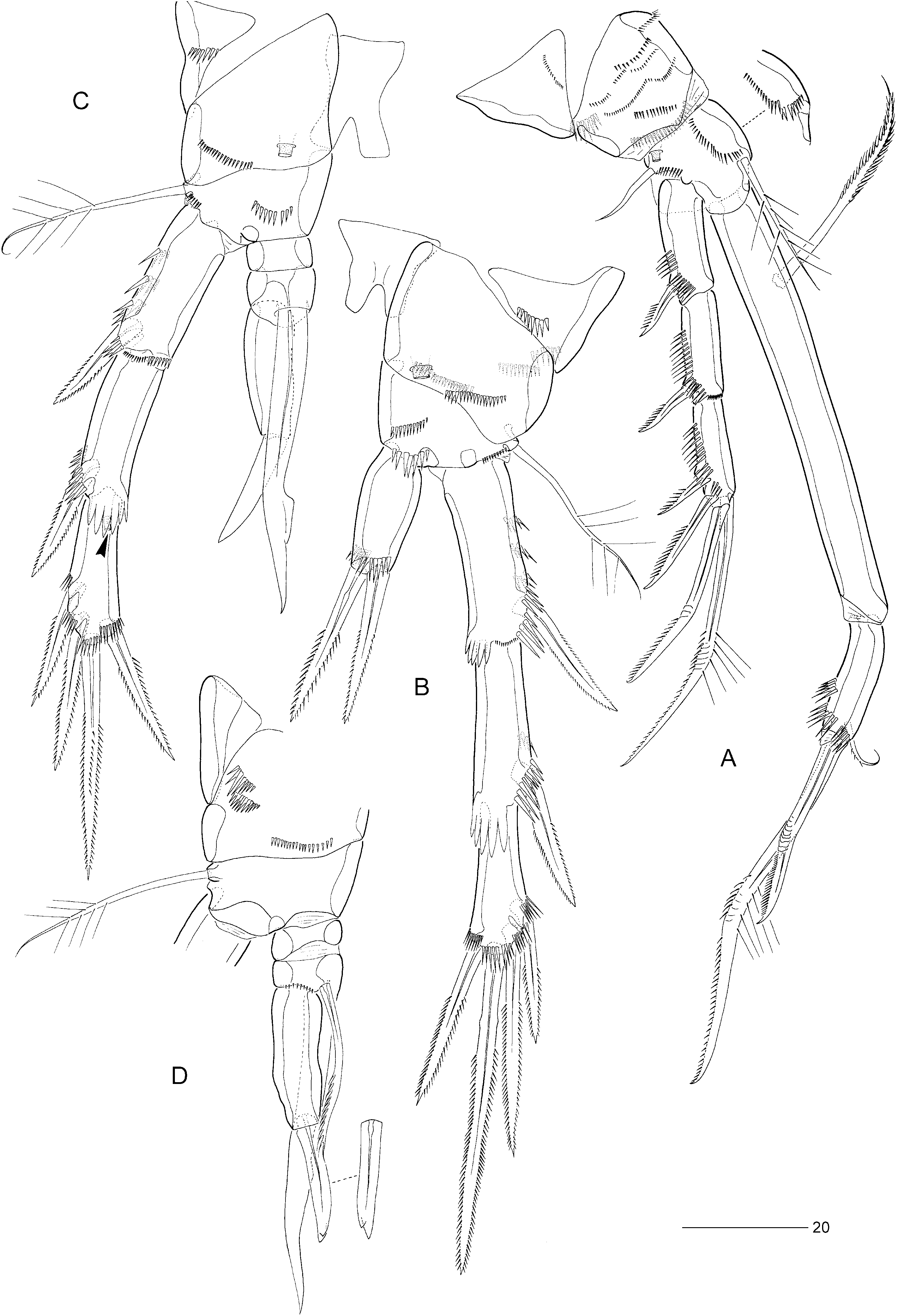
P1 ( Fig. 15A View Figure 15 ). Praecoxa strongly developed, with a row of tiny spinules. Coxa with two spinular rows on posterior surface and eight spinular rows on anterior surface. Basis with one posterior and two anterior spinular rows; with long, plumose, inner seta and short, bare, outer seta; anterior surface with secretory pore. Exopod three-segmented; with two spines and two geniculate setae on exp-3. P1 endopod prehensile, distinctly longer than exopod; proximal segment approximately nine times as long as average width, with pinnate inner seta not plumose in proximal third; distal segment short, with three spinular rows, a subdistal pinnate setule, and two geniculate setae distally (both being distinctly longer than in other species).
Swimming legs P2–P4 ( Figs 15B View Figure 15 , 16A View Figure 16 , 17A View Figure 17 ). P4 distinctly longer than P2–P3. Praecoxae well developed, with spinular row on anterior surface in P2–P4. Coxae with pattern of spinules as in Figures 15B View Figure 15 , 16A View Figure 16 , 17A View Figure 17 ; with large tube-pore on anterior surface of P3 and P4. Bases with outer seta (pinnate and spiniform in P2, long and plumose in P3, long and bare in P4); with spinular rows on anterior surface only ( Figs 15B View Figure 15 , 16A View Figure 16 , 17A View Figure 17 ). Exopods three-segmented, endopods onesegmented. Inner distal spine of P3–P4 exp-3 shorter than outer distal one ( Figs 15B View Figure 15 , 17A View Figure 17 ). Inner setae of P4 endopod and exp-3 serrate. Inner element of P2 endopod spiniform, pinnate, less than half length of distal spine. Armature elements of P3 endopod spiniform; of approximately equal length with inner spine, slightly longer than outer. Seta and spine formulae as for genus.
Fifth pair of legs ( Fig. 16C View Figure 16 ) with baseoendopod and exopod fused into a common elongate plate, tapering distally; apex with strong, articulating spine, approximately as long as the plate and with a flagellate tip; outer margin with three plumose setae (including seta derived from baseoendopod); inner margin with two serrate spines, one plumose seta, and one long, pinnate seta fused to plate and distinctly swollen in proximal half; anterior surface with three large tube-pores.
Sixth legs ( Fig. 14A, D View Figure 14 ) each represented by small operculum closing off gonopore; armature consisting of three setiform elements with outer slightly longer than others. Genital apertures not fused medially; copulatory pore of moderate size, located slightly anterior to gonopores; leading via short chitinized copulatory duct to paired anterior extensions positioned anterior to genital apertures; copulatory pore flanked by two small secretory pores.
Single egg-sac ( Fig. 13A View Figure 13 ) containing approximately 12– 13 eggs arranged biserially, enclosed in a common egg-sac membrane; egg-sac connected with each genital aperture via transparent string.
Male. Body length: 690 µm. Spermatophore 100 µm. Anal somite without spinules on ventral anterior or hind margins ( Fig. 14B View Figure 14 ).
Antennule as in E. incerta .
P2 ( Fig. 16B View Figure 16 ) with inner distal corner of basis not modified into spinous process. Exp-3 modified; outer distal element more setiform than in female; inner distal element transformed into strong claw, directed medially and posteriorly, pinnate along middle third. Endopod slightly shorter than in female; anterior surface with only one spinular row (four in female); apical spine distinctly longer and more slender than in female; inner spine bare.
P3 ( Fig. 15C View Figure 15 ) exp-1 with reduced hyaline frill; exp-3 with secretory pore on anterior surface near joint with exp-2; outer distal spine of exp-3 distinctly shorter than in female. P3 endopod ( Fig. 15C, D View Figure 15 ) distinctly threesegmented, longer than exp-1; enp-1 small, without armature; enp-2 with serrate, posterior seta and long, rigid apophysis arising from anterior surface; distal half of apophysis with flimsy barb; enp-3 tapering distally, with one strong, bare spine apically (bifid at tip).
P4 endopod two-segmented ( Fig. 17C View Figure 17 ); enp-1 with spinular row, without armature; enp-2 with two serrate setae along inner margin and two pinnate spines apically, inner distal spine distinctly shorter than in female.
Fifth legs ( Fig. 17D View Figure 17 ) with baseoendopod and exopod fused into a common, elongate plate, tapering distally towards long, spinous process, which is longer than the plate, slightly curved and bare; inner margin with serrate spine; outer margin with small serrate spine (partly fused to the plate), three naked setae and a sparsely plumose seta derived from baseoendopod; anterior surface with three large tube-pores.
Sixth pair of legs ( Fig. 14B, C View Figure 14 ) asymmetrical, with three setae each, decreasing in length medially; outer seta sparsely plumose. Left or right leg articulating according to sinistral or dextral development of testis and vas deferens. First postgenital somite with transverse spinular row near ventral anterior margin ( Fig. 14B View Figure 14 ).
Caudal ramus ( Fig. 17F View Figure 17 ) as in female, except for length and shape of proximal styliform part of seta V, which is distinctly longer than caudal ramus length.
Variability: Nicholls (1939) described the female P4 endopod as being partly divided into two segments. This partial subdivision, marked by a suture line halfway along the outer margin ( Fig. 17B View Figure 17 ), was found in most female specimens examined. However, in some individuals and sometimes in the same specimen, no such subdivision could be observed ( Fig. 17A View Figure 17 ). Surprisingly, the separation in two distinct segments in the male was overlooked by Nicholls (1939).
Remarks: Nicholls (1939) figures two geniculate setae and only one outer spine on P1 exp-3 of the female, whereas for the male he states ‘... with 3 terminal setae on exopod’. Re-examination of all syntypes revealed that Nicholls’ illustration was based on a damaged or aberrant female.
There is little doubt that Wilson’s (1932) illustrations of E. incerta were based on E. arenicola . Evidence for this is found in his illustrations of the male, which show a two-segmented P4 endopod and agree in most other aspects, and in his statement that the female seta V of the caudal ramus ‘... is stout and spiniform proximally and passes abruptly at an angle into a filiform distal portion’. For the male he characterized the latter seta as being enlarged at the base, which conforms to the sexual dimorphism found in E. arenicola .
Differential diagnosis: Evansula arenicola clearly occupies an isolated position in the genus by virtue of its two-segmented P4 endopod in the male. It is also the only species that displays sexual dimorphism in the caudal seta V (bulbiform in the female) and the inner distal spine of the P4 endopod (reduced in the male). The male P2 endopod is significantly smaller in comparison with other species. The male P3 endopod, on the contrary, is much larger than in its congeners and differs in the presence of a strong, naked apical spine. The general shape of the caudal rami and the P1 (enp-1 inner seta not plumose in proximal part; enp-2 with much longer geniculate elements) provide additional differentiating characters.
Distribution: Canada: St. Lawrence River, Quebec ( Nicholls, 1939).
USA: Katama Bay, Marthas Vineyard, Massachusetts ( Wilson, 1932); it is conceivable that Coull’s (1971, 1977) records of E. incerta from the North Carolina continental shelf also belong to E. arenicola .

No known copyright restrictions apply. See Agosti, D., Egloff, W., 2009. Taxonomic information exchange and copyright: the Plazi approach. BMC Research Notes 2009, 2:53 for further explanation.