
Evansula incerta, (T. SCOTT, 1892), 1932
|
publication ID |
https://doi.org/10.1111/j.1096-3642.2006.00227.x |
|
persistent identifier |
https://treatment.plazi.org/id/03F487A0-FFAD-FFA8-FFC7-FCCDFA1194A4 |
|
treatment provided by |
Felipe |
|
scientific name |
Evansula incerta |
| status |
|
EVANSULA INCERTA (T. SCOTT, 1892)
Synonyms: Tetragoniceps incertus T. Scott, 1892 ; Evansia incerta (T. Scott, 1892) Scott (1906a) .
Original description: Scott (1892: 254–255, plate XII, figs 1–17).
Additional descriptions: All subsequent redescriptions are unreliable, as they were based on another species ( Sars, 1911; Wilson, 1932) or on an amalgam of several species of which E. incerta may or may not have been part of ( Klie, 1929; Kunz, 1935; Scheibel, 1972).
Type locality: Scotland, Firth of Forth, not specified by Scott (1892), but according to a later paper ( Scott, 1906b) the types were dredged off St. Monans.
Material examined: (1) NHM, reg. nos. 44505–507: Firth of Forth, Scotland; 2 ♀♀, 1 ♂ in alcohol (labelled as ‘cotypes’); coll. T. Scott, 9 September 1894; as part of Cannon A. M. Norman collection ( 1911.11.8); 1 ♀ (on 11 slides) and 1 ♂ (on seven slides) dissected. This vial also contained 1 ♂ of E. spinosa sp. nov. (see below); (2) Royal Museum of Scotland, Edinburgh, reg. no. 1955.63.115: Firth of Forth , coll. T. Scott, June 1901 (labelled Tetragoniceps incertus ): vial empty .
Redescription
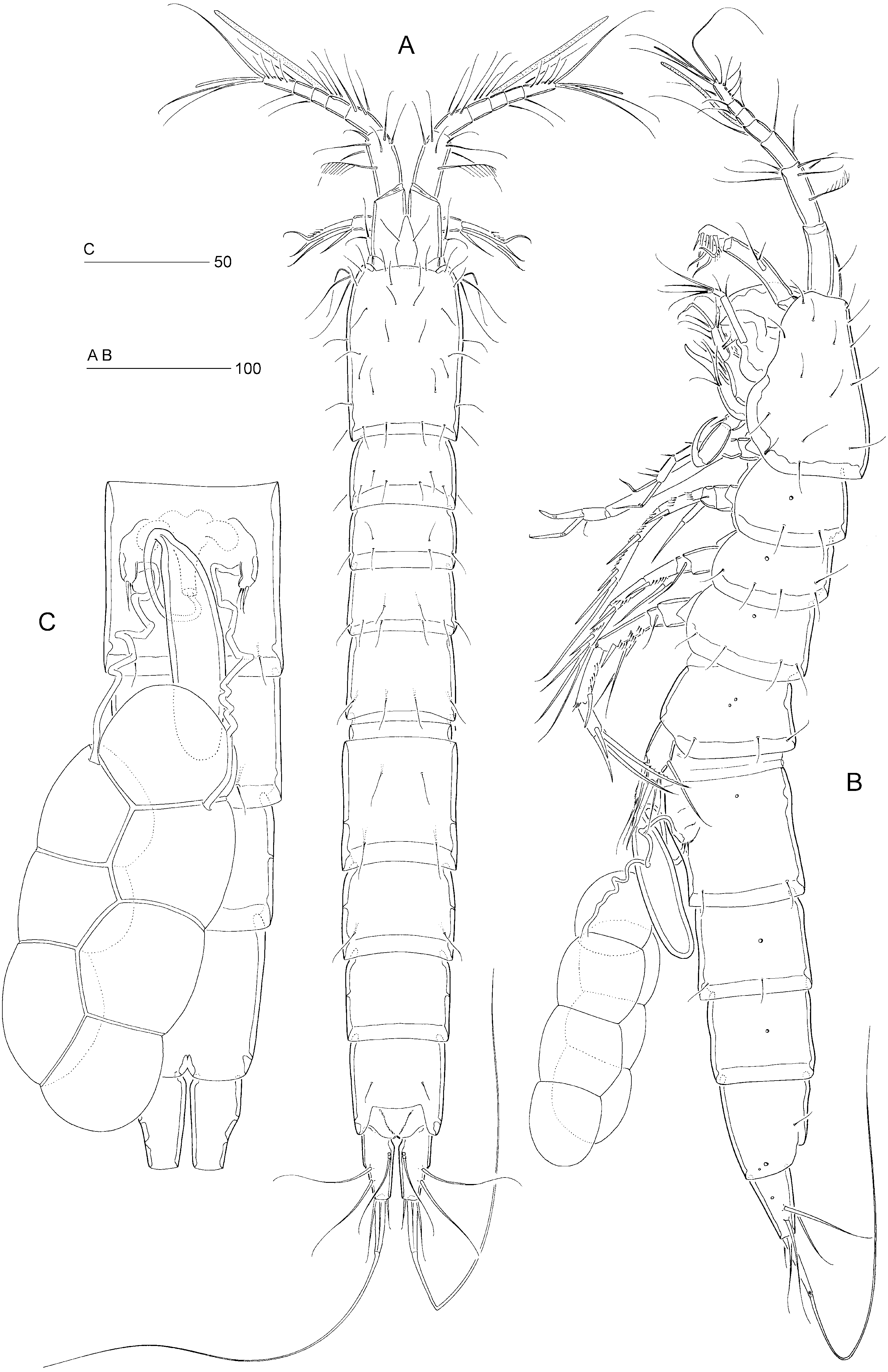
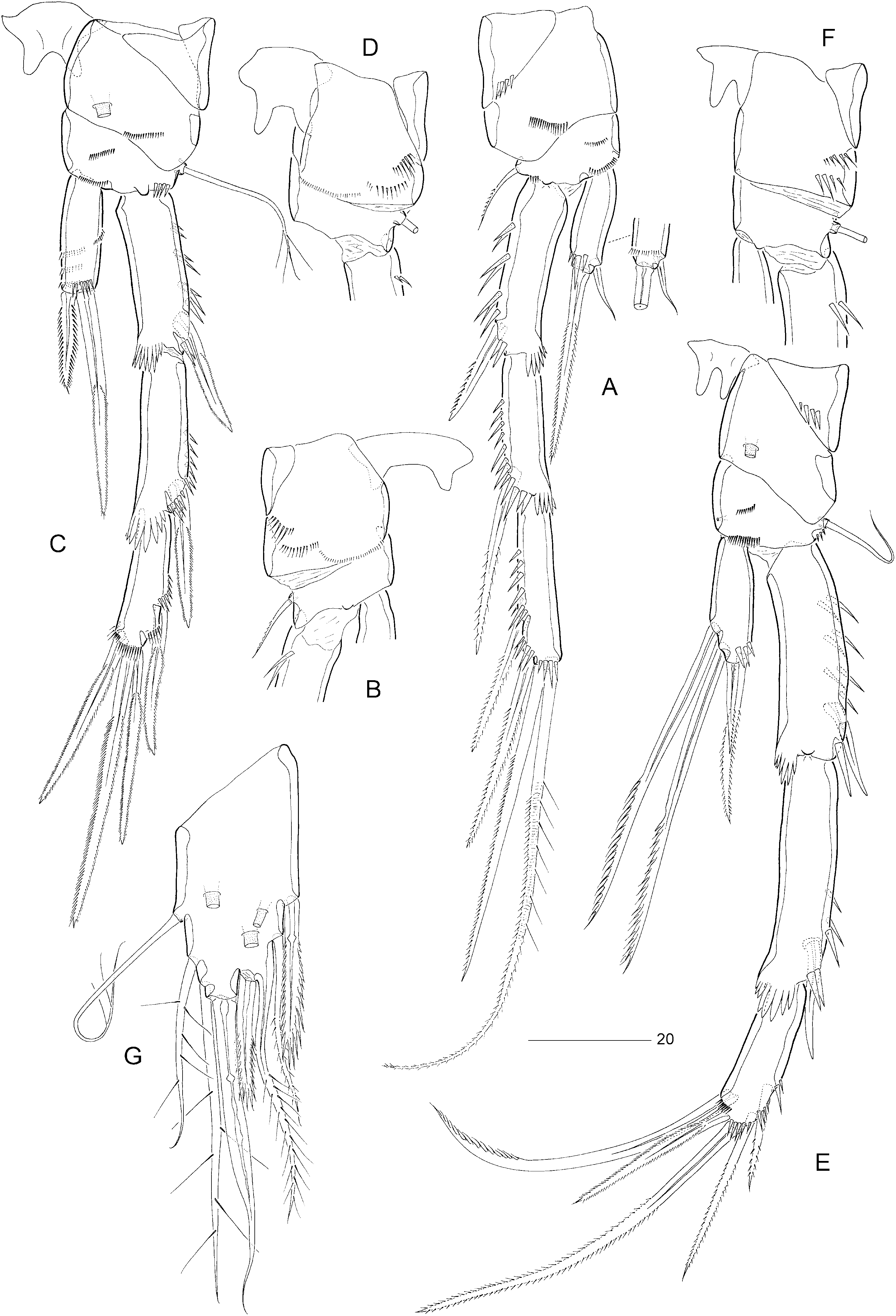
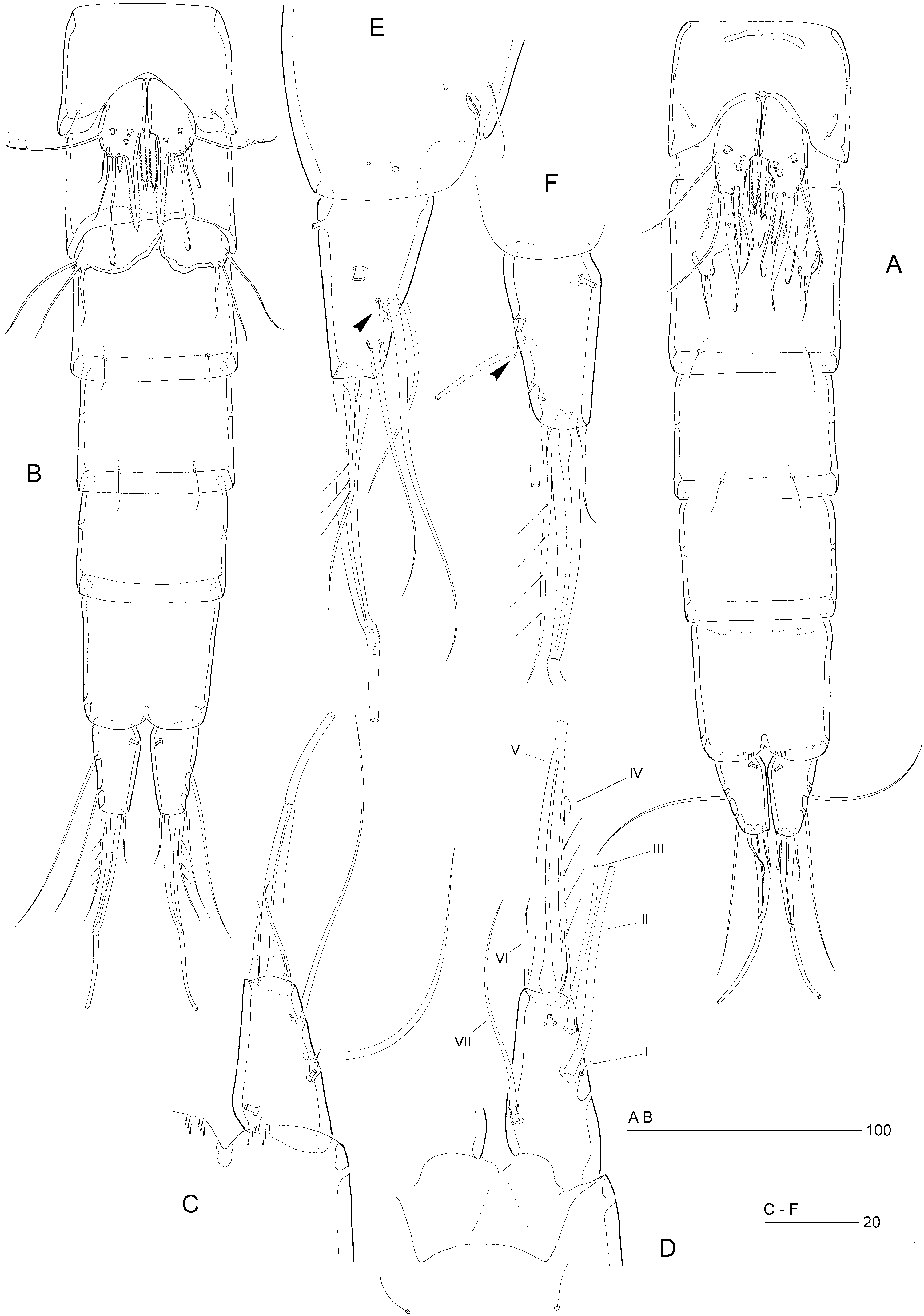
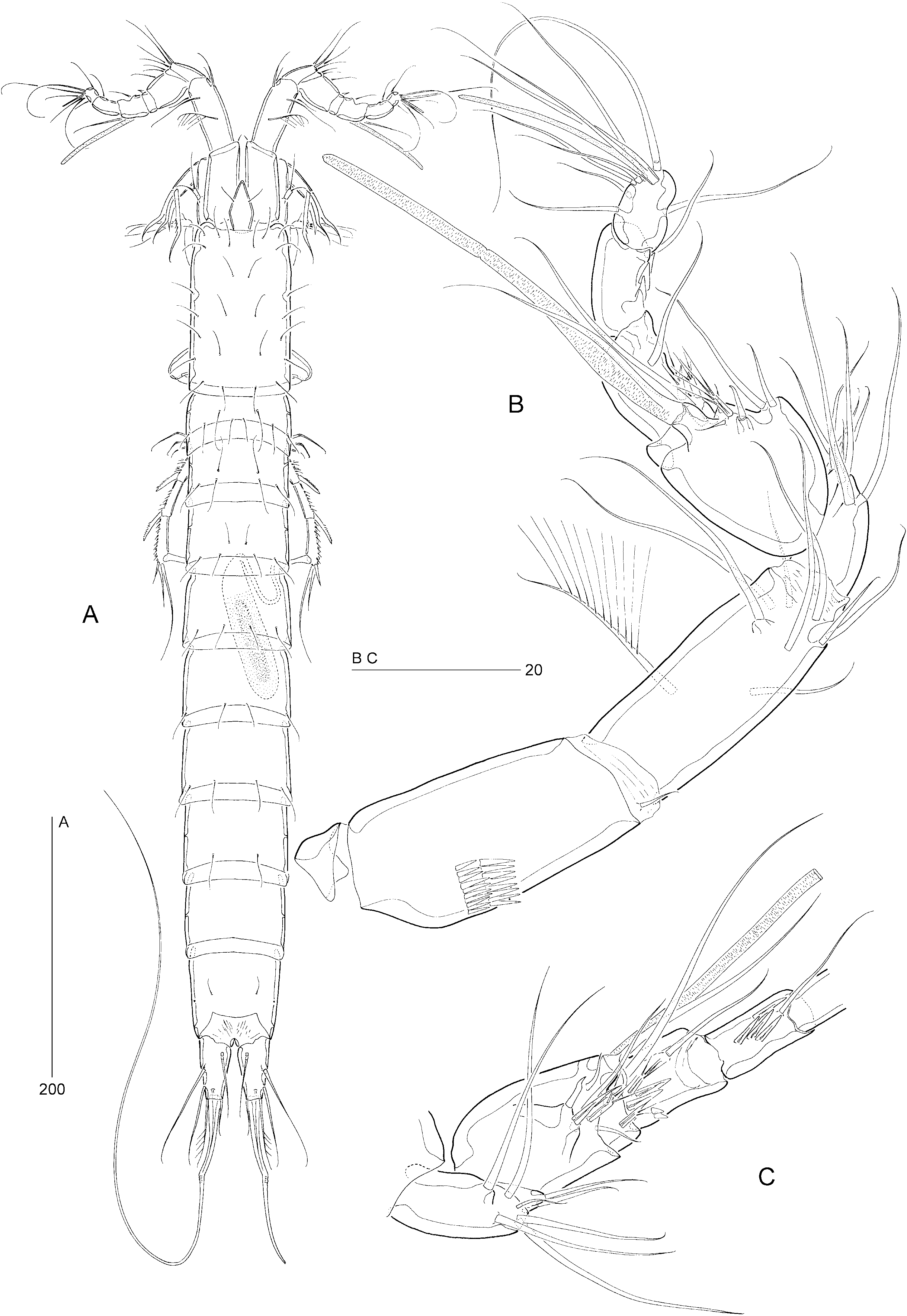
Female: Total body length: 695–710 µm ( N = 2; mean = 702.5 µm). Body slender, cylindrical ( Fig. 1A, B View Figure 1 ), semitransparent, light brown; no distinct separation between prosome and urosome. Genital double-somite completely fused ( Figs 1B, C View Figure 1 , 2D View Figure 2 , 6A View Figure 6 ), original segmentation marked dorsally by paired anterior and posterior sensillae ( Fig. 1A View Figure 1 ). Anal somite only slightly longer than wide (57 × 60 µm), with three pairs of secretory pores laterally ( Fig. 6E View Figure 6 ); posterior margin with two short spinular rows on either side of ventral midline ( Fig. 6A, C View Figure 6 ). Anal operculum weakly developed, unarmed ( Fig. 6D View Figure 6 ).
Caudal ramus conical ( Fig. 6A, C View Figure 6 ), length (measured along the outer margin) approximately 1.9 times the proximal width; dorsal surface without chitinous spur; with seven setae, setae I–VI in distal and seta VII in proximal half ( Fig. 1A View Figure 1 ); seta I diminutive; setae II–III long and bare; seta IV short and bare, approximately half length of styliform part of V ( Fig. 6C View Figure 6 ); seta V long, with distinct flexure zone between styliform part and long distal flagellate part, fused at base with vestigial seta IV; seta VI vestigial; seta VII tri-articulate at base and located along proximal inner margin; ventral surface with one simple and two tube-pores ( Fig. 6C View Figure 6 ), dorsal surface with one tube-pore.
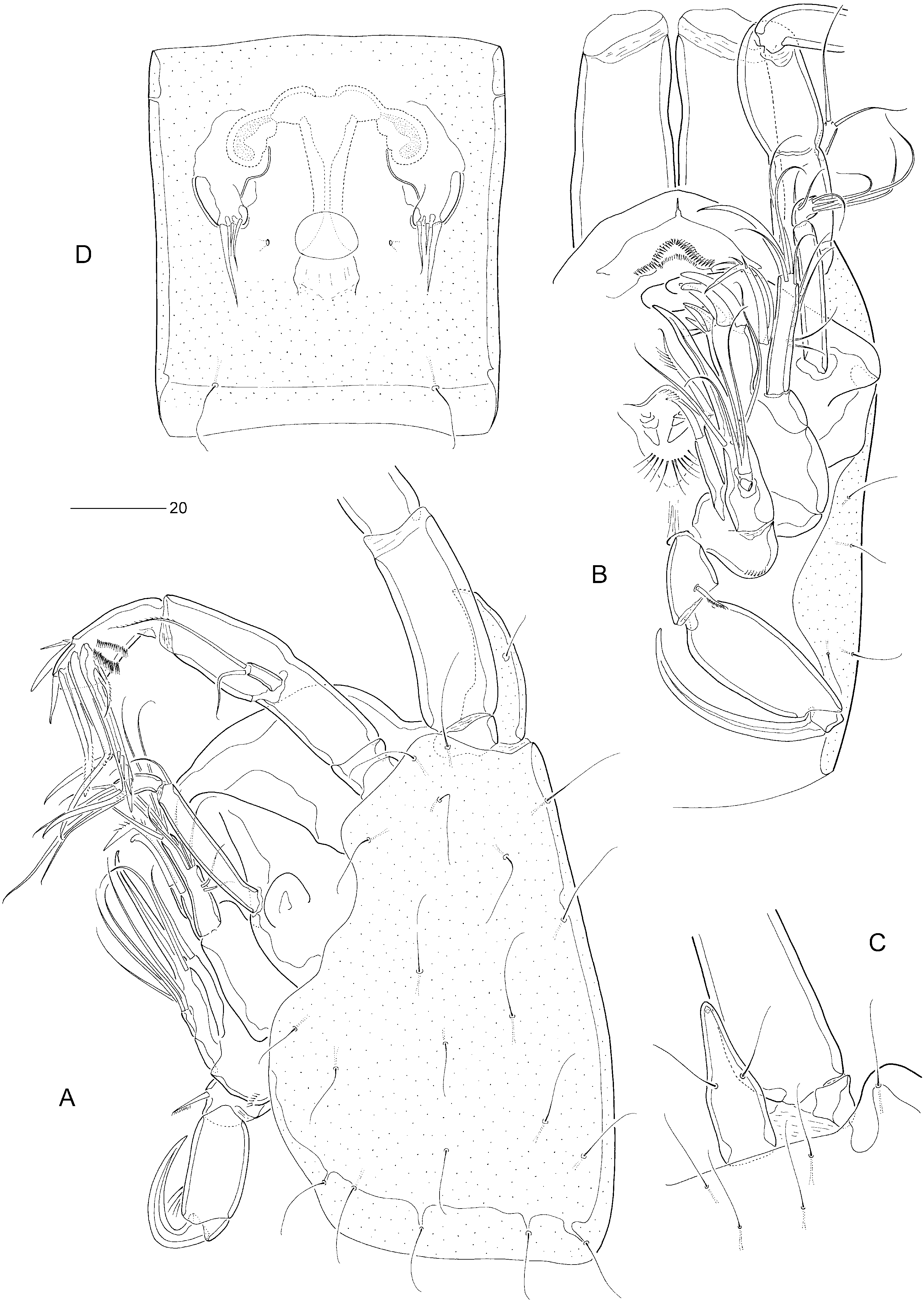
Rostrum elongate ( Fig. 2A, C View Figure 2 ), with parallel margins in proximal half, tapering distally; distinctly shorter than first antennulary segment; demarcated at base; base surrounded by area of flexible integument; with two long sensillae; subapical pore positioned midventrally.
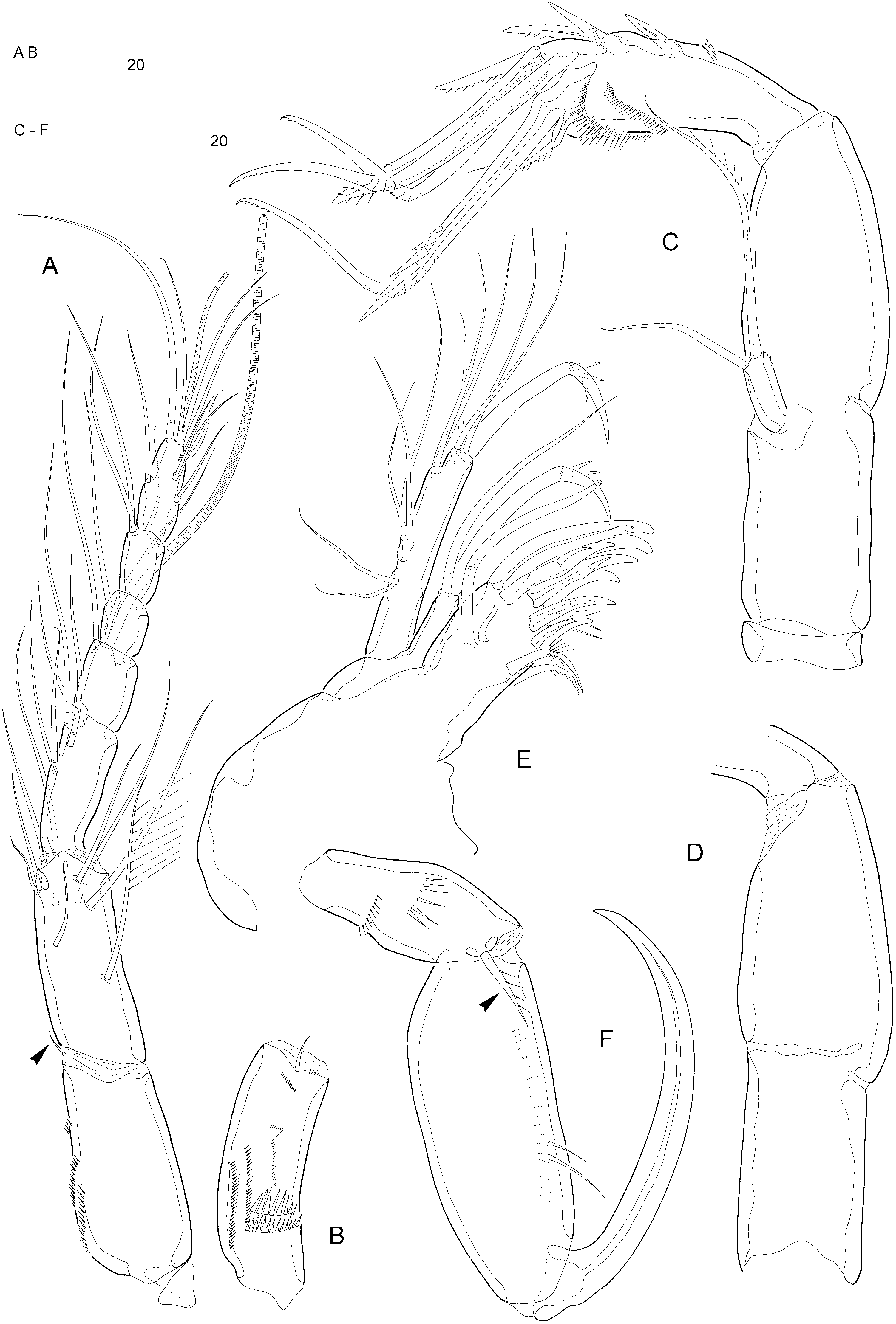
Antennule seven-segmented ( Fig. 3A View Figure 3 ). Segment 1 only slightly shorter than segment 2, with small sclerite around proximal posterior margin, with pattern of spinular rows on anterior surface, as illustrated in Figure 3B View Figure 3 ; segment 2 longest, without secretory pore; segment 4 with distal cylindrical process bearing large aesthetasc (90 µm). Armature formula: 1-[1], 2-[8 + 1 pinnate], 3-[5], 4-[1 + (1 + ae)], 5-[1], 6-[3], 7-[7 + acrothek]. Apical acrothek consisting of two long setae and one slender aesthetasc (35 µm).
Antenna ( Figs 2A View Figure 2 , 3C, D View Figure 3 ) with small unarmed coxa; basis and first endopod segment incompletely fused forming allobasis, abexopodal margin without ornamentation; exopod a narrow segment, with one long, pinnate and one shorter, naked seta apically; endopod with two lateral spines and distal armature consisting of two pinnate spines, two geniculate setae and one large geniculate spine bearing spinules at approximately mid-margin and fused at base with a short, pinnate seta.
Labrum ( Fig. 2A, B View Figure 2 ) a well-developed, ventrally produced extension; distal margin with short, blunt spinules; lateral margins with finer setules.
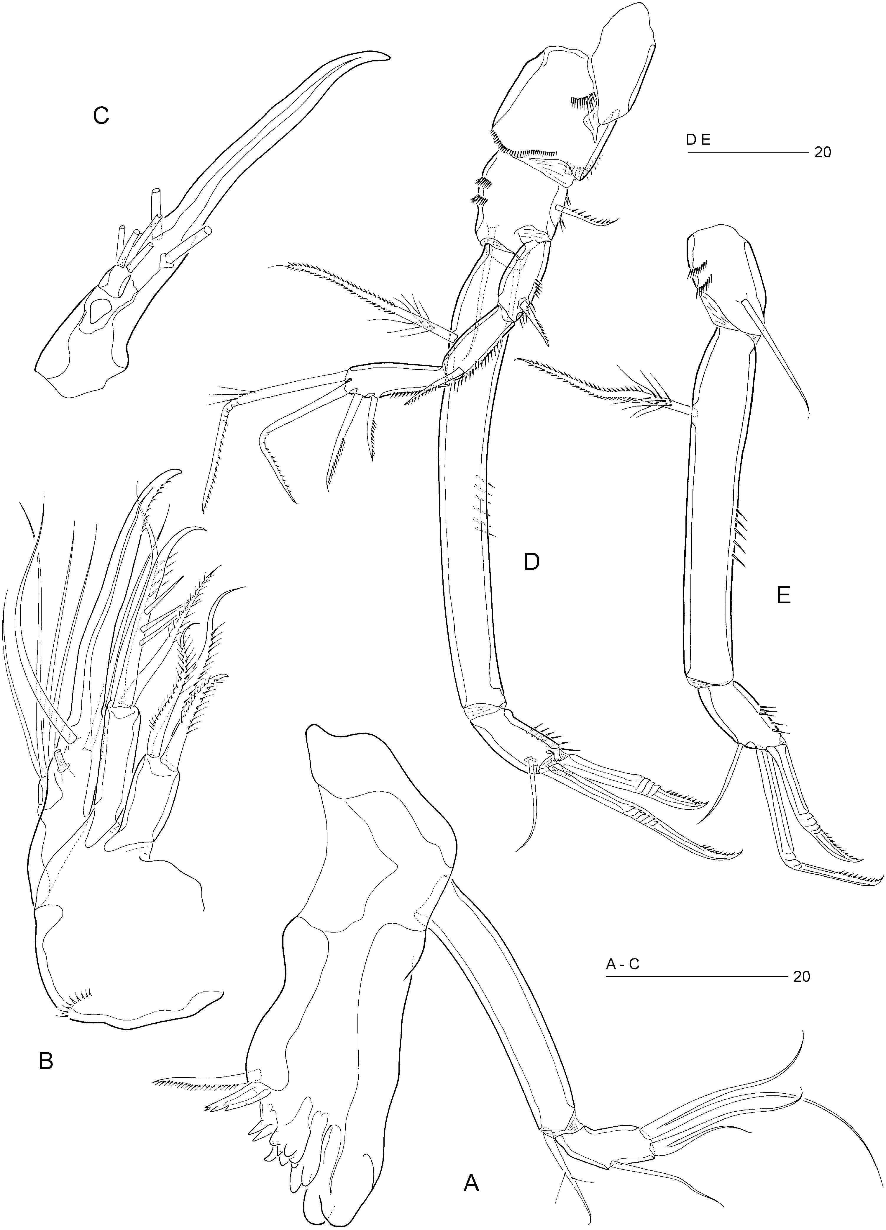
Mandible ( Fig. 4A View Figure 4 ). Gnathobase well developed; uniramous palp, consisting of basis and onesegmented endopod; basis elongate, with one lateral, pinnate seta; endopod with one outer and four apical setae fused in two clusters.
Maxillule ( Fig. 3E View Figure 3 ). Praecoxal arthrite with ten spines/setae around distal margin and two tubular setae (one long, one rudimentary) on anterior surface. Coxal endite with one geniculate claw and one seta. Basis and rami largely fused into elongate palp; basal armature represented by two lateral setae, and three setae plus a geniculate claw apically. Endopod represented by a small semidiscrete segment with three setae, exopod by two small setae.
Maxilla ( Fig. 4B, C View Figure 4 ). Syncoxa with two endites, proximal endite with three articulating setae, distal endite with one pinnate claw and two setae, all articulating. Allobasis drawn out into a claw-like pinnate endite armed with two additional setae; with a distinct tube-pore. Endopod a discrete segment with four long setae and with a small sclerite around its base ( Fig. 4C View Figure 4 ).
Maxilliped ( Figs 2A, B View Figure 2 , 3F View Figure 3 ) well developed, subchelate, directed inwards. Syncoxa well developed, with one pinnate seta and two spinular rows on anterior surface. Basis elongate, with two to three long spinules anteriorly and a spinular row along inner margin posteriorly. Endopod represented by a strong, curved, bare claw.
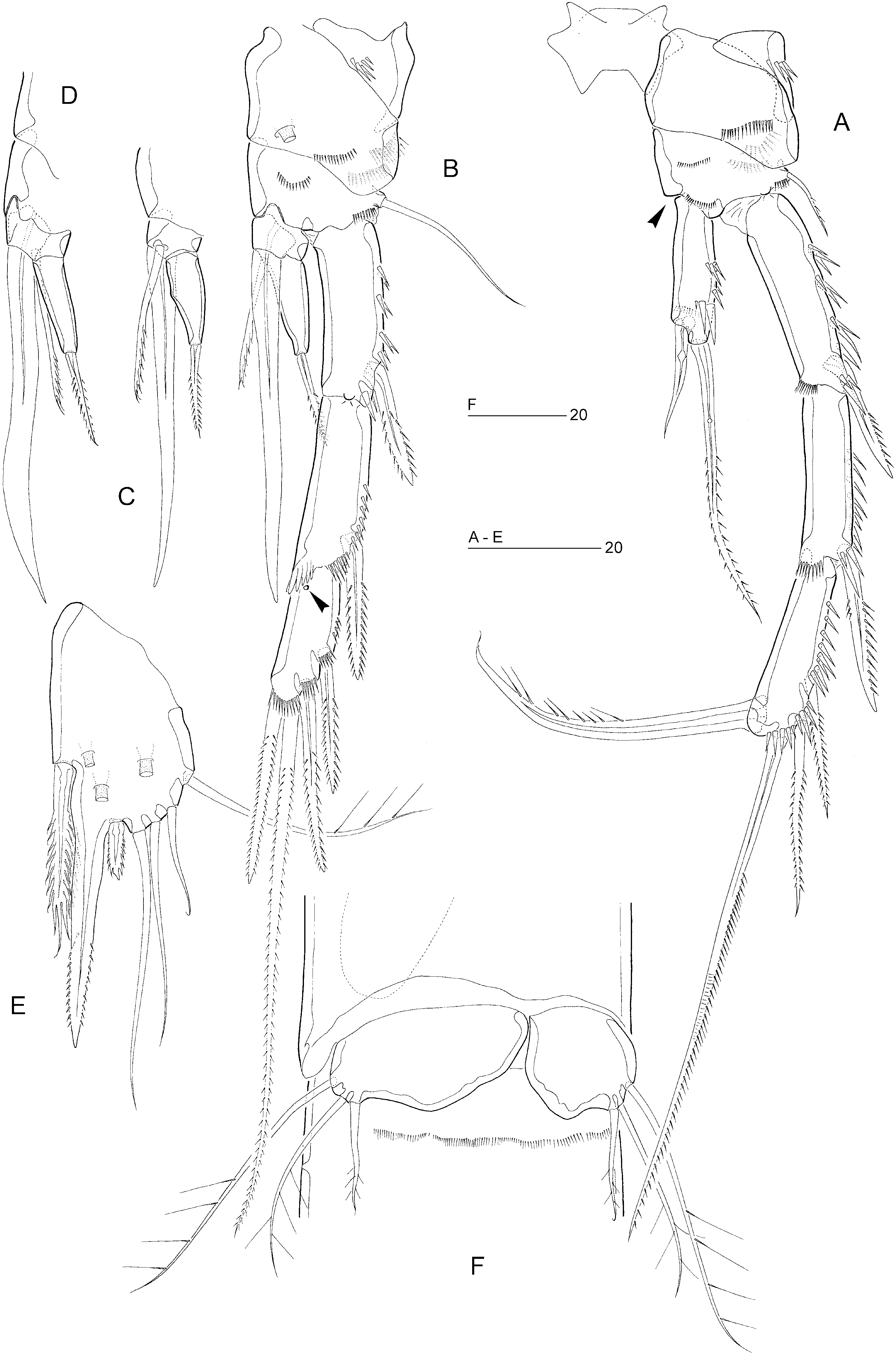
P1 ( Fig. 4D, E View Figure 4 ). Praecoxa strongly developed. Coxa with three spinular rows. Basis with long, naked, inner seta and short, pinnate, outer seta. Exopod three-segmented; with outer spine on exp-2, with two spines and two geniculate setae on exp-3. P1 endopod prehensile, distinctly longer than exopod; proximal segment approximately ten times as long as average width, with pinnate inner seta being plumose in proximal third; distal segment short, with subdistal setule, and two geniculate spines distally.
Swimming legs P2–P4 ( Fig. 5A–F View Figure 5 ). P4 distinctly longer than P2–P3. Width of intercoxal sclerites decreasing in antero-posterior direction ( Fig. 5B, D, E View Figure 5 ). Praecoxae well developed, with spinular row on anterior surface in P2 and P4. Coxae with pattern of spinules, as in Figure 5A,B,C,D,E,F View Figure 5 , with a large tubepore on anterior surface of P3 and P4. Bases with outer seta (short and pinnate in P2, long and plumose in P3, long and bare in P4); with spinular rows on anterior surface only ( Fig. 5A, C, E View Figure 5 ). Exopods threesegmented, endopods one-segmented. Exopodal spines of P3 minutely serrate; inner distal spine shorter than outer distal one ( Fig. 5C View Figure 5 ). Inner setae of P4 endopod and P4 exp-3 serrate. Inner element of P2 endopod setiform, bare, approximately 0.3 times the length of distal spine. Armature elements of P3 endopod spiniform; inner spine less than half length of outer spine. Seta and spine formulae as for genus.
Fifth pair of legs ( Figs 5G View Figure 5 , 6A View Figure 6 ) not fused medially, no distinct intercoxal sclerite. Baseoendopod and exopod fused into a common, elongate plate, tapering distally; apex with strong, articulating spine, distinctly longer than plate and with flagellate tip; outer margin with three sparsely plumose setae (including seta derived from baseoendopod); inner margin with two serrate spines, one pinnate seta and one long, pinnate seta fused to plate; anterior surface with three large tube-pores.
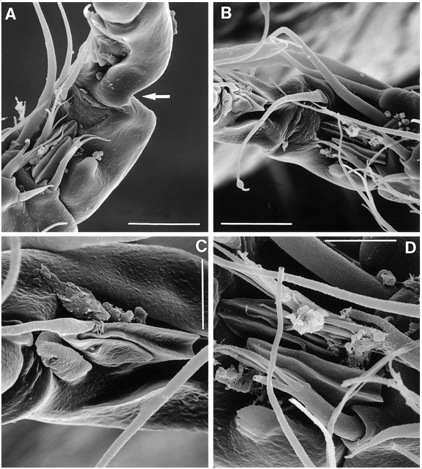
Sixth legs ( Fig. 2D View Figure 2 ) each represented by small operculum closing off gonopore; armature consisting of spiniform outer element and two accessory setules. Genital apertures not fused medially; copulatory pore large, located slightly posterior to gonopores; leading via a short chitinized copulatory duct to paired anterior extensions positioned anterior to genital apertures; copulatory pore flanked by two small secretory pores.
Single egg-sac ( Fig. 1B, C View Figure 1 ) containing approximately seven eggs arranged in a biserial way, enclosed in a common egg-sac membrane; egg-sac connected with each genital aperture via transparent string.
Male: Body length: 705 µm ( Fig. 7A View Figure 7 ). Spermatophore 110 µm. Anal somite without spinules near ventral anterior ( Fig. 6B View Figure 6 ) and hind margins ( Fig. 6B–F View Figure 6 ).
Antennule ( Fig. 7B, C View Figure 7 ) indistinctly nine-segmented; geniculation between segments 7 and 8; segment 1 with one minute seta and spinular pattern as in female; segment 2 longest, with one plumose and eight naked setae; segment 3 with six setae; segment 4 minute, forming an incomplete ring, with two short setae; segment 5 distinctly swollen, with six elements along the anterior margin and with a distal cylindrical process bearing basally fused seta and large, constricted aesthetasc (53 µm); segment 6 with one long seta and one short pinnate spine; segment 7 with three spiniform elements and one seta; segment 8 with one seta and four modified elements (modified as in Figure 24C View Figure 24 for E. cumbraensis ); segment 9 with five posterior setae, one anterior seta and one seta plus an acrothek apically. Apical acrothek consisting of two naked setae and one slender aesthetasc (15 µm).
P2 ( Fig. 8A View Figure 8 ) with inner distal corner of basis not modified into spinous process (as in other cylindropsyllid genera) but lateral margin more chitinized than in female. Endopod slightly larger than in female; outer margin with more spinules; apical seta distinctly longer, extending beyond distal margin of exp- 2, with sparser ornamentation than in female; inner seta larger than in female. Exp-3 modified; outer distal element setiform and distinctly longer than in female; inner distal element transformed into strong claw, directed medially and posteriorly, and with distal half pinnate and tapering to a fine tip.
Spines of P3 exopodal segments with pinnate ornamentation ( Fig. 8B View Figure 8 ); exp-1 without hyaline frill; exp-3 with secretory pore on anterior surface near articulation with exp-2; outer distal element of exp-3 setiform and distinctly longer than in female. P3 endopod ( Fig. 8B–D View Figure 8 ) distinctly two-segmented; enp-1 small, with short, serrate, posterior seta and long, rigid apophysis arising from the anterior surface; distal half of apophysis bilaterally compressed (cf. Fig. 8C, D View Figure 8 ); enp- 2 tapering distally, with one short, pinnate seta apically.
Fifth legs ( Figs 6B View Figure 6 , 8E View Figure 8 ) not fused medially, no distinct intercoxal sclerite. Baseoendopod and exopod fused into a common elongate plate, tapering distally towards long, pinnate, spinous process, which is longer than the plate; inner margin with serrate spine; outer margin with small serrate spine, three naked setae and a sparsely plumose seta derived from baseoendopod; anterior surface with three large tube-pores.
Sixth pair of legs ( Figs 6B View Figure 6 , 8F View Figure 8 ) asymmetrical, with three sparsely pinnate setae each, decreasing in length medially. Left or right leg articulating according to sinistral or dextral development of testis and vas deferens. First postgenital somite with transverse spinular row near ventral anterior margin ( Fig. 8F View Figure 8 ).
Caudal ramus conical ( Fig. 6D–F View Figure 6 ), slightly longer than in female, length (measured along the outer margin) approximately 2.2 times the proximal width; seta IV long and uniplumose, extending to flexure zone of seta V.
Differential diagnosis: Evansula incerta and E. arenicola are the only species in the genus that have retained the syncoxal seta on the maxilliped and that lack the presence of a raised spinular row or spinous process on the dorsal surface of the caudal rami. In both species seta c of the female P5 (cf. Figure 36 View Figure 36 for reference position) is fused to the segment. However, this character is also shared by E. spinosa sp. nov. Females of E. incerta and E. arenicola can be differentiated by the general facies of leg 1, the shape of the caudal rami and seta V, the length of the inner distal seta of the P2 endopod, and the presence/absence of ventral anterior spinule rows on the anal somite. Males can be distinguished by the endopodal segmentation of P3 and P4.
Distribution: Scotland: St. Monans in Firth of Forth ( Scott, 1892, 1906b).
Although many authors have recorded specimens they attribute to E. incerta , there is good reason to believe that in fact they have often mistaken other undescribed species for it. The reasons for this conclusion lie in the frequently repeated statements about the difficulty in differentiating E. incerta , E. pygmaea , and E. arenicola . With the discovery of several new species in the North Sea, the reliability of previously published records from north-western Europe becomes uncertain.
The true state of confusion reigning in the genus is illustrated by the situation in the Firth of Forth – the type locality of both E. incerta (St. Monans) and E. pygmaea (Musselburgh) . Examination of a single, intertidal sandflat sample taken at Elie (near St. Monans) revealed the presence of E. cumbraensis sp. nov. ( type locality Isle of Cumbrae, and widely distributed in the North Sea) and resulted in the discovery of two other new species, which are currently under study. Surprisingly, the sample did not contain any E. incerta or E. pygmaea . A re-examination of T. Scott’s material of the Forth River produced the second record of E. spinosa sp. nov. ( type locality Korshavn, Norway), raising the number of Evansula species in the Firth of Forth to six.
Evansula incerta of Sars (1911) and at least part of Scheibel’s (1972, 1973) material from the Kieler Bucht are in fact E. spinosa sp. nov. Scheibel’s illustrations of the P 5 in different specimens attributed to E. incerta raise the suspicion that he was dealing with an amalgam of species. There is no doubt that the American records of E. incerta by Wilson (1932) and Coull (1971, 1977) are based on misidentifications and probably all pertain to E. arenicola . Scott’s (1903b) record from East Finmark ( Norway) concerns a different species, here described as E. polaris (see below).
All other records provide insufficient information and, consequently, are uncertain at this moment.
Sweden: Hållö ( Por, 1964), Isle of Bonden ( Por, 1964; Swedmark & Teissier, 1967).
Scotland: River Ythan (Hockin, 1981, 1982a, b, c, 1983, 1984; Hockin & Ollason, 1981).
Wales: Porth-y-Post and Port Swtan (Church Bay), Anglesey ( Geddes, 1972).
Germany: Kieler Bucht ( Klie, 1929, 1950; Remane, 1933; Kunz, 1935), off Bottsand and Weisenhaus in Kieler Bucht ( Noodt, 1956, 1957), Boknis Eck in Kieler Bucht ( Scheibel, 1976), Helgoland ( Kunz, 1938; Klie, 1950), Isle of Sylt ( Noodt, 1952, 1957), Amrum ( Noodt, 1957).
Belgium: North Sea coastal zone ( Govaere et al., 1980).
France: Kersaint, Finistère ( Bodin & Boucher, 1981; Bodin, 1988), Charente-Maritime ( Bodin, 1976, 1977), Bassin d’Arcachon, Gironde ( Renaud-Debyser, 1963a, b), Contis-Plage, Landes ( Noodt, 1955a, b; Delamare Deboutteville, Gerlach & Siewing, 1955; Delamare Deboutteville, 1960).
Hockin (1984) recorded the presumed ectocommensal suctorians Thecacineta inclusa Meunier, 1903 and Thecacineta cothurnoides Collin, 1909 from E. incerta in the River Ythan estuary, Aberdeenshire, Scotland.
| T |
Tavera, Department of Geology and Geophysics |
No known copyright restrictions apply. See Agosti, D., Egloff, W., 2009. Taxonomic information exchange and copyright: the Plazi approach. BMC Research Notes 2009, 2:53 for further explanation.
|
Kingdom |
|
|
Phylum |
|
|
Class |
|
|
Order |
|
|
Family |
|
|
Genus |
Evansula incerta
| Huys, Rony & Conroy-Dalton, Sophie 2006 |
E. spinosa
| Huys & Conroy-Dalton 2006 |
E. polaris
| Huys & Conroy-Dalton 2006 |
E. arenicola
| Nicholls 1939 |