
Evansula spinosa, Huys & Conroy-Dalton, 2006
|
publication ID |
https://doi.org/10.1111/j.1096-3642.2006.00227.x |
|
persistent identifier |
https://treatment.plazi.org/id/03F487A0-FF8D-FF8C-FD75-F9BEFA859635 |
|
treatment provided by |
Felipe |
|
scientific name |
Evansula spinosa |
| status |
sp. nov. |
EVANSULA SPINOSA SP. NOV.
Synonym: Evansula incerta (T. Scott, 1892) sensu Sars (1911) .
Original description: Sars (1911: 415–416, suppl., plate 39).
Additional description: Scheibel (1972).
Type locality: Norway, Korshavn , near Lindesnes; at 30–50 fathoms .
Material examined: (1) From type locality: holotype ♀ dissected on eight slides; found among spirit- preserved specimens of Neobradya pectinifera T. Scott, 1892 , deposited in Zoologisk Museum, Oslo; coll. G.O. Sars; deposited in NHM (reg. no. 1995.428); (2) NHM, reg. no. 1995.429: Firth of Forth (no further details specified), Scotland; 1 ♂ paratype [found among spirit-preserved ‘cotypes’ of E. incerta : reg. nos 44505– 507; as part of Cannon A. M. Norman collection ( 1911.11.8)]; coll. T. Scott, 9 September 1894 ; dissected on eight slides; (3) NHM, reg. no. 2005.2069: Southern Bight of North Sea , off Suffolk ( UK), 51°57′24′ N 2°10′57 ′E; 42.7m depth; 1 ♀ paratype in alcohol; coll. R. Huys, 30 March 1992 .
Etymology: The species name is derived from the Latin spina, meaning spine, and refers to the dorsal spinous process on the caudal ramus.
Description
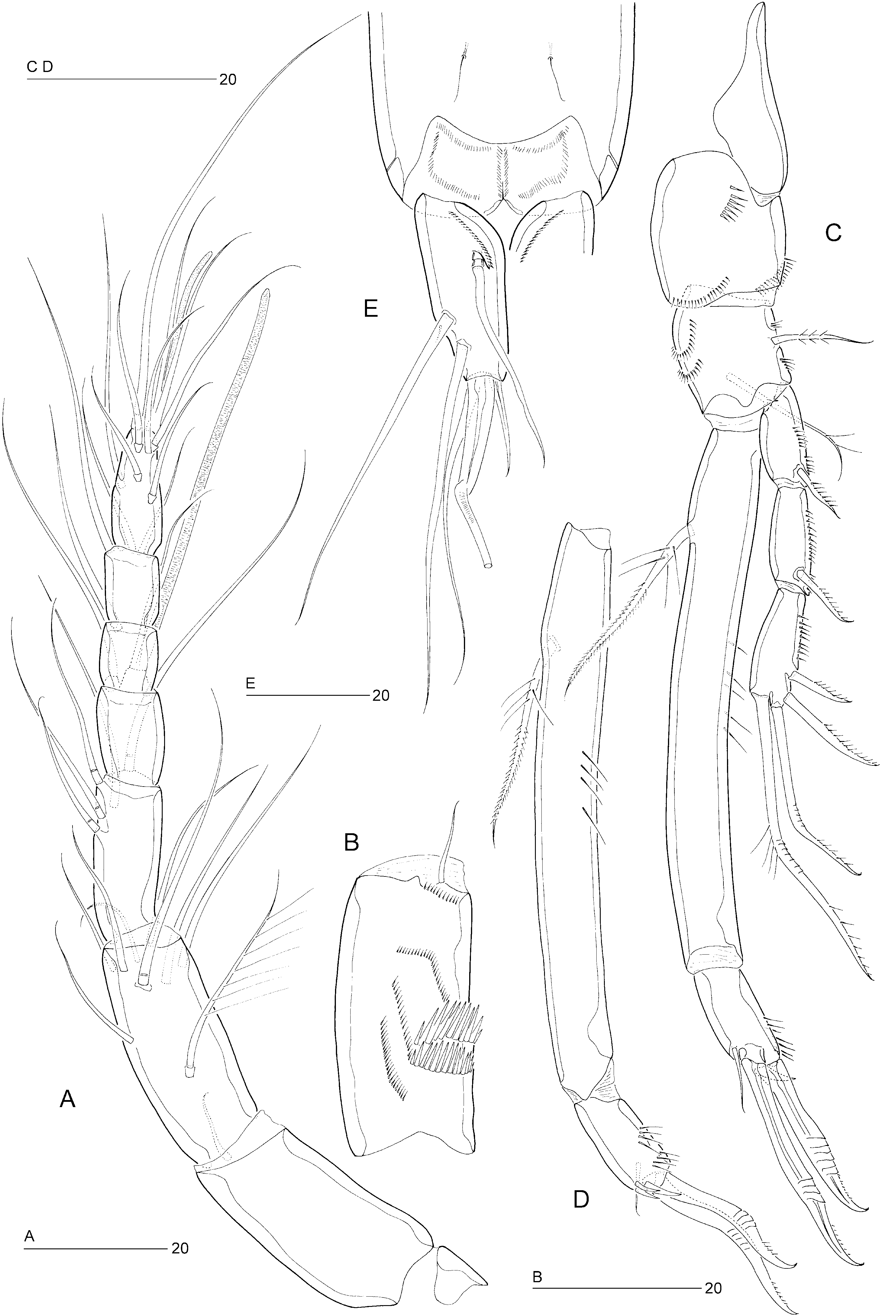
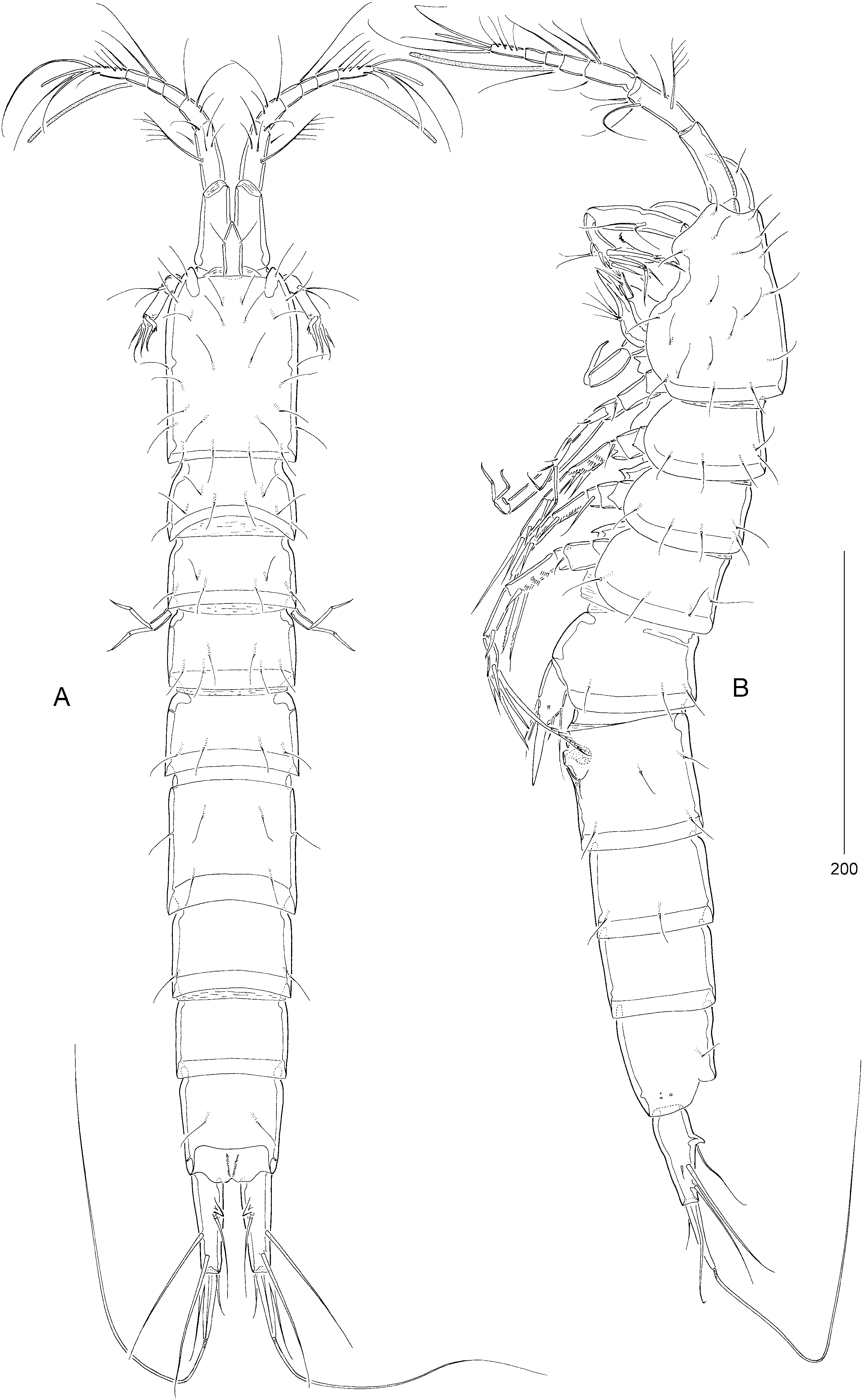
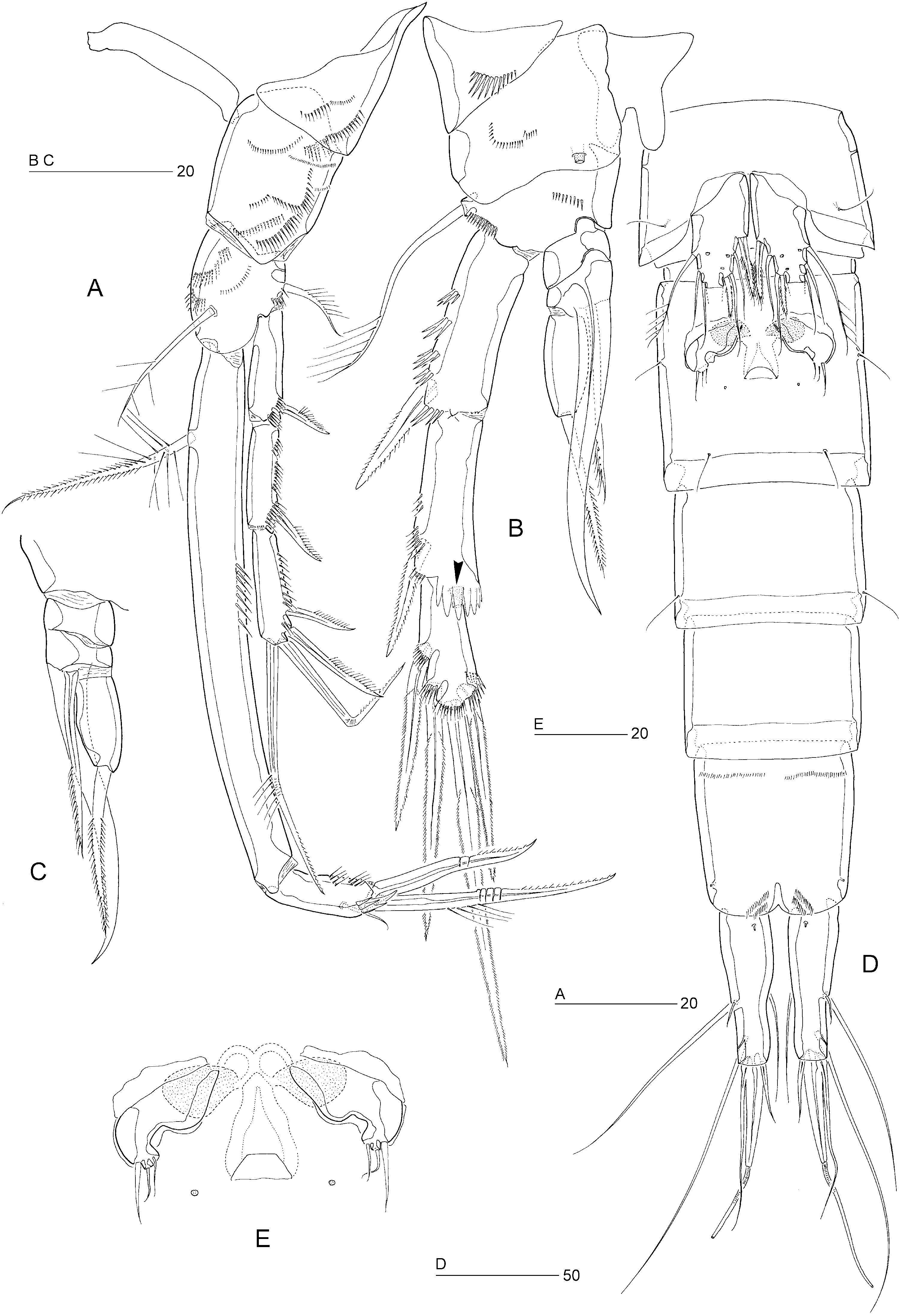
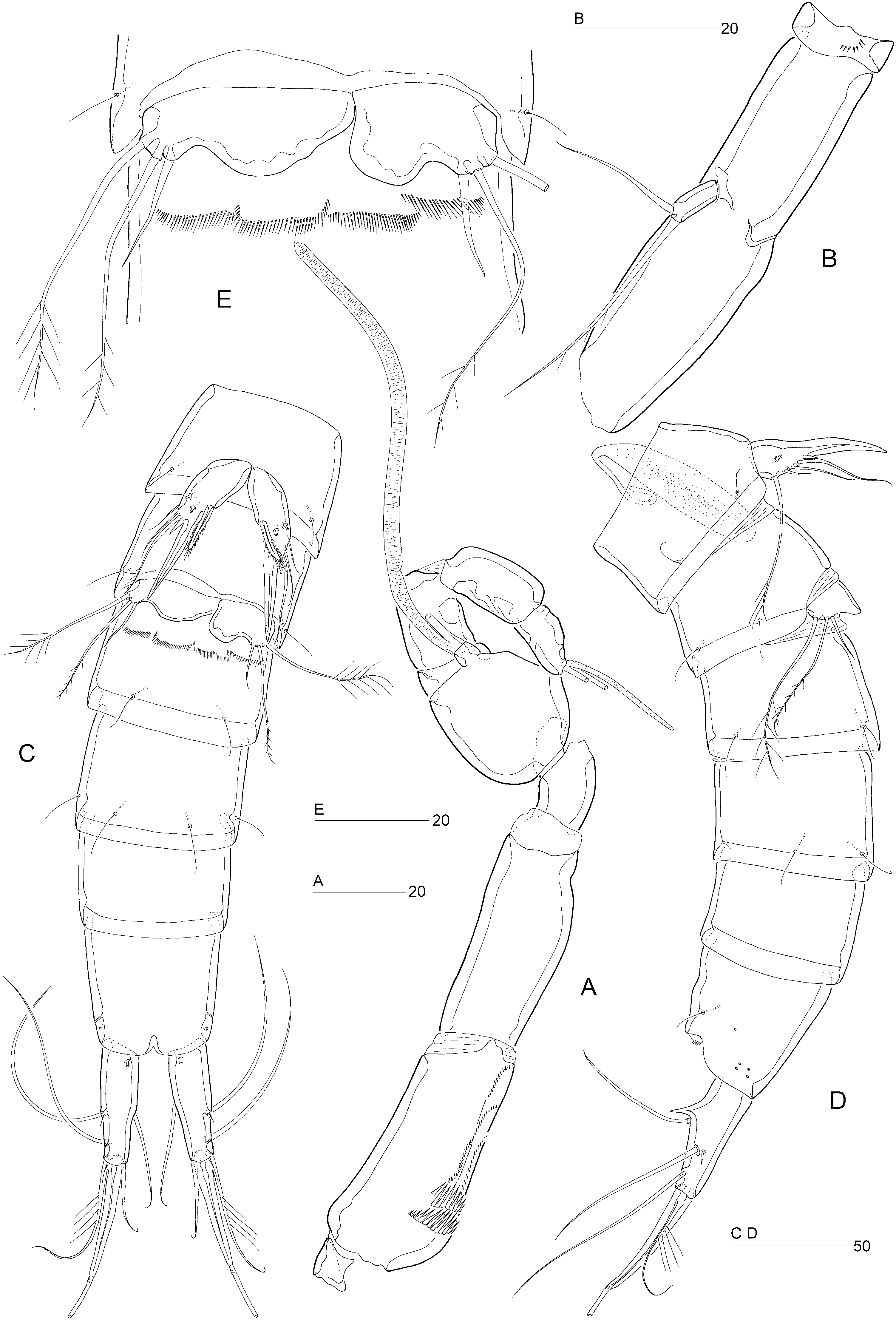
Female: Total body length: 700 µm. Body slender, cylindrical ( Fig. 28A, B View Figure 28 ), semitransparent, light brown; no distinct separation between prosome and urosome. Genital double-somite completely fused ( Figs 28A, B View Figure 28 , 29D View Figure 29 ); original segmentation marked dorsally by paired anterior and posterior sensillae ( Fig. 28A View Figure 28 ). Anal somite only slightly longer than wide (72 × 66 µm), with three pairs of secretory pores laterally ( Fig. 32D View Figure 32 ); anterior margin with ventral transverse row of tiny spinules ( Fig. 29D View Figure 29 ); posterior margin with two short spinular rows on either side of ventral midline ( Figs 29D View Figure 29 , 32E View Figure 32 ). Anal operculum weakly developed, unarmed ( Fig. 32C View Figure 32 ).
Caudal ramus cylindrical ( Figs 29D View Figure 29 , 32C–E View Figure 32 ) with slightly concave inner margin, length (measured along the outer margin) approximately 3.5 times the proximal width; dorsal surface with chitinous spur ( Fig. 32C, D View Figure 32 ); with seven setae, setae I–VI in distal and seta VII in proximal half ( Fig. 32C View Figure 32 ); seta I small, larger than in other species; setae II–III long and bare; seta IV bare, longer than styliform part of V; seta V long, with distinct flexure zone between styliform part and long distal flagellate part, fused at base with seta IV; seta VI vestigial; seta VII tri-articulate at base and located near proximal inner margin; ventral surface with one tube-pore ( Fig. 32E View Figure 32 ), lateral surface with one tubular and one simple pore ( Fig. 32D View Figure 32 ).
Rostrum elongate ( Fig. 28A View Figure 28 ), with parallel margins in proximal half, tapering distally; distinctly shorter than first antennulary segment; with two long sensillae.
Antennule seven-segmented ( Fig. 28A, B View Figure 28 ); armature formula as in E. incerta ; segment 1 slightly longer than segment 2.
Antenna with spinular row on coxa ( Fig. 31B View Figure 31 ); basis and first endopod segment incompletely fused to form allobasis, abexopodal margin without ornamentation; exopod small, with one long, pinnate and one shorter, naked seta.
Antennary endopod, mandible, maxillule, and maxilla as in E. incerta .
Maxilliped as in E. pygmaea . Syncoxa without seta.
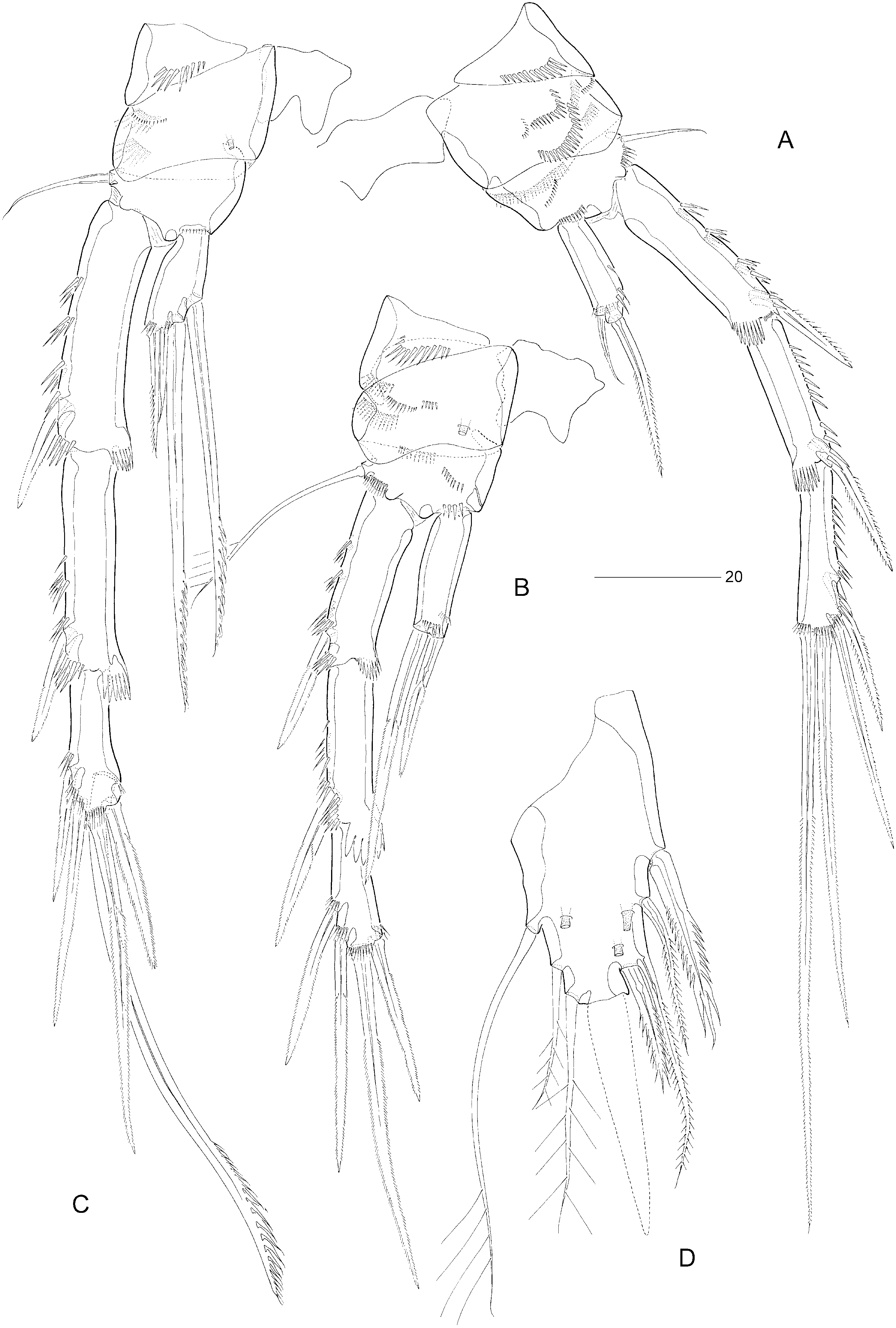
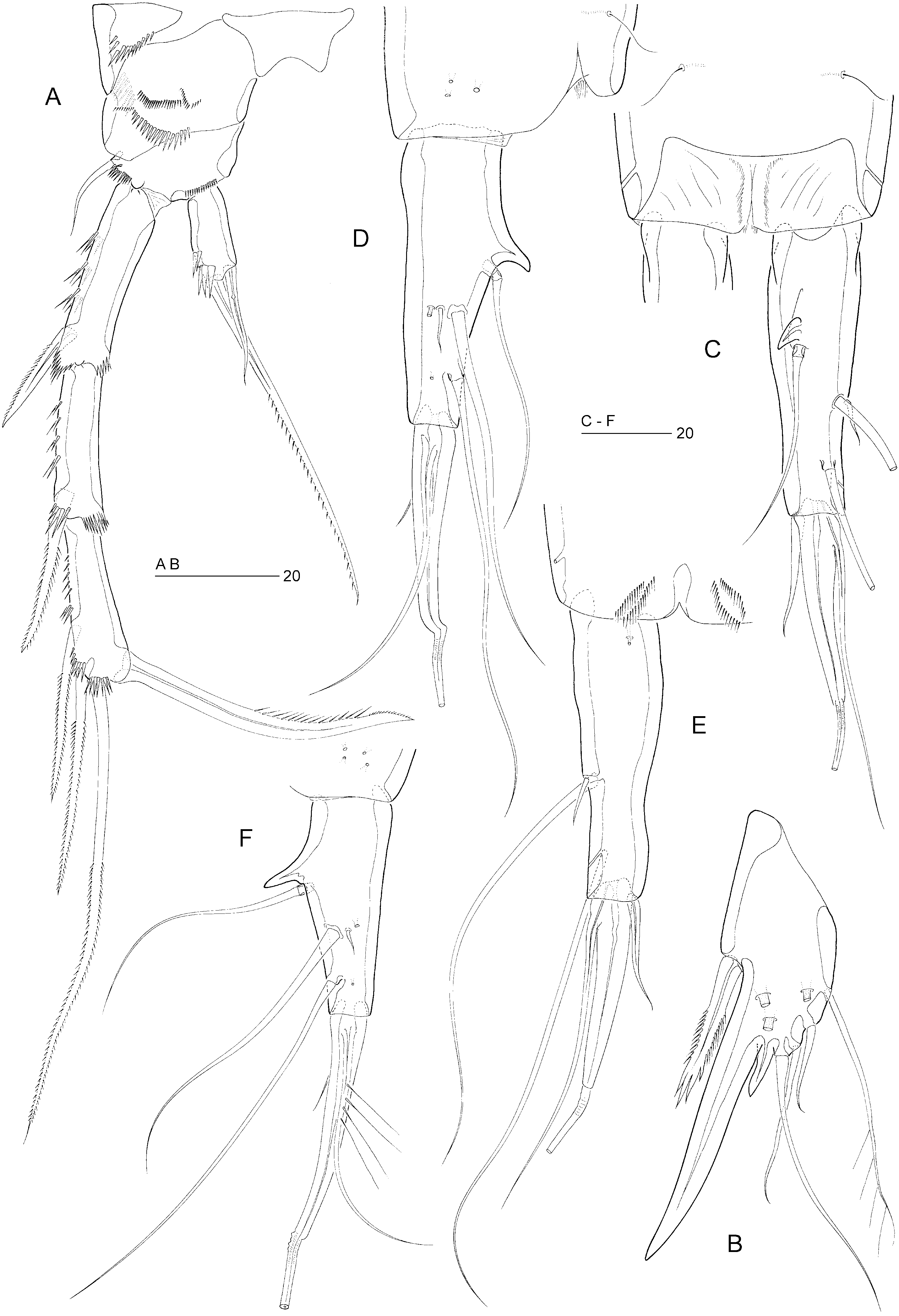
P1 ( Fig. 29A View Figure 29 ). Praecoxa strongly developed, with three spinular rows. Coxa with six spinular rows on anterior and two on posterior surface. Basis with long, sparsely plumose inner seta and shorter, pinnate outer seta. Exopod three-segmented; with two spines and two geniculate setae on exp-3. P1 endopod prehensile, distinctly longer than exopod; proximal segment approximately ten times as long as average width, with pinnate inner seta being plumose in proximal third; distal segment short, with three spinular rows, a subdistal setule, and two geniculate spines distally.
Swimming legs P2–P4 ( Fig. 30A–C View Figure 30 ). P4 distinctly longer than P2–P3. Width of intercoxal sclerites decreasing in antero-posterior direction. Praecoxae well developed, with spinular row on anterior surface in P2–P4. Coxae with pattern of spinules as in Figure 30A,B,C View Figure 30 , with large tube-pore on anterior surface of P3 and P4. Bases with outer seta (short and naked in P2 and P4, long and plumose in P3); with spinular rows on anterior surface only. Exopods three-segmented, endopods one-segmented. Exopodal spines of P3–P4 minutely serrate. Inner distal spine of P3–P4 exp-3 shorter than outer distal one. Inner setae of P4 endopod and exp-3 serrate. Inner element of P2 endopod setiform, pinnate, approximately half length of distal spine. Armature elements of P3 endopod spiniform; inner spine longer than half length of outer spine. Seta and spine formulae as for genus.
Fifth pair of legs ( Figs 29D View Figure 29 , 30D View Figure 30 ) with baseoendopod and exopod fused into a common elongate plate, tapering distally; apex with strong, articulating spine (lost on both sides during dissection), approximately as long as the plate and with a flagellate tip; outer margin with three sparsely plumose setae (including seta derived from baseoendopod); inner margin with two serrate spines, one pinnate seta and one long pinnate seta fused to plate; anterior surface with three large tube-pores.
Sixth legs ( Fig. 29D, E View Figure 29 ) each represented by small operculum closing off gonopore; armature consisting of spiniform outer element and two accessory setules. Genital apertures not fused medially; copulatory pore large, located slightly posterior to gonopores; leading via short chitinized copulatory duct to paired anterior extensions positioned anterior to genital apertures; copulatory pore flanked by two small secretory pores.
Egg-sac not confirmed.
Male: Body length: 670 µm. Spermatophore 75 µm. Anal somite without spinules near ventral anterior and hind margins ( Fig. 31C View Figure 31 ).
Antennule ( Fig. 31A View Figure 31 ) distinctly nine-segmented; geniculation between segments 7 and 8; segment 1 with one minute seta and spinular pattern as in female; slightly longer than segment 2; segment 5 distinctly swollen, with distal cylindrical process bearing basally fused seta and large aesthetasc (100 µm); acrothek on segment 9 with slender aesthetasc (27 µm).
P2 ( Fig. 32A View Figure 32 ) with inner distal corner of basis not modified into spinous process but lateral margin slightly more chitinized. Exp-3 modified; inner distal element transformed into strong claw, directed medially and posteriorly, and with distal half pinnate. Endopod slightly shorter than in female; outer margin with two spinular rows; apical seta distinctly longer, clearly extending beyond distal margin of exp-2, with sparser ornamentation than in female; inner seta slightly larger than in female, bare.
Spines of P3 exopodal segments with pinnate ornamentation ( Fig. 29B View Figure 29 ); exp-1 with reduced hyaline frill; exp-3 with secretory pore on anterior surface near joint with exp-2. P3 endopod ( Fig. 29B, C View Figure 29 ) distinctly three-segmented; enp-1 small, without armature or ornamentation; enp-2 with serrate, posterior seta and long, rigid sigmoid apophysis arising from anterior surface; enp-3 with weakly chitinized inner margin and one pinnate seta apically.
Fifth legs ( Figs 31C, D View Figure 31 , 32B View Figure 32 ) with baseoendopod and exopod fused into a common elongate plate, tapering distally towards long, smooth, spinous process, which is longer than the plate; inner margin with serrate spine; outer margin with small, bare spine completely fused to plate, three naked setae and a sparsely plumose seta derived from baseoendopod; anterior surface with three large tube-pores.
Sixth pair of legs ( Figs 31C, E View Figure 31 ) asymmetrical, with one short, naked and two long, sparsely pinnate setae each. First postgenital somite with transverse spinular row near ventral anterior margin ( Fig. 31E View Figure 31 ).
Caudal ramus cylindrical ( Fig. 32F View Figure 32 ), shorter than in female; seta IV long and uniplumose; styliform part of seta V comparatively longer than in female.
Remarks: Although there are several discrepancies between Scott’s (1892) and Sars’ (1911) descriptions of E. incerta , only the dorsal spinous process has attracted subsequent workers’ attention. The presence or absence of this structure was regarded as part of the intraspecific variability and consequently both the Norwegian and Scottish populations were believed to represent two forms of the same species. Kunz (1938) and Scheibel (1972) claimed that they had found both ‘forms’ in their samples from Helgoland and the Kieler Bucht, respectively. However, at least in Scheibel’s case, there is evidence that E. spinosa occurred in his material (cf. his illustration of the caudal ramus; Scheibel, 1972: tafel XVII, Fig. 10 View Figure 10 ). It should be noted that Sars (1911) inadvertently reversed the female P2 and P 3 in his illustrations.
Differential diagnosis: Evansula spinosa can be readily distinguished by the presence of a dorsal spur on the caudal ramus in both sexes, and by the fused outer spine on the male P5.
Distribution: Norway: Korshavn, near Lindesnes ( Sars, 1911; present account), Troldfjord in Lofoten Islands.
Germany: Kieler Bucht ( Scheibel, 1972), probably also Helgoland ( Kunz, 1938).
Scotland: Firth of Forth (present account).
England: off Suffolk (present account).
| T |
Tavera, Department of Geology and Geophysics |
| R |
Departamento de Geologia, Universidad de Chile |
No known copyright restrictions apply. See Agosti, D., Egloff, W., 2009. Taxonomic information exchange and copyright: the Plazi approach. BMC Research Notes 2009, 2:53 for further explanation.