
Ephemera danica
|
publication ID |
https://doi.org/ 10.5281/zenodo.176552 |
|
DOI |
https://doi.org/10.5281/zenodo.6243212 |
|
persistent identifier |
https://treatment.plazi.org/id/03F31C15-FFEE-B308-B5D5-13FA27C8FBC4 |
|
treatment provided by |
Plazi |
|
scientific name |
Ephemera danica |
| status |
|
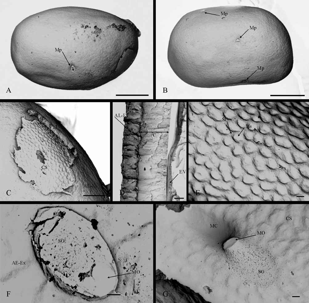
Description of Egg Morphology
General features: Oval-like egg ( Fig. 3 View FIGURE 3 A), although sometimes quadrangular ( Fig. 3 View FIGURE 3 B) or with irregular form. Very simply egg shape, with smooth surface due to the “complex adhesive-extrachorion layer” which completely surrounds the egg ( Fig. 3 View FIGURE 3 A, B). Several micropyles have been observed in the equatorial area, frequently some may be displaced towards the subpolar area ( Fig. 3 View FIGURE 3 A, B). Egg-size between 175–209 µm length and 114–128 µm width.
Attachment structures: An adhesive layer is the only attachment structure that the egg possesses. This layer covers the whole egg, hiding the chorion surface completely, except at the sperm guide of each micropyle ( Fig. 3 View FIGURE 3 B). The adhesive layer is a compact material of granular or undifferentiated appearance ( Fig. 3 View FIGURE 3 C), whose thickness is approximately half that of the chorion layer ( Fig. 3 View FIGURE 3 D). The extrachorion is a very slight film covering the adhesive layer and its differentiation is very difficult, even in a cross-section of the eggshell ( Fig. 3 View FIGURE 3 D), probably because it and the adhesive layer are strongly adhered and form a single unit. We therefore prefer to call this attachment structure as a “complex adhesive-extrachorion layer”.
Chorionic sculpturing: The chorionic features are only observed by SEM when the adhesive layer covering its surface has been removed ( Fig. 3 View FIGURE 3 A, C). Chorionic sculpturing consists of small depressions of circularshape (1.3–2.9 µm diameter), whose distribution and arrangement extend regularly the whole chorion surface and could be related to a small meshed reticulation pattern ( Fig. 3 View FIGURE 3 E). The mesh of net-like sculpturing consists of small depressions and a polygonal reticulum ridge-like strand with hexagonal, pentagonal or irregularly shaped units ( Fig. 3 View FIGURE 3 E)
Micropyles: The micropyle are “tagenoform-type”, with an oval shaped sperm guide ( Fig. 3 View FIGURE 3 F), sometimes almost elliptical, followed by a long intrachorionic tube, the micropylar canal. If the complex extrachorionadhesive layer is present, the sperm guide is the only part of the micropyle that can be differentiated by SEM, because it is perfectly delimited by this attachment structure ( Fig. 3 View FIGURE 3 F). When the complex layer is removed, the sperm guide can be observed since this area does not present the typical chorionic sculpturing ( Fig. 3 View FIGURE 3 G); therefore, the sperm guide is a “suprachorionic-chorionic” type. In these circumstances, the proximal zone of the micropylar canal can also be observed ( Fig. 3 View FIGURE 3 G). Sperm guide size is 7.4–8.5 µm length x 5 µm width.
The number of micropyles appearing in an egg is variable and we have observed a maximum of three ( Fig. 3 View FIGURE 3 B). These are arranged in the equatorial area of the egg, more or less aligned in a cross-sectional direction, and separated by a distance close to half the total width of the egg ( Fig. 3 View FIGURE 3 B). Sometimes, one of these micropyles can be displaced to the subpolar area ( Fig. 3 View FIGURE 3 B)
Partial discussion
The egg morphology of E. danica was described in detail by Degrange (1960), although partial descriptions and isolated data can also be found in Bengtsson (1913), Degrange (1956), Koss and Edmunds (1974) and Haybach (2003). The characteristic morphology of eggs given by Degrange (1960), concerning attachment structures and chorionic sculpturing, is re-described in the study, although our observations of micropyle morphology are closer to the description given by Koss and Edmunds (1974).
We regard the attachment structure in the eggs of E. danica as a complex formed by an adhesive layer and the extrachorion. The adhesive layer is closer to the chorion surface and, in agreement with Degrange (1960) and Koss and Edmunds (1974), it has a granular or amorphous appearance, but never fibrous. The extrachorion is a delicate film covering the adhesive layer surface, almost imperceptible in cross-section, but the smooth appearance of the egg surface is due to its presence. The close relationship between the adhesive layer and the extrachorion is not limited to form a structural complex, but also extends to a physiological function. Probably, the presence of extrachorion would explain the different physiological response of the adhesive layer observed by Degrange (1960) when the eggs were submerged in physiological liquid or merely in water.
This “complex extrachorion-adhesive layer” is a chorionic structure common to all Ephemera eggs, as well as to all Ephemeridae eggs ( Degrange 1960; Koss 1968; Koss & Edmunds 1974). Its presence should be taken into account in SEM studies of egg morphology, because it can hide the chorionic sculpturing or prevent the correct observation of the micropyle. Chorionic sculpturing in Ephemera eggs is very common and its specific features have been described in many species, since this chorionic characteristic can be differentiated easily by light microscopy ( Degrange, 1960; Koss, 1968; Koss & Edmunds, 1974). For these reasons, we think it necessary to treat with caution SEM descriptions of egg morphology that do not consider the presence of an adhesive layer, such as in Balasubramanian et al. (1991) and Kang and Yang (1994). Some eggs, which partially lost their attachment structure during the preparation procedure for SEM analysis, confirmed the observation of Degrange (1960), who mentioned that E. danica chorion presents a net-like sculpturing of small mesh, although reticulation can be polygonous irregular or not.
In agreement with the observations of Koss and Edmunds (1974), the sperm guide is present in E. danica eggs and so is the micropyle, the “tagenoform-type”, but not the “linear-type” or “ type 4” according to Degrange (1956, 1960). The sperm guide leaves traces on both the chorion and “complex extrachorion-adhesive layer” (“suprachorionic-chorionic” type) but, while the trace in the attachment structure is very conspicuous (it is a well delimited hole), on the chorion surface it is poorly defined since it is only differentiable due to the absence of typical chorionic sculpturing. Probably, the sperm guide was less nectly defined in the eggs studied by Degrange (1960) (fresh-eggs and each enveloped by a thick transparent adhesive layer) and he could only observe it when the eggs were stained by aniline blue, differentiating an oblong hole in the adhesive layer just at the micropyle opening. Overlapping of both sperm guide traces could be the reason that Degrange (1960) did not observe the sperm guide trace on the chorion surface when the eggs were stained. On the other hand, the micropylar canal features observed by us are nearer the characteristics described by Degrange (1960) than by Koss and Edmunds (1974). The proximal part of the micropylar canal is slightly raised with respect to the chorion surface, but the absence of chorionic sculpturing in this area means that this feature seems more pronounced. In any case, the proximal part of the micropylar canal does not project above the chorion as it does Caenis eggs ( Malzacher 1982).
From a taxonomic point of view, a non-fibrous adhesive layer as the only attachment structure and a "tagenoform" micropyle seem to be common features of Ephemeridae eggs, although Ephemera eggs could be characterized because the adhesive layer lacks any type of sculpturing. According to Koss and Edmunds (1974), chorionic sculpturing seems to be the only egg characteristic which would allow the identification of species of Ephemera , since its variability could be species-specific. However, although Degrange (1960) found differences in the chorionic sculpturing of eggs from the three species of Ephemera studied, he did not consider them to be of taxonomic use; in fact, his proposed key for differentiating species differentiation based on egg morphology has only one entry covering these three species of Ephemera . Without doubt, a SEM study of the eggs of several Ephemera species would help decide whether chorionic sculpturing is useful for taxonomic purposes. In the Segura river basin, E. danica occurs with other Ephemera species ( E. glaucops Pictet, 1843 and E. lineata Eaton, 1870 ) ( Ubero-Pascal et al. 1998), but, unfortunately, we did not find specimens with eggs of these species. Hopefully, specimens with eggs will be found so that they can be compared with the E. danica data described in this paper.
No known copyright restrictions apply. See Agosti, D., Egloff, W., 2009. Taxonomic information exchange and copyright: the Plazi approach. BMC Research Notes 2009, 2:53 for further explanation.
|
Kingdom |
|
|
Phylum |
|
|
Class |
|
|
Order |
|
|
Family |
|
|
Genus |