
Pseudopyrochroa euryfoveata Zhan & Young, 2023
|
publication ID |
https://doi.org/ 10.11646/zootaxa.5323.4.8 |
|
publication LSID |
lsid:zoobank.org:pub:FAC743C5-5D83-4CD9-B5E4-15604BD64E5C |
|
DOI |
https://doi.org/10.5281/zenodo.8222671 |
|
persistent identifier |
https://treatment.plazi.org/id/03F287FE-945B-FFEB-82F7-F9870C51F8DB |
|
treatment provided by |
Plazi |
|
scientific name |
Pseudopyrochroa euryfoveata Zhan & Young |
| status |
sp. nov. |
Pseudopyrochroa euryfoveata Zhan & Young , sp. nov.
Vernacular Chinese name: ựñfflňffΨ
Type locality: China: Sichuan Province, Ya’an City, Liziping (+ḄDZṃẘ, BȒffi, ƤŦḢ)
Type specimens: Holotype ( MHBU) [adult ♁]: CHINA: Sichuan Province // Ya’an City // [DZṃẘ•BȒ ffi]; Alt. 1810 m; 26 Apr. 2022 // Zhihong Zhan [ḂĖḾ] and // Kaixiang Jing [Ðmḁ] leg.; [2 nd label]: 28.5941°N; 102.1749°E adult // collected beneath bark of dead standing logs; [3 rd label]: Pseudopyrochroa // euryfoveata // Zhan & Young ♁ // det. D. K. Young & Z. Zhan. Allotype ( WIRC) [adult ♀]: CHINA: Sichuan Province // Ya’an City // [DZṃẘ•BȒffi]; Alt. 1810 m; 26 Apr. 2022 // Zhihong Zhan [ḂĖḾ] and // Kaixiang Jing [Ðmḁ] leg.; [2 nd label]: 28.5941°N; 102.1749°E adult // collected beneath bark of dead standing logs; [3 rd label]: Pseudopyrochroa // euryfoveata // Zhan & Young ♀ // det. D. K. Young & Z. Zhan; Paratypes [1 adult ♁ with exuvium] ( DYCC): CHINA: Sichuan Province // Ya’an City // [DZṃẘ•BȒffi]; Alt. 1830 m; 14 Feb. 2022 // Zhihong Zhan [ḂĖ Ḿ] and // Kaixiang Jing [Ðmḁ] leg.; [2 nd label]: 28.5939°N; 102.1744°E larva // collected beneath bark of dead laying logs; [3 rd label]: 03 March 2022, pupated; 20 March // 2022, eclosed adult; [4 th label]: Pseudopyrochroa // euryfoveata // Zhan & Young ♁ // det. D. K. Young & Z. Zhan; [1 adult ♀] ( KXJC): CHINA: Sichuan Province // Ya’an City // [DZṃẘ•BȒffi]; Alt. 1830 m; 14 Feb. 2022 // Zhihong Zhan [ḂĖḾ] and // Kaixiang Jing [Ðm ḁ] leg.; [2 nd label]: 28.5939°N; 102.1744°E larva // collected beneath bark of dead laying logs; [3 rd label]: 01 March 2022, pupated; 16 March // 2022, eclosed adult; [4 th label]: Pseudopyrochroa // euryfoveata // Zhan & Young ♀ // det. D. K. Young & Z. Zhan.
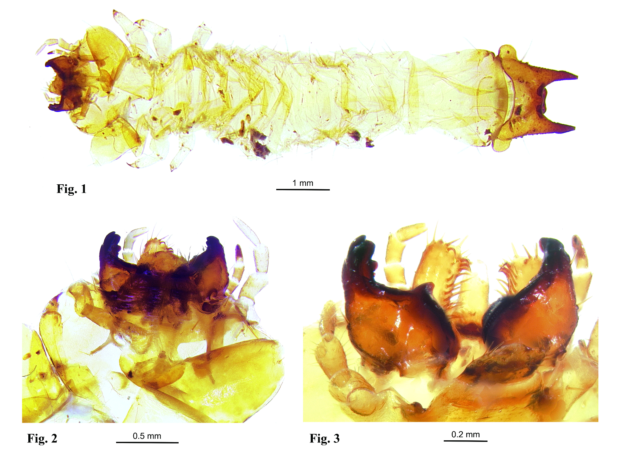
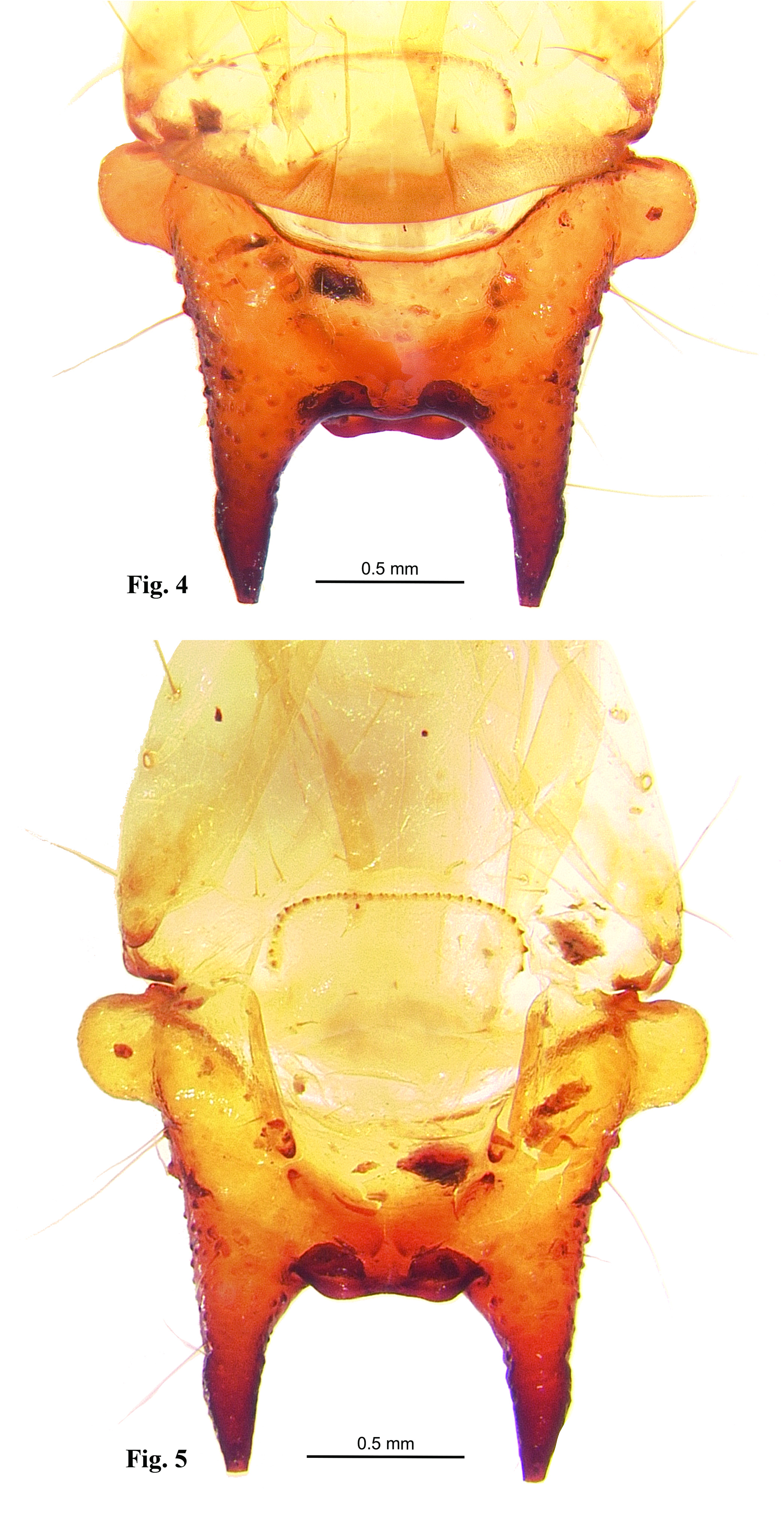
Diagnosis: Larva. Larvae of P. euryfoveata can be characterized as follows: 1) mandibles asymmetrical, with well-developed right mandibular mola and left mandibular tooth; 2) maxillary articulating area well-developed; 3) maxillary mala with uncus; 4) hypostomal rods well developed, divergent ( Fig. 2 View FIGURES 1–3 ); 5) a single arch of asperities along the ventro-anterior margin of the abdominal sternite IX ( Fig. 5 View FIGURES 4–5 ); 6) tergum IX hinged, capable of considerable dorso-longitudinal movement, extending ventrally to form the entire terminal urogomphal plate ( Figs. 1 View FIGURES 1–3 , 4–5 View FIGURES 4–5 ); 7) urogomphal plate with well developed, heavily sclerotized urogomphal lip and paired urogomphal pits ( Fig. 4 View FIGURES 4–5 ).
The other described Pseudopyrochroa larvae from mainland China, P. reni and P. facialis , also lack distinct urogomphal pit microsculpturing. Like that of P. euryfoveata , the larva of P. reni possesses a continuous, well-developed urogomphal lip between the urogomphi supporting the urogomphal pits ( Pan et al. 2021, Fig. 5C View FIGURES 4–5 ), while that of P. facialis is deeply cleft, narrowly but distinctly separating the urogomphal pits ( Zhan et al. 2022, Fig. 6 View FIGURES 6–9 ). The urogomphi of P. euryfoveata are straight and subparallel (urogomphal length:width of separation at bases ≈ 0.79). The urogomphi of P. reni ( Pan et al. 2021, Fig. 5A View FIGURES 4–5 ) are straight but conspicuously divergent (urogomphal length:width of separation at bases ≈ 0.88). The urogomphi of P. facialis ( Zhan et al. 2022, Fig. 4 View FIGURES 4–5 ) are relatively long, narrowly separated, nearly straight and subparallel (length:width of separation at bases ≈ 0.33). The urogomphal lip of P. euryfoveata is shallowly emarginate ( Figs. 4–5 View FIGURES 4–5 ).
Adult. The widely open, shallowly depressed configuration of the complex, interocular cranial pits in P. euryfoveata ( Fig. 8 View FIGURES 6–9 ) is somewhat similar to that of P. satoi Young 2003 ( Fig. 9 View FIGURES 6–9 ), but the pits are more rounded in P. satoi whereas they are elongate-reniform in P. euryfoveata . Also, the broad, slightly asymmetrically triangular antennal pedicel of P. satoi is diagnostic.
Descriptions. Adult. Male. [n = 2] Length 5.5–6.8 mm; humeral width (measured across the elytra at the apex of the scutellum) 1.9–2.4 mm; maximal elytral width 2.7–3.7 mm. Antennae, dorsal cranium anteriad cranial “neck”, venter, including cranium, posterior margin (♁ paratype) or nearly all (♁ holotype) of prosternum, all of meso- and metasterna, legs, and abdomen black; cranial “neck”, pronotum including hypomera, anterior portion of prosternum (♁ paratype), scutellum, and elytra yellowish-orange to reddish-orange.
Head: Dorsal cranial surface generally finely to moderately coarsely, shallowly punctate, punctures of cranial neck more coarse, finely setose, setae along genal regions longer, more conspicuous; frons finely to moderately coarsely, shallowly punctate. Interocular cranial pits ( Figs. 6, 8 View FIGURES 6–9 ) moderately punctate, well developed, paired, each elongate-ovate, broadly, moderately shallowly excavate, each with inwardly decumbent yellowish to golden setae around lateral and anterior margins of pits; lateral margins of pits positioned closely to eyes and antennal insertions, pits completely separated mesally; anterior rim of cranial pits strongly raised, acutely prolonged posteriorly at meson. Vertex ( Figs. 6, 8 View FIGURES 6–9 ) broadly convex, anterior face beset with short, anteriorly and anterolaterally decumbent yellowish to golden setae; vertex acute anteromesally, sloping downward anteromesally. Antennae ( Fig. 6 View FIGURES 6–9 ) 11- segmented, with scape and pedicel shinning, moderately punctate, densely clothed with short, black setae; scape elongate, enlarged distally, approximately twice as long as maximal width, and 3.7× length of pedicel; antennal pedicel short, transversely ovate-triangulate, approximately 0.3× length of flagellomere I; flagellum densely setose, strongly pectinate, rami of flagellomeres with gradually increasing lengths, ramus of flagellomere I short, weakly developed, distinctly shorter than II, ramus of II 0.5× length of III, ramus of III 0.8× length of IV, rami of IV–VII slightly longer than preceding flagellomere, ramus of VIII approximately 0.7× length of flagellomere IX.
Thorax: Pronotum ( Fig. 6 View FIGURES 6–9 ) transversely ovate, width 1.2× length; anterior pronotum well rounded, shining, shallowly and densely punctate, vestiture consisting of a moderately dense covering of fine reddish-orange setae. Thoracic legs black, slender; thoracic femora slightly swollen in distal 2/3, coarsely punctulate distally; legs densely setulose; pretarsal claws yellowish-amber. Elytra long, covering abdomen, slightly explanate beyond basal 1/4, longitudinal costae faint to obsolete; elytral vestiture consisting of short, dense, erect to decumbent yellowish-amber setae.
Abdomen: Ventral surface finely to moderately coarsely, densely punctulate, vestiture consisting of short, general, erect to decumbent yellowish-amber setae. Six ventrites; ventrite VI narrowing distally, apical margin shallowly, obtusely emarginate. Genitalia with parameres elongate, largely fused, narrowly separated apically, each bearing a recurved hook; penis elongate, somewhat dorsoventrally flattened, tapered apically, and produced into a bluntly recurved hook.
Female. [n = 2] Length 5.5–6.9 mm, humeral width 1.9–2.4 mm; maximal elytral width 2.9–3.7 mm (n=2) ( Fig. 7 View FIGURES 6–9 ). Very slightly larger and stouter than male, lacking cranial pits, interocular region broadly, bilaterally, shallowly impressed posteriad antennal insertions. Cranial surface sparsely, shallowly punctulate, with bilateral conspicuous semierect to decumbent yellowish-amber setae. Antennae densely setose, velvet-like in appearance; pedicel small, subquadrate, flagellum pectinate, flagellomeres larger, relatively more robust than those of male; ramus of each flagellomere distinctly shorter than those of male, subequal or slightly longer than that of preceding flagellomere. Abdomen with five exposed ventrites, ventrite five with apical margin entire, broadly rounded.
Larva. A single exuviae ( Fig. 1 View FIGURES 1–3 ) from a last instar larva was available for study. Thus, while significant diagnostic features are able to be described, general measurements (length, head capsule width, etc.) cannot be accurately rendered. Width (across widest portion of abdominal segment VIII) 2.5 mm; maximal width across basal urogomphal lobes 2.5 mm; mesal length of urogpmphal plate from base to apex between urogomphal pits 2.5 mm; width between urogomphal apices 2.5 mm. Body orthosomatic with sides subparallel; moderately sclerotized except parts of cranium, mandibles, and urogomphal plate more heavily sclerotized; body vestiture consisting of short to moderately elongate, scattered setae. Thoracic and abdominal terga I–IX lacking distinct parabasal ridges. Head and body creamy-yellowish to amber, melanization much darker in areas of heavy sclerotization such as tips of mandibles, urogomphi, urogomphal lip, and urogomphal pits.
Head: Prognathous, flattened, exerted from prothorax. Epicranial suture lyriform with stem short, frontal arms complete nearly to antennal insertions; endocarinae absent. Symmetrical labrum anteriad fused frontoclypeal region. Number and configuration of stemmata not clear in exuvial cranium. Antennal insertions fully exposed, antennae filiform, 3-segmented, sensorium of segment II arising dorsolaterally near apex of II, campaniform, maximum basal width 0.3× width of II at its point of origin, sensorium length 2× basal width. Mouthparts retracted. Mandibles ( Figs. 2–3 View FIGURES 1–3 ) heavily sclerotized, movable, asymmetrical, molar area of mandibles well-developed, left mandible bearing a prominent molar tooth; mandibular apices with 3–4 short, bluntly rounded apical and subapical teeth. Maxillae each with 1-segmented cardo which is diagonally folded anteriorly upon itself toward the stipes and thus appearing 2-segmented; a well-developed, undivided, pad like maxillary articulating area; ventral surface of stipes with dense, largely double row of stout setae mesad palpifer along adoral margin; galea and lacinia fused to form maxillary mala; mala bearing stout apical and adoral setae and spinose dentiform uncus at apico-adoral margin, 3–4, stout, elongate-spatulate, retrorsely bent setae associated with uncus ( Fig. 3 View FIGURES 1–3 ); 3-segmented, filiform maxillary palpus, palpomere II approximately 0.8× length of I, III 1.3× length of II, slender, tapering distally, acutely rounded apically. Labium with mentum ovate-subquadrate, submentum elongate with sides shallowly sinuate basally, apical margin slightly more heavily sclerotized, convexly rounded; well-developed, elongate ligula, mesal length 1.7× basal width; each labial palpus short, 2-segmented. Hypopharyngeal sclerome well-developed, heavily sclerotized, molar like, transverse; proximal region of hypopharynx with setal brushes. Hypostomal rods ( Fig. 2 View FIGURES 1–3 ) well developed, divergent; gular sutures separate.
Thorax: Thoracic segmentation well-developed, sides parallel; cervicosternum divided into three plates. Legs well-developed, moderately short, 5-segmented including tarsungulus, vestiture consisting of sparse, short setae; coxae large, separated by 2–2.5 coxal diameters.
Abdomen: Flattened, moderately sclerotized, tergites I–VII subequal in length and width; VIII longitudinally sub-rectangular, approximately combined length of VI+VII, sides parallel. Sides of abdominal segments I–VIII with scattered, elongate setae; setae of tergites I–VIII short, sparse. Sternite VIII ( Fig. 5 View FIGURES 4–5 ) emarginate apically. Tergite IX forming urogomphal plate ( Figs. 1 View FIGURES 1–3 , 4–5 View FIGURES 4–5 ), widest basally where it forms well developed rounded lateral lobes; surface of urogomphal plate bearing numerous, well-developed callosities and several larger, dorsal and lateral setigerous calli; urogomphi heavily sclerotized, moderately stout, nearly straight, lateral margins very slightly convergent; each urogomphus tapering and truncate apically; ventral surface of urogomphal plate shallowly, but sharply excavate basally at articulation with sternites IX and X, excavation narrowing distally to bases of urogomphi and urogomphal lip. Urogomphal plate possessing a heavily sclerotized, shallowly emarginate urogomphal lip mesally, widely and distinctly separating urogomphal pits; paired urogomphal pits ( Fig. 4 View FIGURES 4–5 ) well developed, heavily sclerotized, located distally between the heavily sclerotized, fixed urogomphi; urogomphal pits smooth, without apparent rugulae or microsculpturing. Sternite IX ( Fig. 5 View FIGURES 4–5 ) broadly, transversely U-shaped, rounded anteriorly, largely recessed into emargination of sternite VIII, possessing continuous semicircular arch of approximately 30–35 well-developed asperites along anterior and lateral margins. Segment X strongly reduced, transverse, recessed into emargination of sternite IX, visible ventrally anteriad anal opening. Ventrolateral faces of urogomphi bearing 3–5 clusters of yellowish setae, 2.5–3× longer than setae found on head and thorax.
Spiracles: One pair of well-developed, ovate thoracic spiracles, situated ventrolaterally along anterior end of mesothorax. Paired, ovate abdominal spiracles, subequal in size, located on dorsolateral margin of abdominal tergite I and ventrolateral margins of abdominal laterotergites II–VII; paired, large, annular spiracles of abdominal laterotergite VIII ( Fig. 5 View FIGURES 4–5 ) located ventrolaterally at approximately distal 1/3 of its length.
Etymology. The specific epithet, euryfoveata , is derived from the Greek root, “ eury -” meaning broad or wide and the Latin root, “ fovea -” (= “a pit”). The Chinese vernacular name “ ựñ ” means “dimple”, referring the paired, broadly open, elongate-ovate cranial pits of adult males. Like the Chinese vernacular name, the epithet refers to the characteristically shallow and broadly open excavations, or cranial pits, located dorsally between the compound eyes of adult males.

No known copyright restrictions apply. See Agosti, D., Egloff, W., 2009. Taxonomic information exchange and copyright: the Plazi approach. BMC Research Notes 2009, 2:53 for further explanation.
|
Kingdom |
|
|
Phylum |
|
|
Class |
|
|
Order |
|
|
Family |
|
|
Genus |