
Zavreliella shidai, Cao & Tang, 2017
|
publication ID |
https://doi.org/10.6620/ZS.2017.56-05 |
|
DOI |
https://doi.org/10.5281/zenodo.8060407 |
|
persistent identifier |
https://treatment.plazi.org/id/03F1854F-067E-9F6C-FE8D-FD571A6D8DB0 |
|
treatment provided by |
Valdenar |
|
scientific name |
Zavreliella shidai |
| status |
sp. nov. |
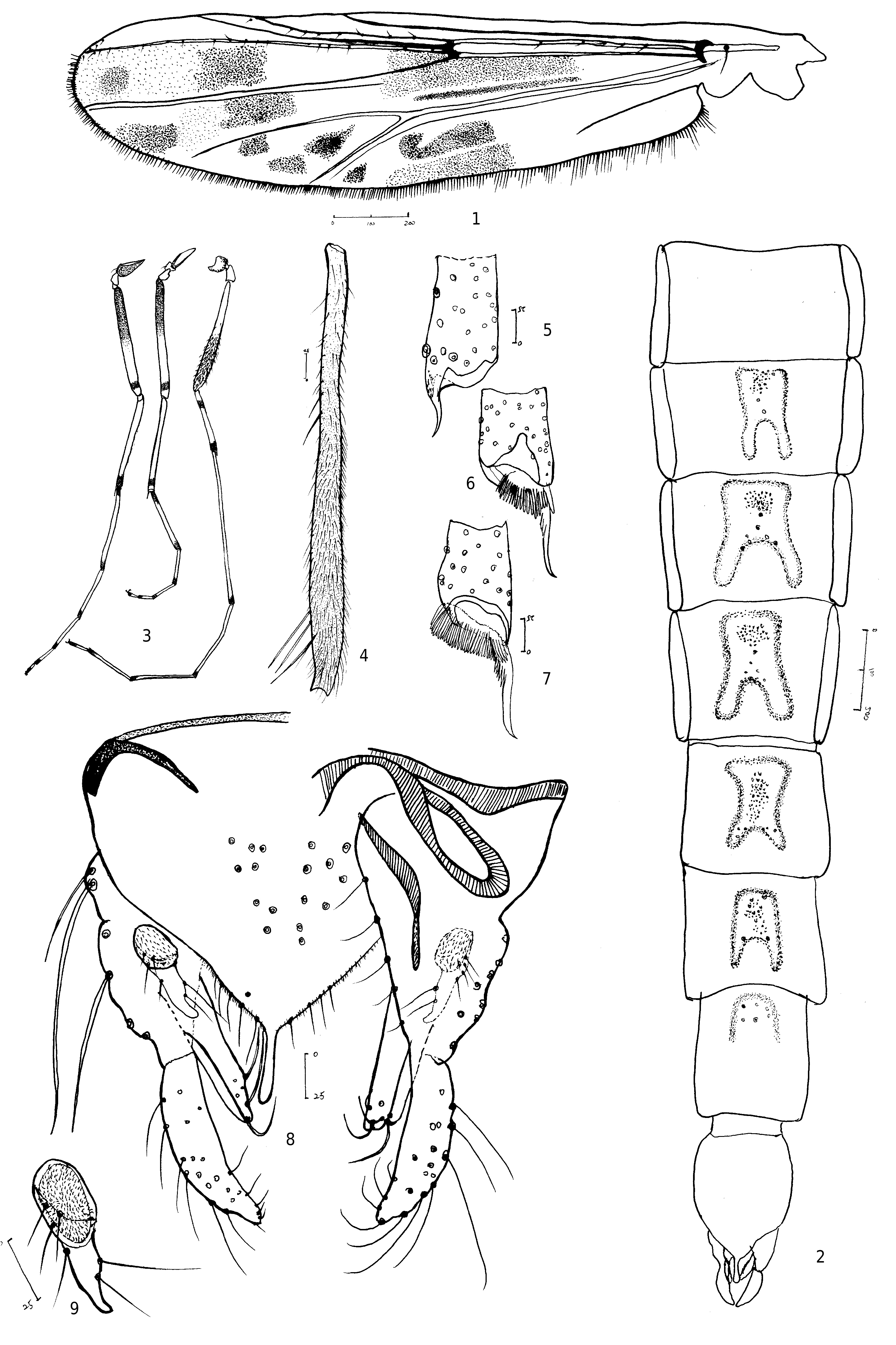
Zavreliella shidai sp. n.
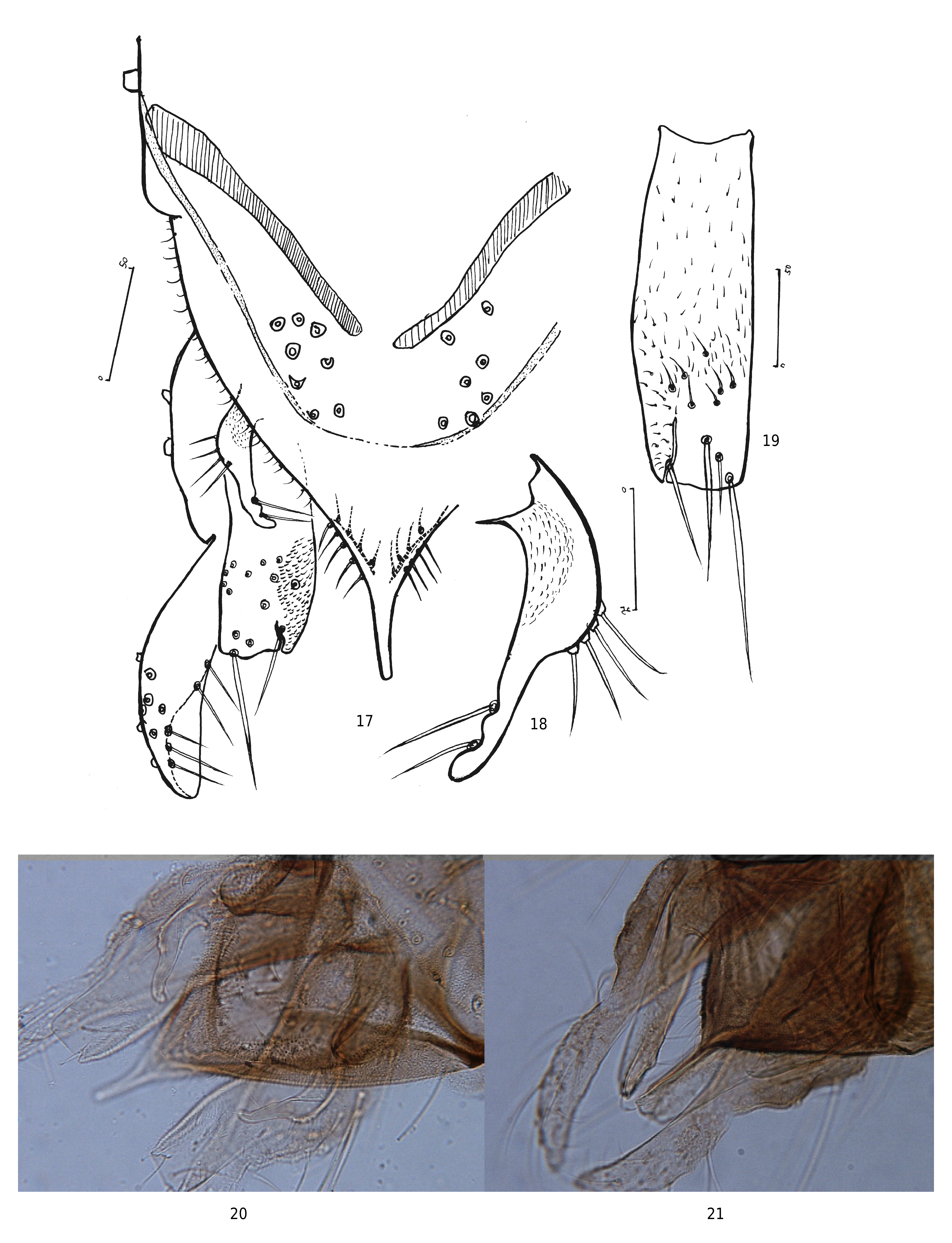
( Figs. 1-16 View Figs View Figs , 21 View Figs )
urn:lsid:zoobank.org:act:
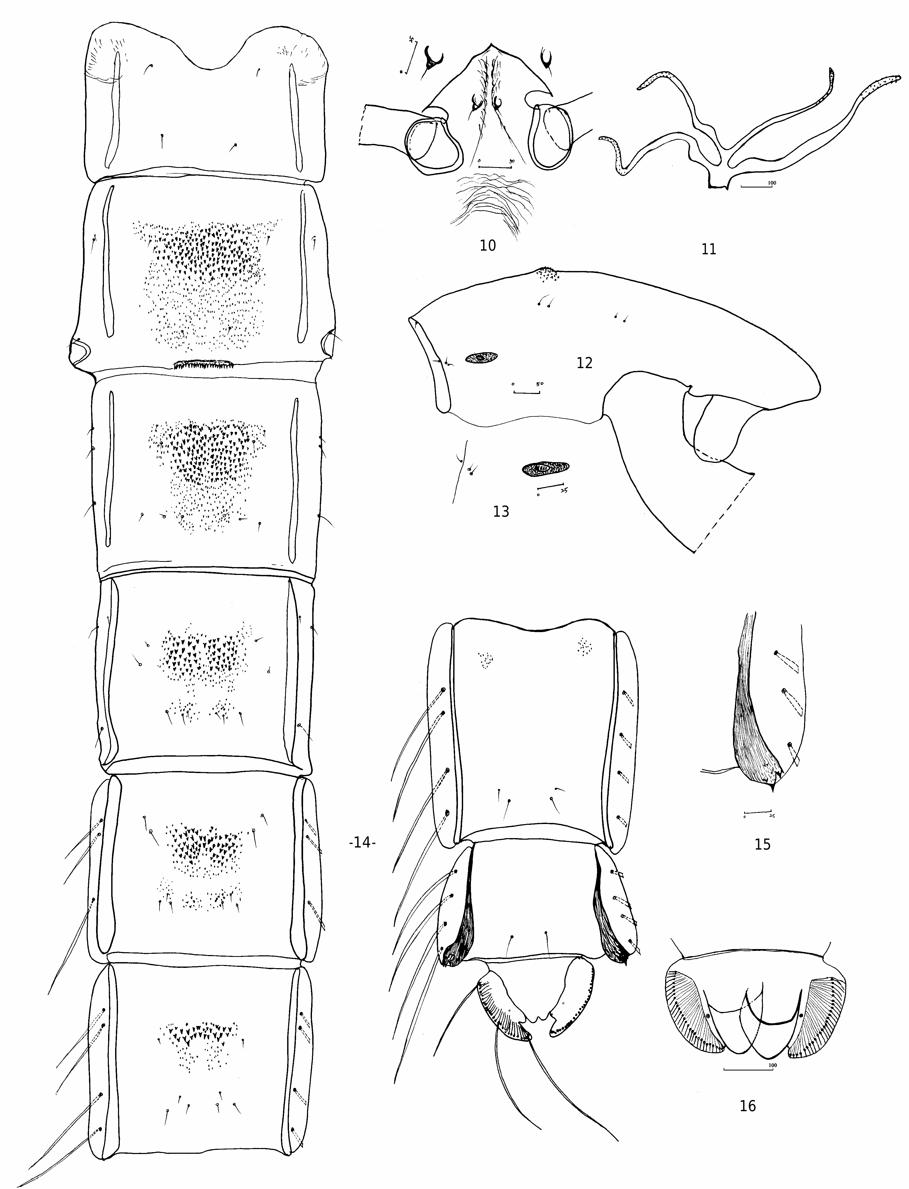
Diagnosis: The male of the new species can be separated from others by the following combination: distinct abdominal markings and mottled wing; inferior volsella with the longitudinal suture laterally; superior volsella with two lateral setae on its inner margin. Pupa can be separated from others by the absence of distinct paired point patches on T II-VI, T VI with 4 LS-setae.
Description: Male ( n = 1) ( Figs. 1-9 View Figs , Table 1)
Total length 3.5 mm. Wing length 1.7 mm. Total length/wing length 2.1. Wing length/length of front femur 1.8.
Coloration: Head and thorax brown to dark brown; wing with distinct dark spots ( Fig. 1 View Figs ), a curved spot present in the anal cell; legs yellow with dark rings ( Figs. 3, 4 View Figs ), color pattern of which are similar to those of North American population of Z. marmorata ( Reiss 1990, figs. 1-2), but basal ring on fore tibia of this new species is distinctly narrower than that of the latter species. Abdomen medium brown, tergites II-VII each with dark saddle-shaped median elevation bearing a tuft of setae ( Fig. 2 View Figs ).
Head: Temporals 10, uniserial, including 2 inner verticals, 5 outer verticals and 3 post orbitals. Frontal tubercle spindle-shaped, 23 µm in high and 13 µm wide in the middle. Frist antennal flagellomere dark brown, other flagellomeres yellow or pale brown, AR 1.5, ultimate flagellomere 683 µm long. Clypeus with 20 setae. Tentorium 150 µm long, 35 µm wide. Lengths of palpomeres 1-5 (in µm): 48, 31, 160, 150, 215.
Thorax: Antepronotums 0; acrostichals 6, beginning some distance from antepronotum, and running to the shallowly rounded tubercle;
dorsocentrals 9, without scutal fossal setae anteriorly; humeral pit minute, but clear; prealars 3; scutellars 9.
Wings: VR 1.25. Brachiolum with 1 seta; R with 15, R 1 with 14, R 4+5 with 22 setae.
Legs. Foretibia apically with curved spur, 50 µm long ( Fig. 5 View Figs ); spur on mid-tibia 73 µm long, inner basal portion with 4-5 side teeth, combs consisting of 25 teeth ( Fig. 6 View Figs ); spur on hind tibia 90 µm long, inner basal portion with 8-10 teeth, combs consisting of 40 teeth ( Fig. 7 View Figs ). Lengths (in µm) and proportions of legs in table 1.
Abdomen: Numbers of setae on saddle-shaped dark areas in T II-VI: 30, 48, 40, 42, and 20 ( Fig. 2 View Figs ).
Hypopygium ( Figs. 8 View Figs , 21 View Figs ): Anal tergite bands short, separated widely. Median anal tergite with 9 setae on each side. Anal point nearly parallel-sided and distally slightly rounded, 45 µm long. Superior volsella ( Fig. 9 View Figs ) broadly digitiform, 40 μm long, with 4-5 setae on its dorsal and 2 long setae on its middle portion of inner margin. Basal lobe with hemispheric projection, dominant, clearly microtrichiose. Inferior volsella 120 μm long, with a distinct longitudinal suture laterally. Phallapodeme 90 μm long. Transverse sternapodeme 70 μm long, lateral sternapodeme 90 μm long. HR 1.0, HV 2.6.
Pupa ( n = 4) ( Figs. 10-16 View Figs ):
Coloration: Largely yellow to pale brown. Abdominal spines and spinules darker than cuticle. Total length 3.9-5.0, 4.3 mm. Abdomen 3.1-4.0, 3.4 mm long.
Cephalothorax: Cephalic tubercle small, conical, 10-12 µm wide and 5 µm high ( Fig. 10 View Figs ). Frontal setae short, 10-12 µm long. Anteromedian thorax smooth, with a distinct tubercle. Thoracic horn with 4 branches, of which the basal two branches are slightly stronger than others ( Fig. View Figs
11). Antepronotals 2. Precorneals minute, only 2 observed. Dorsocentrals divided into 2 groups, Dc 1 and Dc 2 ca. 40 µm long, slightly longer than those of Dc 3 and Dc 4 ( Fig. 12 View Figs ). Basal ring flatly ellipse-shaped ( Fig. 13 View Figs ).
Abdomen: Tergal paired point patches not clearly delimited. Tergite II-VI with obvious anterior transverse bands of points stronger than those of median and posterior patches. T I bare, T II-III with extensive spinules, subquadrate in outline, anterior spinules distinctly stronger than those of the posterior ( Fig. 14 View Figs ). T IV-VI split into anteromedian patch and posterior patches, the anterior bands with somewhat of a tendency to separate into 2 sub-patches if only considering the stronger spines. T VII reduced into two anterolateral patches, T VIII bare. Conjunctive spinules between segments absent. Hook row on T II relatively weak, with 28- 34, 32 hooks, occupying 0.3-0.4, 0.3 of tergal width. Vortex absent, pedes spurii B on segment II, weak. Comb of segment VIII composed of 1 larger and 2-3 small teeth, the longest spur 15-20, 18 µm ( Fig. 15 View Figs ). Lateral taeniae of segments V-VIII: 3, 4, 4, 4. Anal lobe with 26-30, 29 taeniae. Male genitalia sac extending slightly past the distal margin of anal lobe ( Fig. 16 View Figs ).
Material examined: Holotype. male with pupal exuviae. CHINA: Guangdong Province, Guangzhou City, Conghua District, Dongkeng Reservoir , 23-iii-2015 (emerged 02-iv-2015), coll. HQ Tang. Paratypes. 2 pupal exuviae, as holotype except 20-viii-2015; 1 pupal exuviae, as holotype except 27-vii-2015.
Etymology: Named after Prof. Shida Wang who first reported the related genus from China and contributed to elucidation of Chinese midge fauna.
Distribution: China ( Guangdong Province)
Remarks: The pupa was collected from a small clean reservoir with several submerged macrophytes, and successfully reared to the adult stage. The adult undoubtedly belongs to the Z. marmorata group because the basal lateral lobe of superior volsella has a hemispherical microtrichiose projection, and the inferior volsella has a longitudinal suture laterally. The male most resembles that of Z. marmorata in the inferior volsella with long apical lobes and the digitiform superior volsella with two short setae along the inner margin, but differs from it in the hemispherical lateral lobe of superior volsella and the wing without any marking around the anal lobe. In Z. marmorata , the superior volsella has a weakly developed lateral lobe and the wing bears dark markings on the base of anal cell ( Reiss 1990; Cranston et al. 1989).
The pupa is unique in the homogeneous spinulation on the abdominal tergites II-VI, while all the known pupae are armed with paired point patches on each of these tergites ( Reiss 1990; Pinder and Reiss 1986).
| T |
Tavera, Department of Geology and Geophysics |
| VI |
Mykotektet, National Veterinary Institute |
| R |
Departamento de Geologia, Universidad de Chile |
No known copyright restrictions apply. See Agosti, D., Egloff, W., 2009. Taxonomic information exchange and copyright: the Plazi approach. BMC Research Notes 2009, 2:53 for further explanation.
|
Kingdom |
|
|
Phylum |
|
|
Class |
|
|
Order |
|
|
Family |
|
|
SubFamily |
Chironominae |
|
Tribe |
Chironomini |
|
Genus |