
Tokyosoma flexuosum, Mikhaljova, Elena V. & Korsós, Zoltán, 2015
|
publication ID |
https://doi.org/10.11646/zootaxa.3914.5.5 |
|
publication LSID |
lsid:zoobank.org:pub:8DC3C263-63B4-4206-8FE3-82ACA768DA8D |
|
DOI |
https://doi.org/10.5281/zenodo.6094967 |
|
persistent identifier |
https://treatment.plazi.org/id/03F087F6-FFCF-4F08-6FFD-F8AE557F4F57 |
|
treatment provided by |
Plazi |
|
scientific name |
Tokyosoma flexuosum |
| status |
sp. nov. |
Tokyosoma flexuosum sp. n.
Figs 1–4 View FIGURE 1 View FIGURES 2 – 4 , Map.
Material examined. Holotype: 1 male ( HNHM), Japan, Central Ryukyus, Okinawa Island, Katsuren Peninsula, next to White Beach, secondary forest, 26°18’43” N, 127°53’59” E, 60 m, 22 October 2010, leg. Z. Korsós.
Diagnosis. Differs from congeners mainly by the sinuous shape of the lateral coxal branch of the posterior gonopod, by the form of the posterior gonopod colpocoxite, which carries a lateral large spinous process, as well as in a flattened, concave, setose process of the coxa of male leg 10.
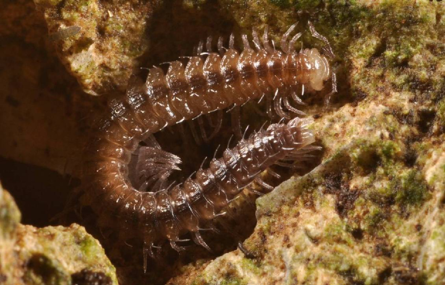
Description. Male ( Fig. 1 View FIGURE 1 ). Length about 15 mm, width with paraterga about 1.5 mm. Coloration in alcohol pale grayish, with broad dark brown band along axial line. Paraterga light brown ventrally. Venter and lower portion of pleura pale. Collum, genae pale. Anterior portion of head light brown. Legs with marbled brown distal parts. Ocellaria black. Antennae dark brown.
Body with 31 or 32 segments. The specimen is broken, thus it is impossible to accurately count the number of body segments, but it is most likely the true number is 32. Head covered with both relatively long and short setae. Eye patches composed of at least 24–26 ocelli. Collum semi-circular. Body width gradually increasing until somite 7, parallel-sided on somites 8–20(21), thereafter gradually tapering. Beginning from somite 2, paraterga normally developed, rounded, increasingly less distinct towards hind part of body, strongly reduced on somite 27, absent from somites 28–31. Metazonital macrochaetae in a transverse row on somites 30–31, like an elongate (to different degrees) triangle on preceding somites. Macrochaetae in anterior and hindmost parts of body relatively long, pointed apically, but in middle and posterior parts of body short (excluding caudolateral macrochaetae) and blunt. Anterolateral macrochaetae in posterior part of body clubbed. Metazonites with two very low longitudinal projections placed along axial suture on each side and with a diagonally positioned projection on each paratergite.
Leg pairs 3–7 not enlarged. Legs 3–5 without tarsal papillae. Legs 6–7 with a small group of funnel-shaped tarsal papillae apically near claw. Claws of pregonopodal legs at base dorsally with two small additional claws, and ventrally with a filament. Postgonopodal legs (including leg pairs 10 and 11) without tarsal papillae. Claws of postgonopodal legs (including leg pairs 10 and 11) at base dorsally with two small additional claws and ventrally with a long filament.
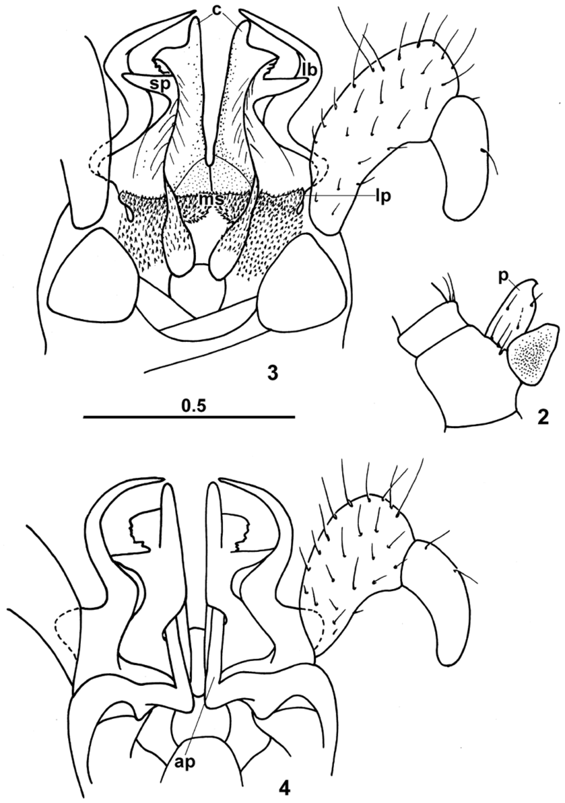
Legs 10 and 11 with coxal glands. Coxa 10 caudoventrally with a flattened concave setose process ( p) with an anteriad curved apex ( Fig. 2 View FIGURES 2 – 4 ). Trochanter 10 with a tiny ventral outgrowth setose apically. Coxa 11 with low ventral prominence. Trochanter 11 with a caudal setose process rounded apically.
Anterior gonopod telopodite 1-segmented, flagelliform, beset with cuticular spinules, its distal part positioned inside sheaths with elevated edges ( Fig. 3 View FIGURES 2 – 4 ). Telopodite base and distal part of coxosternum tightly attached to adjacent mesal portion of posterior gonopod. Posterior gonopod colpocoxites fused sub-basally. Colpocoxites distally with short processes ( c) and, laterally, with large flat spinous processes ( sp). Mesal sheath processes of posterior gonopod colpocoxites fused medially into a single cup-shaped low structure ( ms) covered with pointed spinules. Lateral sheath processes of colpocoxites ( lp) cup-shaped, low, carrying pointed spinules caudally.
Posterior gonopod angiocoxite with a subconical globule, but without process in posterior view. Posterior gonopod coxal part with a long lateral flat branch ( lb) curved mesally, laterally and mesally. Basal part of this branch fused with both colpocoxite and anterior angiocoxite. Angiocoxite depressed centrally in anterior view ( Fig. 4 View FIGURES 2 – 4 ), supplied with a long process ( ap); distal portion of this process penetrating colpocoxite. Posterior gonopod telopodite 2-segmented; femur short and thin.
Female unknown.
Name. The specific epithet refers to the sinuous lateral coxal branch of the posterior gonopod.
Remarks. Tokyosoma flexuosum sp. n. is the first record of the family Diplomaragnidae on the entire Ryukyu Archipelago. Up to now, only one chordeumatid species, Nipponothrix yuwandake Shear & Tanabe, 1994 , belonging to the family Metopidiotrichidae , was known from Amami-o-shima Island, Central Ryukyus ( Nakamura & Korsós 2010).
| HNHM |
Hungarian Natural History Museum (Termeszettudomanyi Muzeum) |
No known copyright restrictions apply. See Agosti, D., Egloff, W., 2009. Taxonomic information exchange and copyright: the Plazi approach. BMC Research Notes 2009, 2:53 for further explanation.
|
Kingdom |
|
|
Phylum |
|
|
Class |
|
|
Order |
|
|
Family |
|
|
Genus |