
Stictothrips
|
publication ID |
https://doi.org/ 10.11646/zootaxa.4772.2.9 |
|
publication LSID |
lsid:zoobank.org:pub:FC6DC72F-BBB8-4CC8-9BBF-44B9E5B0F579 |
|
DOI |
https://doi.org/10.5281/zenodo.3818680 |
|
persistent identifier |
https://treatment.plazi.org/id/03EF3E08-AA05-FF80-E3BE-CCE4EFC0FA0A |
|
treatment provided by |
Plazi |
|
scientific name |
Stictothrips |
| status |
|
Key to species of Stictothrips View in CoL
(leopardinus not examined)
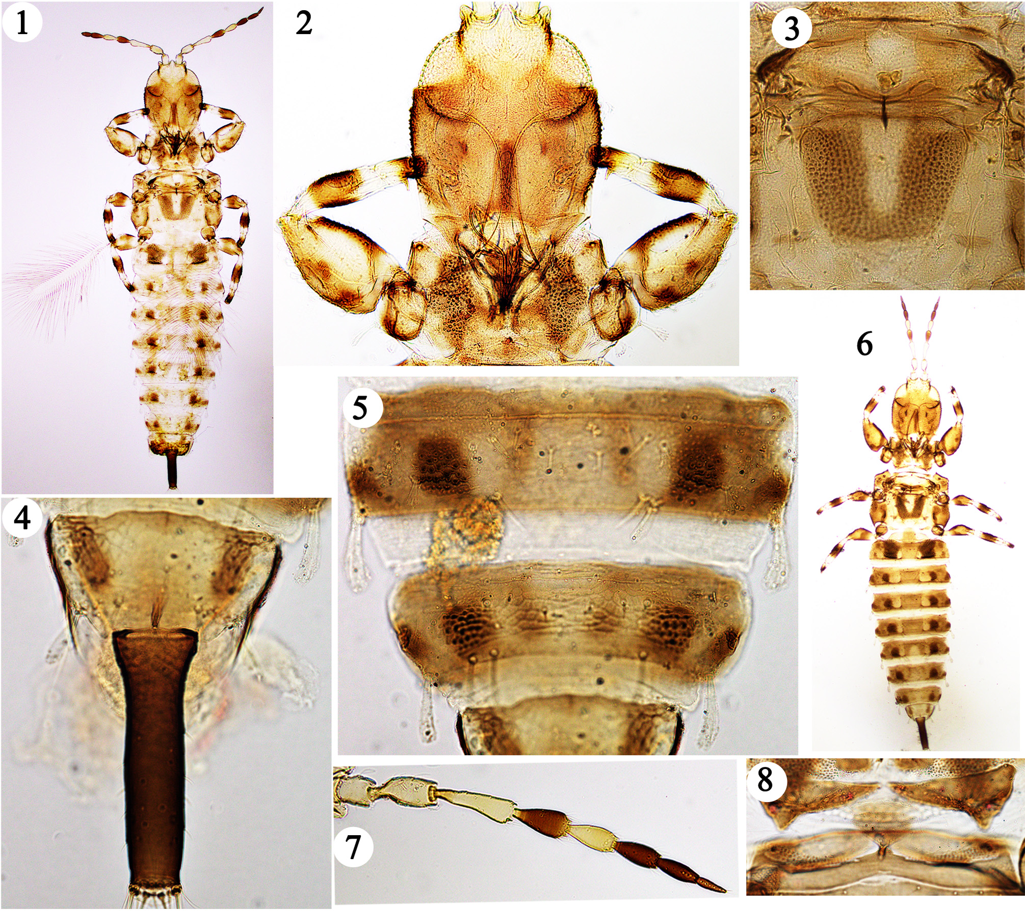
1. Body brown ( Fig. 14 View FIGURES 13–18 ).................................................................................. 2
-. Body bicoloured ( Figs 1, 6 View FIGURES 1–8 , 13 View FIGURES 13–18 ).......................................................................... 3
2. Antennal segment VIII broadly joined to VII; notopleural sutures complete.................................. aoristus View in CoL
-. Antennal segment VIII lanceolate ( Fig. 18 View FIGURES 13–18 ); notopleural sutures incomplete................................ maculatus View in CoL
3. Post ocular setae developed but not extending beyond hind margin of eye ( Fig. 15 View FIGURES 13–18 ); notopleural sutures complete; sternite VIII of male with pair of round pore plates ( Fig. 16 View FIGURES 13–18 )........................................................... farsi View in CoL
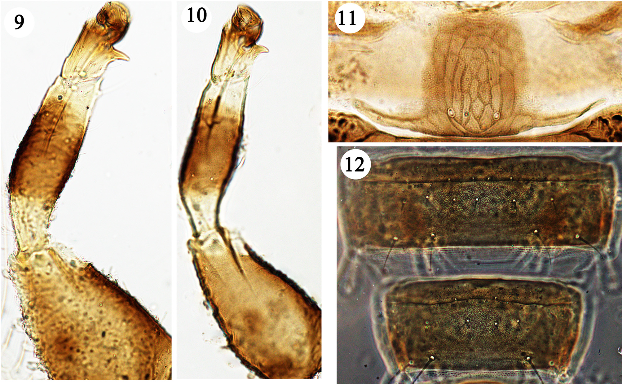
-. Post ocular setae not developed; notopleural sutures incomplete; sternite VIII of male with one median pore plate ( Fig. 12 View FIGURES 9–12 ). 4
4. Head with maxillary stylets about 50% of head width apart ( Fig. 17 View FIGURES 13–18 ); fore tarsal tooth not developed in either sex ( Fig. 17 View FIGURES 13–18 )................................................................................................ namadji View in CoL
-. Head with maxillary stylets close together medially ( Figs 2 View FIGURES 1–8 , 15 View FIGURES 13–18 , 19 View FIGURES 19–21 ); fore tarsal tooth developed in both sexes ( Figs 2 View FIGURES 1–8 , 9, 10 View FIGURES 9–12 , 19 View FIGURES 19–21 )................................................................................................. 5
5. Antennal segment VII as uniformly brown as VI and VIII ( Fig. 7 View FIGURES 1–8 ); tube uniformly brown ( Fig. 4 View FIGURES 1–8 )............ denaeus View in CoL sp.n.
-. Antennal segment VII sharply paler than VI and VIII ( Fig. 20 View FIGURES 19–21 ); tube brown only on distal half ( Fig. 21 View FIGURES 19–21 )................. 6
6. Head with cheeks strongly convex and swollen ( Fig. 19 View FIGURES 19–21 ); antennal segment III at least 2.5 times as long as wide ( Fig. 20 View FIGURES 19–21 ).................................................................................................. faurei View in CoL
-. Head with genae not swollen but weakly convex (cf. Fig. 15 View FIGURES 13–18 ); antennal segment III about 2.0 times as long as wide................................................................................................... leopardinus


No known copyright restrictions apply. See Agosti, D., Egloff, W., 2009. Taxonomic information exchange and copyright: the Plazi approach. BMC Research Notes 2009, 2:53 for further explanation.
|
Kingdom |
|
|
Phylum |
|
|
Class |
|
|
Order |
|
|
Family |