
Pristocnemis caipira, Dasilva, Marcio Bernardino & Pinto-Da-Rocha, Ricardo, 2012
|
publication ID |
https://doi.org/10.5281/zenodo.281134 |
|
DOI |
https://doi.org/10.5281/zenodo.6167366 |
|
persistent identifier |
https://treatment.plazi.org/id/03ED8793-E543-0F3C-8FA4-FDFD95F36C8B |
|
treatment provided by |
Plazi |
|
scientific name |
Pristocnemis caipira |
| status |
sp. nov. |
Pristocnemis caipira sp. nov.
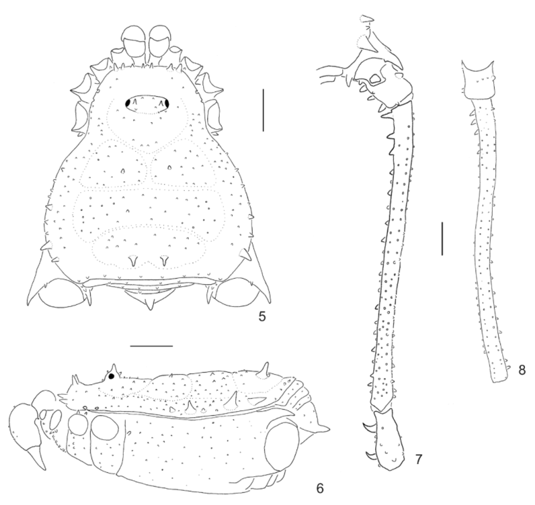
( Figs. 5–8 View FIGURES 5 – 8 , 11–12 View FIGURES 9 – 14 )
Diagnosis. Pristocnemis caipira resembles P. perlatus by the retrolateral armature on male coxa IV and low spines on area III. It differs from other species of the genus by shallow grooves on dorsal scutum, lower number of tarsal segments III–IV (up to 11), presence of a spine on anal operculum, large, pointed tubercles on lateral margins of dorsal scutum, and absence of white patches on dorsal scutum.
Type-material: BRAZIL. Minas Gerais: (Parque Nacional da Serra da Canastra), R. Sawaya leg., male holotype and one female paratype (MZSP 29242); idem, one male and two female paratypes (MZSP 29237).
Etymology. Caipira is a Portuguese noun in apposition referring to people living in the countryside of the southeastern states of Brazil, since this species occurs in the most interior regions of Brazil among all the Caelopyginae .
Male description (holotype):
Measurements. Dorsal scutum: length 5.4; maximal width 5.1. Prosoma: length 2.8; width 2.94. Femur IV length 8.8.
Dorsum ( Figs. 5, 6 View FIGURES 5 – 8 ). Anterior margin of dorsal scutum with two median tubercles, two–three tubercles on each side. Prosoma with two pairs of tubercles near the anterior corners, 18 laterally and behind ocularium. Ocularium with two anterior small tubercles, two median wide and high (about eye diameter) and two posterior small tubercles. Lateral margin of dorsal scutum with tubercles irregularly arranged from ozopores to posterior margin of dorsal scutum, with four enlarged tubercles on external margin (posteriormost largest). Grooves shallow. Area I with 18–20 tubercles on each side (paramedian pair widest); II with 40 (a paramedian pair widest); III with 10 tubercles on anterior half, one pair of spines slightly directed backwards, each one with a tubercle at base. Posterior margin of dorsal scutum with 10 tubercles. Free tergite I with four small tubercles; II with three; III with two tubercles. Anal operculum with six small tubercles, and one conical, large tubercle near posterior margin.
Venter. Coxa I with two anterior tubercles, a median row with five (highest ones on this podomere), six posterior, four apical tubercles; II with two anterior tubercles, median row with nine, six posterior, three apical tubercles; III–IV irregularly tuberculate. Posterior margin of dorsal scutum and free sternites each with a row of tubercles. Anal operculum with tubercles on posterior margin.
Chelicera. Segment I with two tubercles on bulla; II–III with four and three small tubercles, respectively.
Pedipalp. Coxa with two ventral tubercles. Trochanter with three ventral and three dorsal tubercles (median one largest on both sides). Femur with one basal ventral tubercle, without subapical prolateral seta. Patella smooth. Tibial setation: ectal IIi, mesal IiIi. Tarsal setation: mesal and ectal II.
Legs ( Figs. 7, 8 View FIGURES 5 – 8 ). Coxa I with one anterior and one posterior tubercles; II with one anterior, one posterior bifid tubercle; III with only sparse minute tubercles; IV with oblique prolateral apical apophysis, twice longer than the retrolateral one, with subasal tubercle. Trochanter I with four ventral, two retrolateral tubercles; II with one prolateral, two retrolateral and five ventral tubercles; III with two prolateral, three retrolateral, five ventral and two dorsal tubercles; IV with two prolateral, three retrolateral (two–three times larger than wide), five ventral and four dorsal tubercles. Femora I–IV straight; IV with one retrolateral row of tubercles (basal three largest), two ventral rows (largest ones on 1/5 apical), one prolateral row and two dorsal rows of tubercles. Patella IV with two hook-like ventral tubercles. Tibia IV small-tuberculate. Tarsal segmentation: 7, 13, 8, 10–11. Distitarsus I three-segmented; II four-segmented. Basal segment of tarsus I slightly swollen. Claws III–IV pectinate, slightly curved.
Penis ( Figs. 11, 12 View FIGURES 9 – 14 ). Stylus thin and long, with only two ventral subapical projections. Ventral process of glans depressed and enlarged with the apex lance-like shaped. Ventral plate with moderate-size U-shaped cleft in apical margin; with a set of three pairs of setae on subapical part; a set of four pairs of spatulate setae on basal lobe (basalmost longest); one pair of short setae between subapical and basal set of setae and one small pair of setae ventral to the basalmost pair of subapical set of setae. Basal lobe directed dorsally. Dorsal apical projection of truncus enlarged (base of glans almost at the half of ventral plate length).
Coloration. Yellowish brown. Lateral of ocularium with irregular stripe from anterior margin (widest) to groove I (narrowest). Tubercles black, except for the large ones on lateral margin of dorsal scutum and ocularium that are yellow. Enlarged tubercles of areas I–II, base of spines of area III and lateral of areas II–III brownish. Corner of lateral and posterior margins of dorsal scutum and leg IV dark brown. Chelicerae and pedipalps yellowish brown with reticulate pattern. Without white stripes or patches on body. Legs I–III brownish.
Female description (MZSP 2942):
Measurements. Dorsal scutum: length 5.1; maximal width 4.9. Prosoma: length 2.1; width 2.7.
Anterior margin of dorsal scutum with three large tubercles on each side. Lateral margin of dorsal scutum with less tubercles than in male, three large tubercles on external margin. Area I with 32 tubercles, II with 30; III with 18; posterior margin of dorsal scutum with eight tubercles. Free tergite I with seven tubercles; II–III with four tubercles. Anal operculum with a large tubercle on posterior margin. Pedipalpal femur with seven ventral tubercles. Prolateral apical apophysis of coxa IV almost as large as retrolateral one. Femur IV small-tuberculate, with four– five retrolateral basal tubercles, two ventroapical tubercles. Patella and tibia small-tuberculate. Tarsal segmentation: 8–9, 15–18, 15, 16.
Geographical distribution ( Fig. 15 View FIGURE 15 ): Known only from type locality.


No known copyright restrictions apply. See Agosti, D., Egloff, W., 2009. Taxonomic information exchange and copyright: the Plazi approach. BMC Research Notes 2009, 2:53 for further explanation.
|
Kingdom |
|
|
Phylum |
|
|
Class |
|
|
Order |
|
|
SubOrder |
Laniatores |
|
Family |
|
|
SubFamily |
Caelopyginae |
|
Genus |