
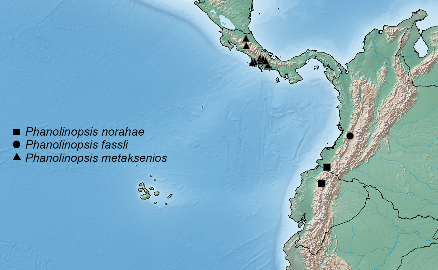
Phanolinopsis Scheerpeltz, 1968
|
publication ID |
https://doi.org/ 10.11646/zootaxa.4323.1.4 |
|
publication LSID |
lsid:zoobank.org:pub:C21944C7-F089-4185-Bd36-7A81870D039F |
|
DOI |
https://doi.org/10.5281/zenodo.6041887 |
|
persistent identifier |
https://treatment.plazi.org/id/03ED6514-6305-F97C-BB81-FD17FD47FDE0 |
|
treatment provided by |
Plazi |
|
scientific name |
Phanolinopsis Scheerpeltz, 1968 |
| status |
|
Phanolinopsis Scheerpeltz, 1968 View in CoL
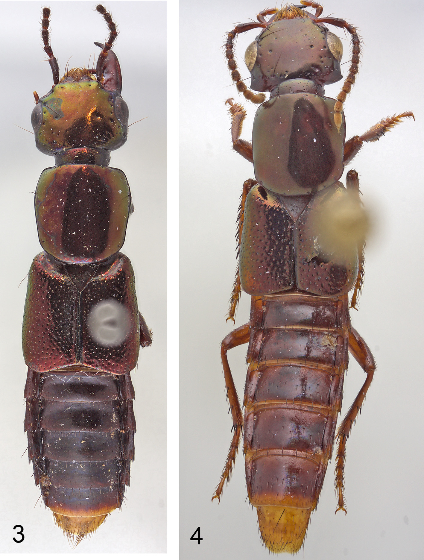
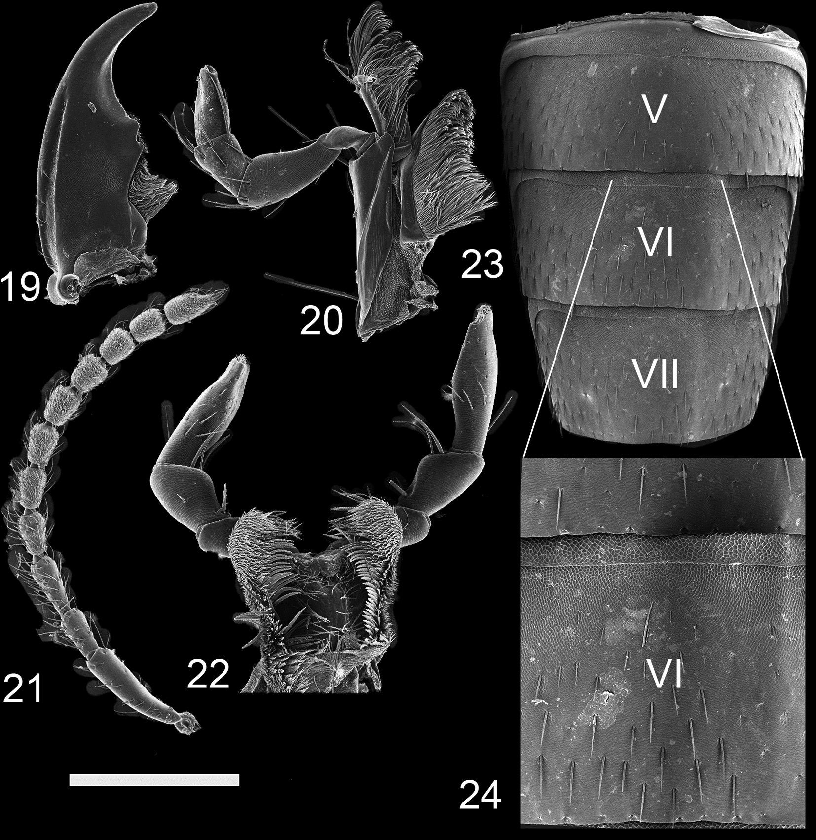
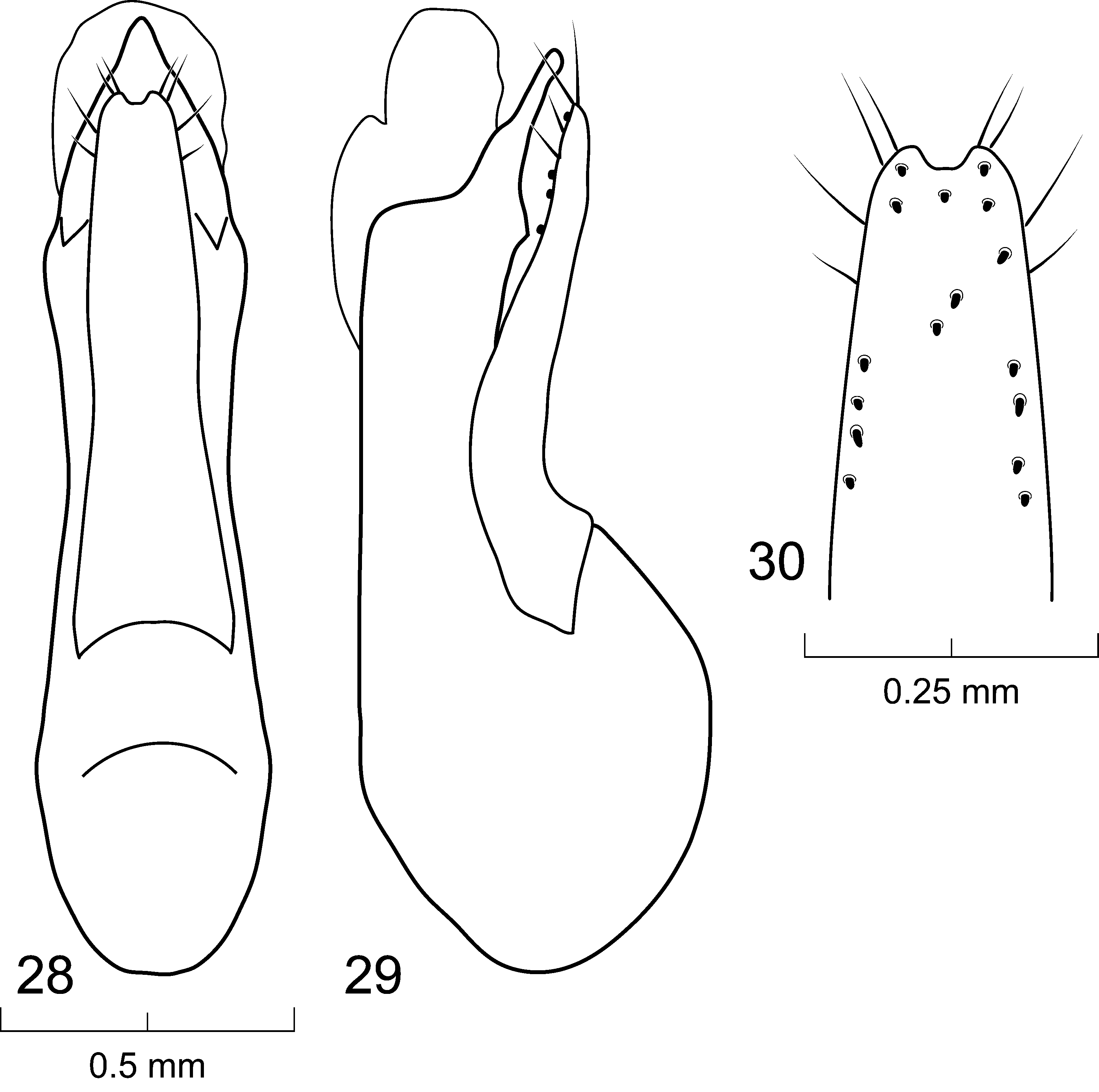
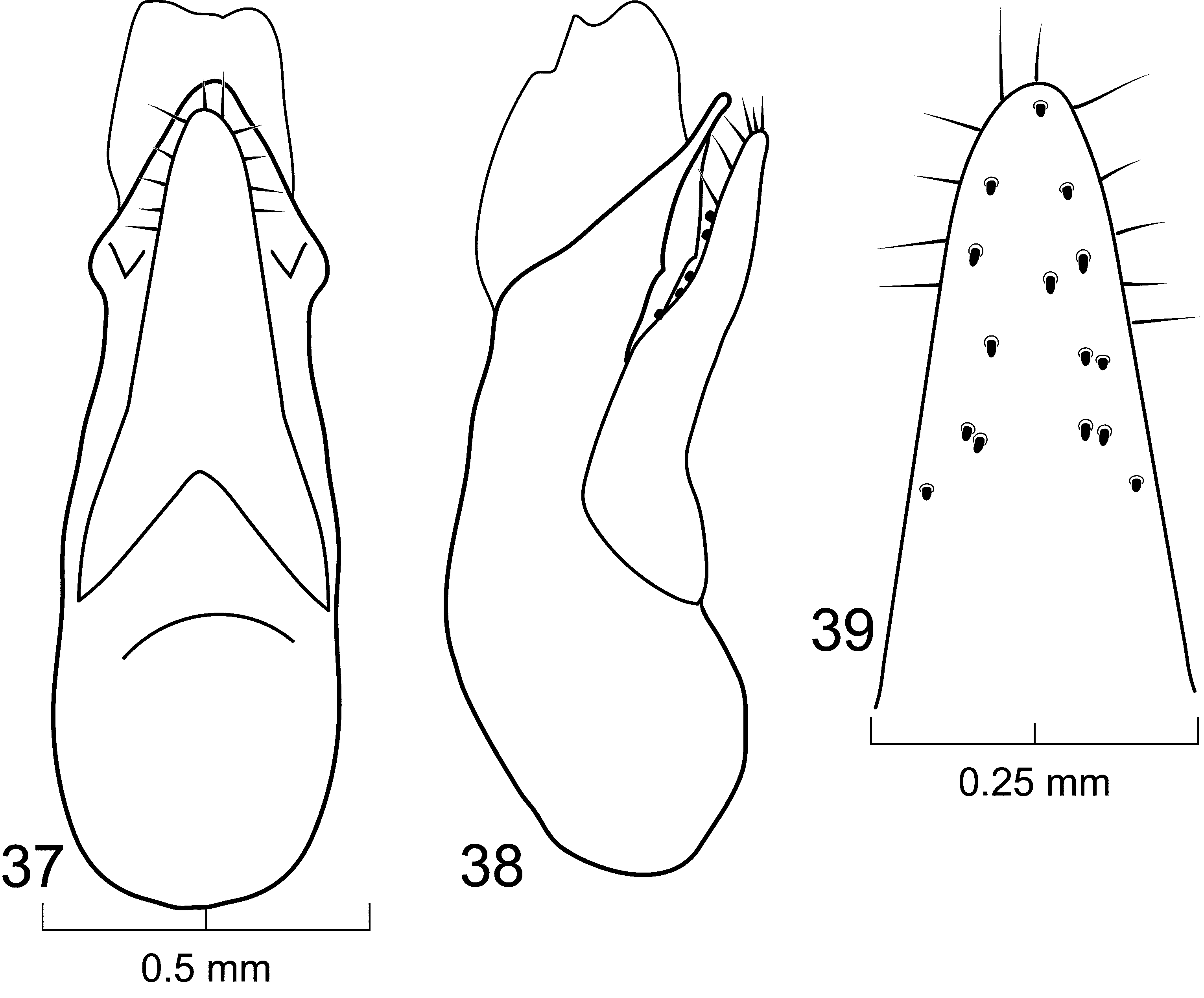
( Figs. 1–44 View FIGURES 1 – 2 View FIGURES 3 – 4 View FIGURES 5 – 6 View FIGURES 7 – 12 View FIGURES 13 – 18 View FIGURES 19 – 24 View FIGURES 25 – 27 View FIGURES 28 – 30 View FIGURES 31 – 33 View FIGURES 34 – 36 View FIGURES 37 – 39 View FIGURES 40 – 42 View FIGURE 43 View FIGURE 44 )
Type species. Phanolinus discedens Sharp, 1884 , fixed by original designation and monotypy.
Diagnosis. Phanolinopsis stands out among other xanthopygines because it is one of few genera that has an impunctate or nearly impunctate pronotum ( Figs. 7–12 View FIGURES 7 – 12 ); with the exception of punctures around the margins of pronotum, species have at most four (or on rare occasions five) punctures at the center of the pronotum, each one delimiting the corner of a square. The only other xanthopygines with similar punctation patterns on the pronotum to Phanolinopsis are Ocyolinus amethystinus Sharp ( Chatzimanolis & Ashe 2009; Chatzimanolis In press), and some species of Torobus Herman. A second key diagnostic characteristic of Phanolinopsis is the presence of an extended and punctate postmandibular ridge, visible both in dorsal ( Figs. 7–12 View FIGURES 7 – 12 ) and lateral ( Figs. 13–18 View FIGURES 13 – 18 ) views of the head that makes the posterolateral corners of the head appears ‘angular’. This feature easily distinguishes Phanolinopsis from Ocyolinus amethystinus (in addition to the lack of long mandibles present in Ocyolinus amethystinus ). Unfortunately, some species of Torobus have both a pronotum punctation pattern and a postmandibular ridge similar to Phanolinopsis . Thankfully, these taxa in Torobus can be easily distinguished from Phanolinopsis by the presence of a postcoxal process (absent in Phanolinopsis ), and the presence of a porose structure on abdominal sternum VII in males (absent in Phanolinopsis ). Other diagnostic features of Phanolinopsis include the following: head transverse (not subquadrate or trapezoid); antennomeres longer than wide or subquadrate but not transverse; mandibles short, blunt, with small tooth; neck prominent, with punctures; elytra shiny with medium to large punctures; and terga V–VI without subbasal (arch-like) carinae.
Despite the name, Phanolinopsis is probably not closely related to Phanolinus . Besides the metallic coloration of the head and pronotum, these two genera share few other characteristics. Based on the structure of the antennae, and the morphology of the head and pronotum, Phanolinopsis is probably closely related to the genus Isanopus Sharp ( Chatzimanolis 2008) . The molecular phylogenetic analysis of Xanthopygina ( Chatzimanolis 2014b) did not include any specimens of Phanolinopsis , however, a forthcoming analysis (Chatzimanolis in prep.) that will incorporate morphological data (in addition to the molecular dataset) and a more comprehensive species list will provide more definitive answers.
Description. Habitus as in Figs. 1–6 View FIGURES 1 – 2 View FIGURES 3 – 4 View FIGURES 5 – 6 , body medium sized, 9.5–15.7 mm in total length. Color of head and pronotum metallic green-brown, purple-brown, brown, blue, green or red; elytra metallic blue-green, blue-purple, purple-brown, brown-green, blue or green; mouthparts, antennae, mesoscutellum, ventral surface of body and legs, reddish brown to brown; abdomen reddish brown to brown except posterior part of VII and VIII orange.
Head transverse, with medium-sized to large setose punctures around margin of head and microsculpture; with two large punctures anteriorly, each adjacent to antenna; surface of epicranium matte due to micropunctures and microsculpture. Clypeus slightly emarginate; anteclypeus not expanded. Eyes medium to large, prominent, occupying 1/2 to 3/4 of lateral margins of head. Ventral surface of head with transverse microsculpture and large sparse punctures; postoccipital suture and ventral basal ridge present; infraorbital ridge pronounced posteriorly; postmandibular ridge present, prominent, extending from near mandible to lateral side of head, delineated by multiple setose punctures ( Figs. 13–18 View FIGURES 13 – 18 ); gular sutures separated throughout length with narrowest point between them medio-posteriorly; nuchal depression prominent forming well defined neck; neck with microsculpture, micropunctures and multiple small punctures.
Antenna ( Fig. 21 View FIGURES 19 – 24 ) with antennomeres 1–3 with multiple rows of macrosetae; antennomeres 4–11 with few macrosetae but covered with microtrichiae; antennomeres 1–9, 11 longer than wide; antennomere 10 longer than wide or subquadrate; antennomere 1 twice as long as antennomere 2; antennomere 3 longer than 2; antennomeres 4–7 subequal in size; antennomeres 8–10 subequal in size, typically shorter than antennomeres 4–7.
Mouthparts with labrum medially emarginate to its base. Mandibles as in Fig. 19 View FIGURES 19 – 24 ; small curved, blunt; left mandible with small bicuspid molar; right mandible with small tooth medially; mandibles with dorsolateral groove extending from condyle to just above tooth; prostheca setose. Maxilla as in Fig. 20 View FIGURES 19 – 24 ; galea and lacinia densely setose; maxillary palpi 4-segmented; P1 small, about 1/3 as long as P2; P2 curved, elongate, longer than P3; P2–P3 with large setae apically; P4 elongate, subequal in length to P3. Hypopharynx as in Fig. 22 View FIGURES 19 – 24 . Submentum with one long and one shorter anterolateral setae in each end; labial palpi 3-segmented; with transverse microsculpture; P1 subequal in length to P2; both P1 and P2 with several long setae; P3 with distal end slightly dilated, not securiform.
Pronotum subquadrate to slightly longer than wide; lateral margins of pronotum concave in dorsal aspect; pronotum broadest in apical 1/3 and narrower at basal angles. Hypomeron expanded, with transverse microsculpture and few micropunctures; superior and inferior marginal lines of hypomeron separate throughout their lengths; superior line fully visible from above, extending around anterolateral margin of pronotum and contacting inferior line at neck fossa; no portion of dorsum of pronotum visible from below. Surface of pronotum matte due to microsculpture and micropunctures; with scattered large setose punctures around margins but with at maximum 4–5 punctures on pronotum disc; margins of pronotum with several large setae. Postcoxal process absent. Mesoscutellum prominent, with dense polygon-shaped microsculpture and micropunctures; with multiple rows of small punctures. Basisternum with dense polygon-shaped microsculpture and carina; anterior marginal depression present; furcasternum with medial carina pointed vertically; furcasternum without polygon-shaped microsculpture.
Elytra slightly shorter than pronotum; with multiple rows of nearly confluent punctures and large setae; with sparse longitudinal microsculpture; elytra appearing shining due to lack of extensive microsculpture. Hind wings fully developed. Mesoventrite with anterior margin forming “lip”; with dense polygon-shaped microsculpture and few punctures along edges; without median carina. Metaventrite with dense uniform medium-sized punctures; metaventral process small, rounded, with v-shaped emargination.
Legs with tarsal segmentation 5-5-5; meso- and metatibia with multiple rows of spurs; protibia without multiple rows of spurs but with single row of spurs apically. Protarsus enlarged in both sexes, with spatulate setae ventrally; meso- and metatarsus not enlarged. Empodium with two small setae.
Abdomen with paired prototergal glands present; abdomen expanding from segment III to segment V (widest) and then becoming narrower towards segment VIII. Abdominal terga III–V with tergal basal and without subbasal (arch-like) carina. Segments with distinctive microsculpture ( Figs. 23–24 View FIGURES 19 – 24 ) anteriorly and on anterolateral corners; sterna with deep elongate uniform punctation in multiple rows; terga V–VII with elongate punctures containing microsculpture. Males with secondary sexual structures of sternites VIII–IX; without porose structure on abdominal sternum VII; sternum VIII with emargination posteriorly; sternite IX with U or V-shaped emargination. Lateral tergal sclerites of the abdominal segment IX long and straight, covered with long macrosetae.
Male genitalia with aedeagus typical of Xanthopygina ( Figs. 25–42 View FIGURES 25 – 27 View FIGURES 28 – 30 View FIGURES 31 – 33 View FIGURES 34 – 36 View FIGURES 37 – 39 View FIGURES 40 – 42 ); with median lobe longer and wider than paramere; median lobe with two dorsal teeth; paramere long, not divided into lobes. Paramere with peg setae and short apical setae. Spermatheca not sclerotized.


No known copyright restrictions apply. See Agosti, D., Egloff, W., 2009. Taxonomic information exchange and copyright: the Plazi approach. BMC Research Notes 2009, 2:53 for further explanation.
|
Kingdom |
|
|
Phylum |
|
|
Class |
|
|
Order |
|
|
Family |