
TEIIDAE GRAY, 1827
|
publication ID |
https://doi.org/ 10.1206/0003-0090(2000)249<0001:TCASOL>2.0.CO;2 |
|
persistent identifier |
https://treatment.plazi.org/id/03EC87AC-FF8B-FFC0-9A12-F27DA084FDF1 |
|
treatment provided by |
Felipe |
|
scientific name |
TEIIDAE GRAY, 1827 |
| status |
|
POLYGLYPHANODONTINAE GILMORE, 1942 (Estes, 1983)
Several taxa described below represent a group of scincomorphs whose classification
has been controversial. These taxa were placed in several different familial groups ( Macrocephalosauridae , Polyglyphanodontidae , Adamisauridae , Teiidae ) by different authors. More recently, Alifanov (1996) classified these in the Macrocephalosauridae —a familial group the name of which was synonymized with Polyglyphanodontinae by Estes (1983). The monophyly of the Macrocephalosauridae needs to be demonstrated before the familial name can be validated; therefore, we tentatively follow Estes (1983) by placing these problematic taxa in the Polyglyphanodontinae ( Teiidae ).
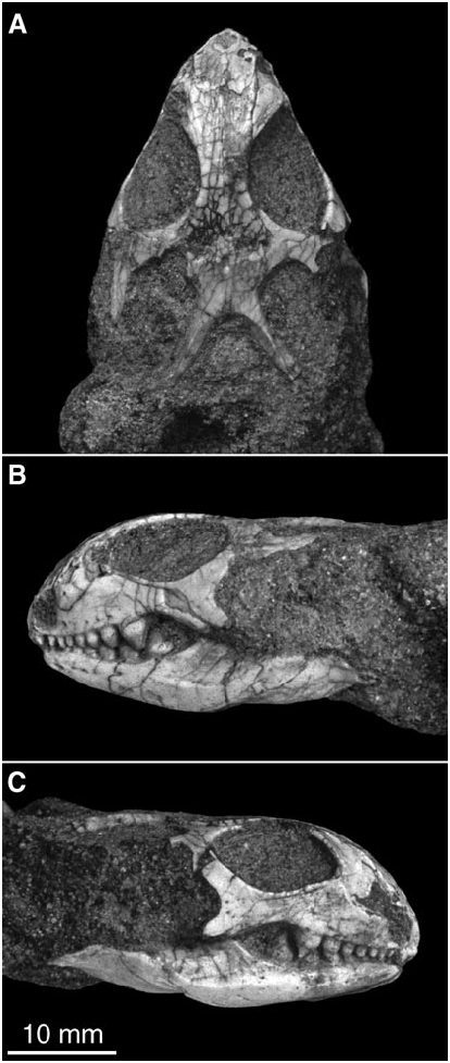
Adamisaurus magnidentatus Sulimski, 1972 Figures 15 View Fig , 16 View Fig
HOLOTYPE: ZPAL MgRII/80, incomplete skull with mandibles.
TYPE LOCALITY AND HORIZON: Bayn Dzak, Mongolian Gobi Desert; Upper Cretaceous Djadokhta Formation.
KNOWN DISTRIBUTION: Djadokhta Formation—Bayn Dzak, Bayan Mandahu, and Tugrugeen Shireh localities (Sulimski, 1972; Gao and Hou, 1996; this paper); Barun Goyot Formation—Khermeen Tsav and Khulsan localities (Sulimski, 1978; this paper).
REVISED DIAGNOSIS: A polyglyphanodontine differing from other members of the group in having the following derived character states: premaxillary spine widened and spatulate; parietal anteromedially develops a rectangular tab overlapping frontals; parietal foramen opens close to frontoparietal suture but on parietal side; jugal expanded with enlarged posteroventral process; ectopterygoid significantly enlarged, with robust ventral process; strong posterior extension of dentary covering large part of surangular, and extending close to posterior surangular foramen; posterior extension of angular bone surpassing posterior surangular foramen and terminating below craniomandibular joint; marginal teeth strongly expanded and bulbous, subacrodont; replacement teeth emerge in crypts below functional tooth row.
REFERRED SPECIMENS: Ukhaa Tolgod— IGM 3/96–3/101 (MAE 9578, 31/93164, 9545, 9410, 45/93163, 48/93163), all incomplete skulls with mandibles from Zophies Hill; IGM 3/102, 3/103 (MAE 32/93 91, 38/9390), incomplete skulls with mandibles from Small Exposure; IGM 3/104–3/ 106 (MAE 117/9393, 118/9393, 122/ 9393), fragmentary skulls with mandibles from Zofia Exposure; IGM 3/107, 3/108 (MAE 449/93124, 9436), fragmentary skull with mandibles from First Strike; IGM 3/109 (MAE 9562), fragmentary skull with mandibles from Camel Humps; IGM 3/110 (MAE 138/93140), fragmentary skull with mandible from Camel Tits Hot Spot; IGM 3/ 111–3/115 (MAE 64/9345, 56/93110, 119/ 9393, 107/9386, and MAE 23), fragmentary skulls with mandibles form Ukhaa Tolgod without sublocality information.
Tugrugeen Shireh—IGM 3/116, nearly complete skull with mandibles articulated with partial postcranial skeleton; IGM 3/ 117–3/122 (MAE 252/9214, 18/932, 167/ 935, 168/935, 226/933, 227/9327), all incomplete skulls with mandibles.
Khermeen Tsav—IGM 3/123–3/125 (MAE 196/9225, 209/9239, 211/9239), all fragmentary skull with mandibles.
REMARKS: Sulimski ( 1972) first described and referred Adamisaurus magnidentatus to the ‘‘? Agamidae ,’’ but later (Sulimski, 1978) erected a monotypic family ( Adamisauridae ) and placed it in the Scincomorpha. Estes (1983) reviewed the available evidence and placed the species in the Polyglyphanodontinae, based on its shared similarities with macrocephalosaurs and Polyglyphanodon .
There is much confusion regarding the cranial morphology of this lizard, and several key sutures were incorrectly identified. First, the premaxilla was described as having only two teeth, but wellpreserved specimens clearly show five to six teeth (e.g., IGM 3/ 116, 3/117). Second, the parietal foramen was described and figured as opening at the frontoparietal suture (Sulimski, 1972: fig. 1, 1978: fig. 1), but much better preserved new specimens (e.g., IGM 3/99, 3/115, 3/116) show that the foramen penetrates the parietal, which anteromedially develops a rectangular tab overlapping the frontals (fig. 15A). Third, the dentary was misinterpreted as having a shorter posterodorsal process overlapping the lateral surface of the coronoid and a slightly longer posteroventral process below the anterior surangular foramen (Sulimski, 1978: fig. 1). The new specimens (e.g., IGM 3/116, 3/117), however, undoubtedly show a much stronger posterior process covering a large part of the lateral surface of the surangular bone and extending to the level of the posterior surangular foramen (fig. 15B, C) Fourth, the angular bone was incorrectly reconstracted as terminating far anterior to the posterior surangular foramen (Sulimski 1978: fig. 1A; contra fig. 1D, F), but wellpreserved specimens (IGM 2/116, 3/117) clearly show that the angular extends posterior to the foramen and terminates at the level of the craniomandibular joint.
Before its skull morphology can be clarified, the phylogenetic position of this bizarre lizard cannot be assessed satisfactorily. An extensive description of the skull osteology based on wellpreserved specimens and reassessment of the phylogenetic relationships of this problematic taxon will be published in a separate paper, but at this stage, we tentatively classify Adamisaurus in the Polyglyphanodontinae ( Teiidae ) as it apparently shares many character states with Macrocephalosaurus and Polyglyphanodon (Estes 1983) .
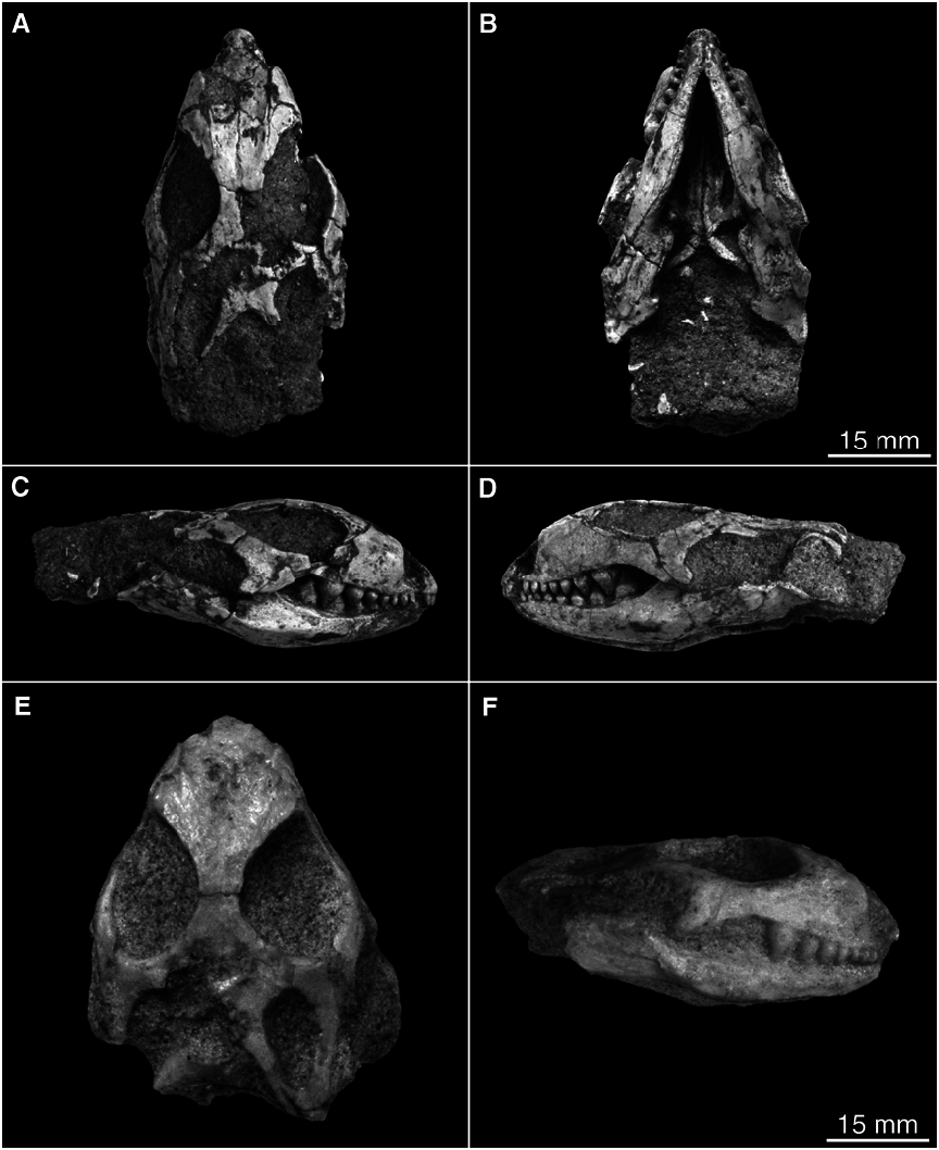
Gobinatus arenosus Alifanov, 1993 Figures 17 View Fig , 18 View Fig
HOLOTYPE: PIN No. 3142/308, incomplete skull with mandibles.
TYPE LOCALITY AND HORIZON: Khermeen Tsav, Nemegt Basin, Mongolian Gobi Desert; Upper Cretaceous Barun Goyot Formation.
KNOWN DISTRIBUTION: Khermeen Tsav (Alifanov, 1993b); Khulsan and Ukhaa Tolgod localities (this paper); Upper Cretaceous Barun Goyot and Djadokhta formations.
REVISED DIAGNOSIS: Differing from other closely related scincomorphs in having the following derived character states: Skull strongly narrow and elongate; small osteodermal ornamentation present on skull roof; premaxillary spine elongate and spatulate basipterygoid process widened as short and roughly squared plate; ventral surface of braincase floor marked with three distinct depressions, one on basisphenoid and two on basioccipital; quadrate foramen present as small pocket with three openings in it; posterior border of angular bone slightly
notched; subdental shelf reduced and sulcus dentalis entirely lost; anterior inferior alveolar foramen enlarged; angular extends to level of posterior surangular foramen; retroarticular process slender and strictly directed posteriorly; marginal teeth bulbous but widely spaced along tooth row; tooth crowns unicuspid and slightly recurved.
REFERRED SPECIMENS: Khulsan—IGM 3/ 126 (MAE 6191), nearly complete skull with mandibles; IGM 3/127 (MAE 212/92 67), incomplete skull with partial right mandible. Ukhaa Tolgod—IGM 3/128 (MAE 268/9365), incomplete skull with mandibles.
DESCRIPTION
Although slightly crushed dorsoventrally, IGM 3/126 is the best preserved material known for Gobinatus arenosus . This specimen is described below, as it reveals important features that are unknown from the holotype.
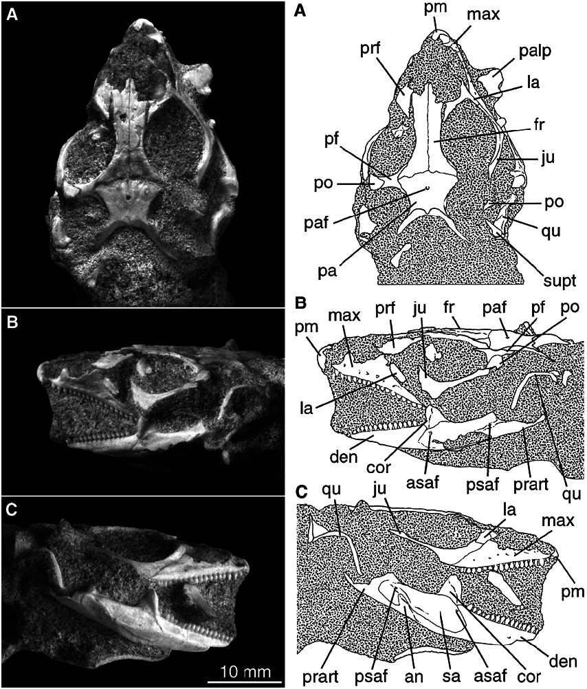
SKULL ROOF: The premaxillae are fused as a single unit, which carries seven teeth (determined from the broken bases). The ante rior surface of the element is smooth and lacks any foramina (anterodorsal premaxillary foramina). The premaxillary spine is extremely elongate; it has a narrow shaft at the base, but the distal part is spatulate (fig 17C). The nasal process extends to such a posterior position that it separates the nasals for about half of their length along the midline of the skull.
The nasals are paired. Each side of the element has a slender anterior process extending along the lateral border of the premaxillary spine (nasal process of premaxilla), and a short posterior process as a thin plate overlapping the anterior shelf of the frontal. The nasal laterally contacts the dorsal process of the maxilla, and is well separated from the prefrontal by an anterolateral process of the frontal (scincomorph synapomorphy, see Estes et al., 1988).
The frontals are paired, with a clear midline suture. The dorsal surface of the element is ornamented with osteodermal rugosities the complexity of which increases posteriorly close to the frontoparietal suture. On IGM 3/
126, a short and shallow groove is seen posteriorly along the midline suture, but such a groove is not illustrated for the holotype (Alifanov, 1993b: fig. 3), and the comparable part on the referred specimens is either not preserved (IGM 3/127) or damaged (IGM 3/ 128). Whether this groove represents a natural condition or an artifact cannot be determined with the available sample. The frontoparietal suture shows a slight undulation, rather than being a simply straight suture.
The parietal table is trapezoidal in shape, having its maximum width at the anterior border, a slightly constricted waist and a narrow posterior width. The lateral border of the table is flanged for the lateral origin of the temporal muscles, and the posterior border is also flanged with a poorly defined median ridge. In dorsal view, the anterior part of the table is ornamented with osteodermal rugosities, but the posterior part of the table is smooth (fig. 17C). The parietal foramen is relatively large in proportion to the table, and it opens anteriorly close to the frontoparietal suture. The supratemporal process is slender and long, having a welldefined dorsal crest. The process posterolaterally contacts the supratemporal bone, posteroventrally contacts the paroccipital process of the occiput, and has no contact with the quadrate.
The maxillae are well preserved on both sides of the specimen. Each element carries 15 teeth to form a complete tooth row (see below). In lateral view, the maxilla is elongate and roughly triangular, having a low nasal process with its apex at the midlevel of the tooth row (fig. 17A, B). The lateral surface of the element is smooth with no osteodermal ornamentation and is penetrated by six lateral superior alveolar foramina (or maxillary foramina) along the ventral border of the bone. The first alveolar foramen is significantly larger than the others on the same specimen. Anteriorly, the premaxillary process overlaps the premaxilla without development of any kind of foramen or aperture at the suture. The anterior palatal process is a welldeveloped medial extension, but has no midline contact with its opposite element. This process has a concave dorsal surface, forming the floor of the narial opening. The posterior wall of the narial opening is penetrated by a single foramen, representing the anterior opening of the superior alveolar canal. The foramen is as large as the anteriormost lateral superior alveolar foramen.
The prefrontal is dorsally exposed as a triangular table. It is anteriorly articulated with the dorsal process of the maxilla, medially with the frontals, but has no contact with the nasals. Posteroventral to the maxillaryprefrontal contact, the lacrimal is well developed and forms about the anterior one third of the ventral border of the orbit. Having no posteroventral process, the jugal is shaped like a hockey stick: it has a short ‘‘blade’’ that forms the ventral rim of the orbit together with the lacrimal and a slender and long ‘‘handle.’’ The posterodorsal process of the bone has a pointed end that is not extended to contact the squamosal (fig. 17A– C).
The postfrontal is slender, and is clearly sutured to the postorbital laterally. Having slender and elongate anterior and posterior processes, the postfrontal is deeply forked medially to clasp the frontoparietal suture. The postorbital has a triangular base and an extremely slender and long posterior process, which fits in the medial groove of the squamosal and forms the anterior part of the supratemporal arch. The anterior border of the postorbital forms the posterior rim of the orbit, together with the postfrontal. Its posterolateral border, however, has an articular surface for the posterodorsal process of the jugal.
The squamosal is greatly reduced in thickness, with a thin and very lightly built base. It bears a welldefined dorsal process hooked anteriorly, and a notched anterior border forming the posterior rim of the supratemporal fenestra (fig. 17C). The squamosal lacks a lateral process, but the anterior process is slender and elongate, and is medially grooved for the posterior process of the postorbital. The supratemporal bone is quite well developed. It attaches to the lateral side of the supratemporal process of the parietal, as normally seen in other lizards; but it extends more posteriorly than the latter process to contact the cephalic condyle of the quadrate. Therefore, the supratemporal is involved in quadrate suspension together with the squamosal.
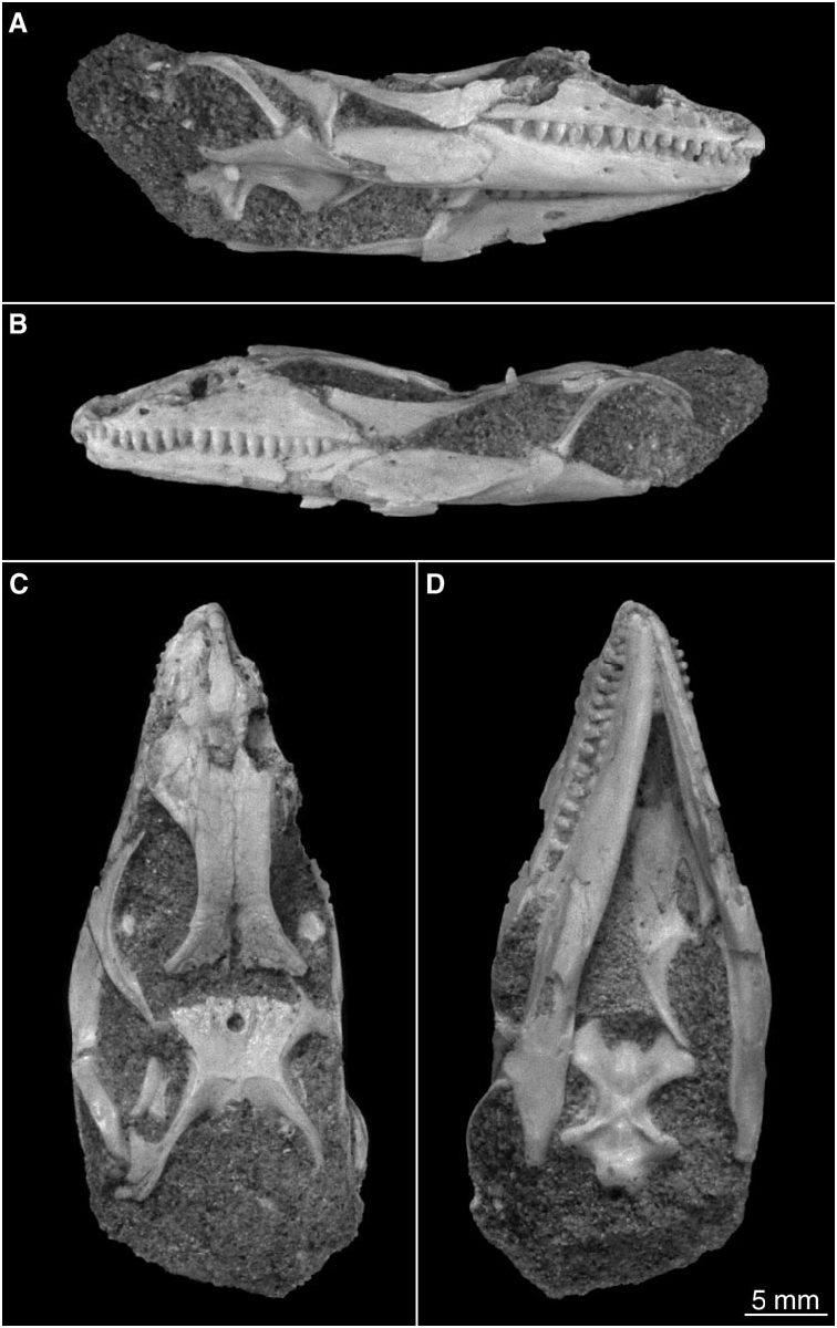
The quadrate is completely preserved on both sides, and is in articulation with the skull and mandibles. The element is slenderly built and relatively straight, lacking the strong arching seen in some other lizards. The anterior surface of the quadrate has a low ridge, running from the cephalic condyle toward the ventral condyle. This vertical ridge separates the narrow tympanic crest from the much wider medial conch, and may served as the origin of the 3a portion of the MAME muscles (Oelrich, 1956; Rieppel, 1980c). Medial to the vertical ridge, the quadrate is strongly widened, concave, and is much posterior in relation to the tympanic crest. The medial border of the quadrate is straight, lacking a pterygoid lappet. The distal condyle is typically saddleshaped with a shallow ventral notch. Above the notch is a small pocketlike depression, in which opens three small foramina for the anastomotic branch of the anterior tympanic vein and posterior condylar artery (Oelrich, 1956). A single quadrate foramen is the common condition in most lizards, and the presence of three foramina in a small pocket may well be a unique condition for Gobinatus .
PALATAL ELEMENTS: Preparation of the skull revealed a well preserved palate (fig. 17D). The vomer (left side shown on the specimen) is wide and strongly elongate, with its posterior extension approaching the level of the posterior end of the maxillary tooth row (see discussion below). Concealed by the right dentary, the right side of the vomer cannot be exposed for observation, but it must have a midline suture contact with the left vomer as indicated by the width of the left element. Most of the element is an elongate thin plate with a welldefined posteromedial process that extends between the palatines and contacts the anterior process of the pterygoid. The ventral surface of the vomer is slightly convex and smooth, lacking any trace of vomerine teeth.
The palatine is also wide and toothless. Anteriorly it has an extensive sutural contact with the vomer, and a spikelike lateral process for articulation with the posterior palatal process of the maxilla. Posteriorly, the palatine has a triangular extension, the medial border of which overlaps the lateral border of the anteromedial process of the pterygoid. The tip of this process fits in a V shaped notch of the pterygoid. Because of the vomerpterygoid contact, the palatine does not form the border of the interpterygoid vacuity (pyriform recess), and the vacuity is extremely narrow as a consequence of the width of the palatal elements.
The pterygoid is not a simple Y shaped element, differing from most of other scincomorphs except some macrocephalosaurs and some lacertiforms. Besides the palatal and ectopterygoid processes normally seen in other lizards, it has a welldeveloped anterolateral process that forms part of the medial rim of the suborbital fenestra. The anteromedial (palatal) process is long and anteriorly abuts the vomer. The lateral (ectopterygoid) process is very short, as commonly seen in other lizards, contacting the ectopterygoid and forming the posterior rim of the suborbital fenestra. Although not completely exposed on the specimen, the posterior (quadrate) process medially has an articular fossa at the base for the basipterygoid process. Remnant pterygoid teeth are developed in the middle part of the element and extend close to the border of the interpterygoid vacuity.
The ectopterygoid is weakly developed and it lacks a robust ventral process as seen in some other macrocephalosaurs (e.g., Adamisaurus ). However, it has a slender anterior process contacting the palatine, and consequently excluding the maxilla from entering the suborbital fenestra. The fenestra itself is greatly reduced to a narrow slit, in keeping with the widening of the palatine and the pterygoid.
BRAINCASE: The braincase floor is well exposed in ventral view on IGM 3/126. Unfortunately the lateral wall and occipital aspect of the braincase cannot be observed as preserved. The braincase floor is marked with three distinct depressions (or pits): a single anterior one in the middle part of the basisphenoid and a pair posteriorly on the basioccipital (fig. 17D). Functionally, the development of these depressions probably increases attachment surfaces for the ventral axial musculature (Oelrich, 1956). The basipterygoid process is wide and platelike. Anteromedially, the parasphenoid process has a slightly widened base, but lacks a wellossified rostrum. The basisphenoid/basioccipital suture is medially irregular and laterally diagonal. The basisphenoid symmetrically has on each side a very slender process extending posteriorly to form the anterior part of the sphenooccipital tubercle. The tubercle itself is slender but prominent, and is more ventrally than laterally directed. The occipital condyle is well exposed on the specimen. The basioccipital forms most of the condyle, with the exoccipitals laterally each contributing slightly less than one third of the condyle.
MANDIBLE: The mandible is well preserved on both sides, with only the ventral border slightly damaged on the left mandible. The lateral surface of the dentary is smooth, and is penetrated by seven mental foramina. The anterior four foramina are close to one another, but the posterior three are irregularly spaced and are far apart from one another. The last foramen opens at the midlevel of the tooth row. The posterior border of the dentary is clearly notched for the surangular and a small part of the angular bone. Both the posterodorsal and the posteroventral processes are slender and short, terminating roughly at the same level below the coronoid summit.
The surangular bone occupies most of the lateral surface of the postdentary part of the jaw. The ventral border of the surangular bone forms a prominent ridge (adductor crest) for attachment of the external mandibular adductor muscles. This crest extends along the surangularangular suture, curving up posteriorly to the posterior surangular foramen anterolateral to the craniomandibular joint. The anterior surangular foramen is relatively large and is located posterior to the dentarysurangular suture, below the coronoid apex. Most of the lateral surface of the bone is smooth, but the ventral adductor crest is slightly ornamented with scarlike sculpture for attachment of the adductor muscles.
The angular bone is incompletely preserved on both sides, but clear impressions allow confident interpretation of the shape and extent of this element. Anteriorly in ventral view, the angular is narrowly wedged by the slender posteroventral process of the dentary, so that it is bifurcated as exposed: a small lateral process intervenes between the surangular and the dentary, and a similar process medially intervenes between the dentary and the splenial. The medial process is penetrated by a small posterior mylohyoid foramen below the posteroventral process of the coronoid. The posterior part of the angular widens slightly and turns dorsolaterally, terminating at the same level, and immediately below, the posterior surangular foramen (fig. 17B). The posterior end of the angular is slightly notched (shown by clear impressions), differing from the strongly bifurcated condition seen in Tchingisaurus multivagus (see below). A much stronger extension is seen in Adamisaurus magnidentatus , in which the angular terminates at the level of the craniomandibular joint (see above).
The articular and prearticular are entirely fused as in many other lizards. The retroarticular process has a wide base at the level of the articular fossa of the jaw, but narrows posteriorly and ends with a slender tubercle that is strictly posteriorly directed.
In medial view, the subdental shelf is significantly reduced to a very slender structure, and the sulcus dentalis medial to the shelf is entirely lost. The splenial is slender and elongate. It anteriorly extends to a point close to the mandibular symphysis and posteriorly terminates at the level of the posterior mylohyoid foramen, where it may contact the posteroventral process of the coronoid bone. The anterior inferior alveolar foramen is significantly enlarged and located below the fourth tooth position from the back (fig. 17A). The anterior mylohyoid foramen is much smaller and is very close to, and right below, the former foramen.
DENTITION: As mentioned above, the premaxillae bear seven teeth counting from the broken bases. The maxillary and dentary teeth are well preserved. These teeth are characteristically thick and bulbous, and the crowns are unicuspid and posteromedially curved. The tooth implantation is subpleurodont, having about one third of the teeth attached to the low lateral parapet of the jaws. The complete maxillary tooth row contains 15 positions as shown on both sides of the upper jaws. The first five teeth are similar in size and are more strongly recurved than other teeth; the sixth tooth is significantly smaller than others, creating a ‘‘step’’ in the tooth row. Those in the middle and posterior part of the tooth row have their crowns more inwardly curved than posteriorly, but the last four teeth are slightly smaller than those in front of them.
The complete dentary tooth row contains 18 positions. The lower dentition shows a similar pattern to the upper dentition, but increase in tooth size from the middle to the posterior part of the tooth row is more pronounced. Despite the thickened tooth shafts, both the maxillary and dentary tooth rows have teeth that are widely spaced from one another. No teeth show development of replacement pits, and this may reflect suppression of tooth replacement in adult individuals (MacLean, 1974).
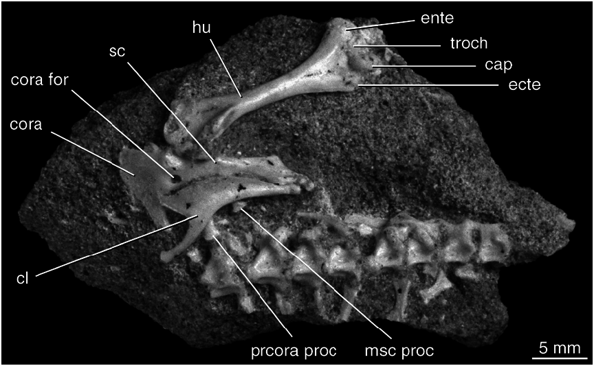
POSTCRANIAL SKELETON: A partial postcranial skeleton of the same individual is preserved in association with the cranial material. Exposed in ventral view, the preserved part of the skeleton includes eight articulated vertebrae, the right clavicle in articulation with the scapulacoracoid complex, and the right humerus in articulation with the pectoral girdle (fig. 18). The interclavicle is not preserved.
The eight vertebrae include the posterior four or five cervicals, as these bear short and expanded ribs that could not have connected with the sternum. In articulation with these cervicals are the first three or four anterior dorsal vertebrae. All these vertebrae have a procoelous centrum, which is triangular in ventral view. As the vertebrae are exposed in ventral view, the morphology of the neural arch cannot be examined. The centrum of all these vertebrae is ventrally crested, but the sharpness of the crest is reduced posteriorly. The intercentra remain separate from the centrum and are in an intervertebral position.
The clavicle is slightly shifted laterally, and so is in articulation with the anterior bor der of the scapula blade. The element is angulated, but has no clavicle fenestra. The proximal end is a narrow, slender rod. The main part of the clavicle is a wide blade with a sharp edge that dorsally becomes slender and extends to the dorsal end of the scapula to presumably contact the suprascapula (an unossified element not preserved).
The scapula and the coracoid plate are completely fused, as no suture can be delimited. The scapula is thick and narrow, more rodlike than bladelike. The anterior border of the scapula is largely covered by the clavicle but two barlike structures exposed represent the mesoscapular process and the procoracoid process, respectively (fig. 18). The rounded coracoid foramen is located between the glenoid fossa and the notched border of the anterior coracoid emargination. No posterior coracoid emargination is developed.
The right humerus is completely preserved and is exposed in ventral view. It has a slen der shaft with expanded proximal and distal ends capped by ossified epiphyses. Proximally, it has an anterior crest for the attachment of M. subcoracoscapularis , and posteroventral to the crest is a triangular depression for M. coracobrachialis brevis (Romer 1956). The distal end of the bone is slightly wider than the proximal end. The entepicondyle is more robustly developed than the ectepicondyle, but neither an entepicondylar nor an ectepicondylar foramen can be identified on this specimen. Between the condyles are the capitellum (or radial condyle) and the trochlea (or ulnar condyle) as seen in other lizards. Differing from the common condition seen in most lizards, however, a welldefined triangular fossa is developed above the two condyles, and a foramen opens in the fossa. A similar condition is seen in extant Tupinambis teguixin (AMNH 141941), but the actual nature of this similarity needs further investigation.
COMPARISON AND DISCUSSION
In tooth morphology and jaw structures all three referred specimens clearly show diagnostic features of Gobinatus arenosus ; accordingly, they are referred to this particular species. One specimen (IGM 3/126) represents the bestpreserved material known for this lizard, allowing us to revise the diagnosis of the species (see above). Alifanov’s (1993b) diagnosis included two other features: trigeminal notch anteriorly closed, and pterygoid strongly bent to the level below the maxillary tooth row; however, the type specimen (PIN No. 3142/308) as figured appears to show that both the braincase and the pterygoid have been distorted (see Alifanov 1993b: fig. 3c). Therefore, the two features
in question are more likely to be artifacts than to be diagnostic of the species.
The tooth number of this species is individually variable. The holotype was described as having 20 maxillary and 24 dentary teeth (Alifanov, 1993b), while IGM 3/ 126 and 3/128 both have 15 maxillary teeth, and the former specimen has 18 dentary teeth. Therefore, the variation range is about five or six positions.
The relationships of this scincomorph lizard are uncertain, although it was classified in the Mongolochamopinae ( Macrocephalosauridae ) by Alifanov (1993b). In light of this uncertainty, it is worthwhile to discuss a few features that may bear on the relationships of this lizard.
(1) The vomer is strongly elongate, with its posterior extension approaching the level of the posterior end of the tooth row. Such a condition of vomer elongation is similar to that in many advanced anguimorphs (see Rieppel, 1980a). However, in Adamisaurus the vomer is also extremely wide, as is typical of other scincomorphs. Anguimorphs have an extremely narrow vomer associated with the elongation of the fenestra exochoanalis (Rieppel, 1980a).
(2) As shown on both the holotype (Alifanov, 1993b) and the new specimens, the posterior end of the angular bone in Gobinatus is slightly notched. The notch condition differs from the deeply bifurcated condition in Tchingisaurus multivagus (see below), and the angular extends to the posterior surangular foramen. Pending the evaluation of this character (i.e., possible homology), the condition in Gobinatus arenosus may represent a more plesiomorphic state than that in Tchingisaurus multivagus . Our observation of a new specimen of Pyramicephalosaurus cherminicus (see below) indicates that the latter species may also share this condition with Gobinatus arenosus .
(3) In spite of their significant difference in size (30 mm vs. 50 mm), Gobinatus arenosus and Dzhadochtosaurus giganteus as figured (compare Alifanov, 1993b: figs. 2, 3) are astonishingly similar to one another in general configuration and several specific features. They both have slender processes of the forked postfrontal, both have the suborbital fenestra reduced to a narrow slit, and both have bulbous teeth with unicuspid crowns. The structural differences recognized by comparison of the figures are that Dzhadochtosaurus giganteus has substantially longer nasals and lacks a posterior notch of the angular. A slender spikelike premaxillary spine is figured for the holotype of the species; however, this morphology is highly doubtful as the same specimen (PIN No. 3143/103) is figured as having a complete premaxilla in dorsal view, but in lateral view it is figured as missing most of this element (Alifanov, 1993b: fig. 2).
Nonetheless, Gobinatus arenosus and Dzhadochtosaurus giganteus are probably more closely related to one another than to any other scincomorphs, as indicated by their astonishing similarities in skull configuration, jaw structure, and tooth morphology.
Tchingisaurus multivagus Alifanov, 1993 Figure 19 View Fig
HOLOTYPE: PIN No. 3142/309, nearly complete left mandible with wellpreserved teeth.
TYPE LOCALITY AND HORIZON: Khermeen Tsav, Nemegt Basin, Mongolia; Upper Cretaceous Barun Goyot Formation.
KNOWN DISTRIBUTION: Barun Goyot Formation—Khermeen Tsav (Alifanov, 1993b); Djadokhta Formation—Ukhaa Tolgod (this paper).
REVISED DIAGNOSIS: Polyglyphanodontine differing from other members of the group in having the following derived character states: Parietal foramen greatly reduced to a minute opening on parietal table; marginal teeth have cylindrical basal part of shaft with abruptly expanded and bulbous crowns; cuspbearing part of crowns laterally compressed and symmetrically tricuspid with higher central cusp and equally developed lateral accessory cuspules; angular bone enlarged and posteriorly bifurcated; prominent adductor crest present on lateral surface of surangular; retroarticular process straight, slender, and pointed.
REFERRED SPECIMEN: IGM 3/129 (MAE 9592), incomplete skull articulated with mandibles from Ukhaa Tolgod (Camel Humps sublocality).
DESCRIPTION
This species is previously known from a single mandible only. IGM 3/129 is the first skull material known for this species; there fore, it is described in detail. Although fairly complete, the specimen is preserved with an openjaw position, and this makes it difficult to expose the palatal region without removal of the lower jaws.
SKULL ROOF: The skull has a pointed snout with a laterally bulging cheek region, giving it a subtriangular shape anteriorly (fig. 19A). The orbit is large and subcircular comprising a large part of the facial region of the skull. The premaxillae are fused, and bear seven teeth. The anterior surface of this element is smooth, without premaxillary foramina. The spine is broken and largely missing; hence, the length and shape of the spine cannot be determined. The nasals are not preserved, but impressions indicate that they were paired and would laterally contact the dorsal process of the maxilla.
The frontals are paired and slightly constricted between the orbits. These elements are anteriorly broken and their suture pattern with the nasals, and possibly with the maxilla, cannot be identified; however, they laterally have an extensive sutural contact with the prefrontal. The dorsal surfaces of the bones are lightly ornamented with osteodermal rugosities. The posterior borders of the frontals each have a shelf underlying the anterior border of the parietal.
The parietal is short and trapezoidal. Its anterior border slightly overlaps the frontals and has an irregular wavy suture with the frontals. The parietal foramen is greatly reduced as a small, round opening located close to the center of the parietal table. The lateral flange of the table is deep and nearly vertical, indicating a lateral origin of the temporal muscles. The table sharply narrows posteriorly, and the posterior flange of the table is more sloped than the lateral flange (fig. 19A). The supratemporal process of the parietal is slender, and is much longer than the parietal table. The articulation pattern of the supratemporal process with the quadrate is unknown, owing to the lateral dislocation of the quadrate on both sides of the skull.
The maxilla is incompletely preserved on both sides (fig. 19B, C). The dorsal process is high, rising above the anterior two thirds of the tooth row. The lateral surface of the process is smooth, slightly concave, and is ventrally penetrated by a row of small lateral superior alveolar foramina. The premaxillary process is a very short spike, attached to the lateral surface of the premaxillae. The anteromedial process is so strongly developed that it extends medially behind the base of the premaxilla, nearly contacting the opposite element. No aperture is developed at the premaxillarymaxillary suture. The posterior process of the maxilla is a short triangle. Along the dorsal margin of the process are the welldeveloped lacrimal and anteroventral processes of the jugal, which together form the ventral border of the large orbit. Like in Gobinatus arenosus (see above), the jugal has an extremely slender posterodorsal process and lacks a posteroventral process, but the jugal of this species is more angulat ed than in the former species. The dorsal process of the jugal contacts the postorbital along a short suture. It can not be determined whether the jugal contacted the squamosal, because of the incomplete preservation of the latter element on the specimen.
The prefrontal is well developed, forming the anterior rim of the orbit. It is laterally articulated with the maxilla, medially with the frontal, but is possibly separated from the nasal by the anterolateral process of the frontal (not preserved). The frontal process of the prefrontal is very short, terminating far anterior to the midlevel of the orbit. The postfrontal and the postorbital are clearly separate elements, and the two together form a short bar separating the orbit from the supratemporal fenestra. The postfrontal is medially well forked to clasp the frontoparietal suture, and is laterally notched to receive a small triangular wedge of the postorbital. The latter element has a very short anterior process (shown on the left side), but the posterior process (preserved on the right side) is slender and long.
Only the right squamosal is partially preserved. It is slightly dislocated vertically and is closely associated with another small splint bone, which is probably the supratemporal. The base of the squamosal is widened slightly, and has a weakly developed dorsal process. The anterior process of the squamosal is slender and is medially grooved for reception of the posterior process of the postorbital.
The quadrate is well preserved on both sides. The element lacks an anterior arching, with a straightly vertical tympanic crest (except for the lateral edge of the cephalic condyle). Anteriorly, the quadrate has an extremely narrow and convex lateral part, but a much wider and strongly concave medial extension. The medial crest is oblique, running from the medial side of the cephalic condyle to the distal condyle. The crest lacks a pterygoid lappet of the quadrate. The distal condyle is ventrally notched, with a welldefined lateral epicondyle. Above the notch, a single quadrate foramen is present on the anterior surface as seen in most of other lizards generally.
MANDIBLE: The mandible is robust, having a strongly convex lateral surface. The posterior border of the dentary is deeply notched to a level slightly anterior to the posterior end of the dentary tooth row (contra Alifanov, 1993b: fig. 7). The coronoid process of the dentary does not extend onto the anterior surface of the coronoid, but on its ventral edge it fully articulates to the surangular bone, leaving no space for the coronoid to wedge in. The dentarysurangular suture in this specimen ends dorsally at the anteroventral tip of the coronoid dorsal process, rather than at the midlevel of the latter process as figured for the holotype (Alifanov, 1993b: fig. 7). The posteroventral process of the dentary has a similar extension, and terminates at the same level as the posterodorsal process.
Anteriorly, the surangular has a blunt process that fits into a notch of the dentary. The anterior surangular foramen is small and close to the surangulardentary suture, below the anterior tip of the coronoid summit. The posterior surangular foramen is even smaller, and is located posterodorsally, close to the glenoid fossa of the jaw. Running between the two foramina is a ventrally curved adductor ridge on the lateral surface of the surangular. Ventral to this ridge, the angular bone is proportionally wide and has extensive exposure on both the medial and lateral side of the jaw. As in the holotype specimen, the angular is posteriorly bifurcated and terminates anterior to the posterior surangular foramen.
DENTITION: The premaxillae are fused into a single bone with seven teeth. The crown pattern of the premaxillary teeth cannot be observed because of erosion. The complete maxillary tooth row contains 18 teeth (shown on both sides of the specimen). The first three maxillary teeth are larger than the rest, and are caniniform with pointed, unicuspid crowns. The other 15 teeth are conspicuously bulbous and tricuspid; however, the base of the crown is very narrow (see fig. 19B, C) The central cusp is well developed, and far more prominent than the lateral accessory cuspules. The lateral cusps are actually not well defined, instead, they are miniature horizontal crests that are not well separated from the main cusp. This crown pattern is different from that in Pyramicephalosaurus cherminicus (see below), in which the three cusps are similar in height and the lateral cusps are clearly separated from the main cusp by a welldeveloped groove.
The dentary tooth row is incompletely preserved on both sides, because the anterior tip of both jaws is missing. The left side has 14 teeth and the right side has 16 teeth preserved. The total number of dentary teeth can be estimated as about 19–20 on comparison with the upper dentition. This estimation is very close to the holotype mandible (PIN No. 3142/309), which is illustrated as having 19 teeth (but described as having 17, see Alifanov, 1993b). All the marginal teeth are subpleurodont, with slightly less than half of the tooth attached to the lateral parapet of the tooth row. This description is different from the figure of the holotype mandible (Alifanov, 1993b: fig. 7), in which the dentary teeth are figured as acrodont (see comments below).
COMPARISON AND DISCUSSION
Alifanov (1993b) named Tchingisaurus multivagus on the basis of a single left mandible (PIN No. 3142/309) from Khermeen Tsav (Barun Goyot Formation), and referred the species to the Macrocephalosauridae Sulimski, 1975 . Because the family Macrocephalosauridae is inadequately diagnosed and the monophyly of the group is highly questionable (Estes, 1983), the referral of Tchingisaurus multivagus to the family is problematic. We tentatively classify the taxon in the Teiidae , pending wholesale revision of this and allied taxa.
The new specimen from Ukhaa Tolgod is identical to the holotype of Tchingisaurus multivagus in jaw configuration, crown pattern of the marginal teeth, and in having an angular that is posteriorly bifurcated. On the basis of these similarities, the new specimen is referred to this species. The deceptive differences in number and implantation of the teeth in the two specimens (see above) are probably the result of a descriptive or illustration error in the original description of the holotype specimen. Alifanov (1993b: 89) described the teeth on the holotype as ‘‘high above level of upper margin of subdental crest,’’ but figured them as acrodont (Alifanov, 1993b: fig. 7).
In terms of tooth morphology, the closest similarity of Tchingisaurus multivagus is to Pyramicephalosaurus cherminicus Alifanov, 1988 . The two forms share similarities in having bulbous teeth with strongly constrict ed crown bases, but are clearly different from one another in detailed crown pattern and the mode of tooth implantation as described above (see also description of P. cherminicus below). In addition, these two species (together with Gobinatus ) differ from the socalled macrocephalosaurs in lacking a prearticular crest medially at the base of the retroarticular process. Such a crest (which differs from the fingerlike process in iguanians) occurs in most lacertids, xantusiids, and teiids (see Estes et al., 1988: character 73). Functionally, the crest is for attachment of the pterygoideus muscle (Rieppel, 1980c), and lack of such a crest in Tchingisaurus multivagus may indicate a fundamental difference in muscle attachment from the macrocephalosaurs.
If Alifanov’s (1993b) observation is correct, Pyramicephalosaurus cherminicus lacks a posterior bifurcation of the angular (but see below) that is characteristic of Tchingisaurus multivagus . Other differences that Alifanov (1993b) mentioned are either ambiguous or invalid. For example, Tchingisaurus multivagus was described as differing from Pyramicephalosaurus cherminicus in having a ‘‘larger number of teeth,’’ but the holotype of the former taxon is ambiguously described as having 17 teeth and figured as having 19. Furthermore, the lower dentition of Pyramicephalosaurus cherminicus on the holotype is incomplete (see Alifanov, 1988: fig. 3); thus, the total number of lower teeth on the specimen is, in fact, unknown (a new speci men from Khulsan has 16 dentary teeth, see below).
As alluded to above, the new specimen from Ukhaa Tolgod has subpleurodont teeth, differing from the ‘‘acrodont’’ condition of the holotype mandible figured by Alifanov (1993b: fig. 7). This supposed difference is probably not individual variation, but is likely to be observational. The holotype specimen is unavailable for this study, and the uncertainty about its tooth implantation cannot be clarified until the specimen is reexamined.
Pyramicephalosaurus cherminicus Alifanov,
No known copyright restrictions apply. See Agosti, D., Egloff, W., 2009. Taxonomic information exchange and copyright: the Plazi approach. BMC Research Notes 2009, 2:53 for further explanation.