
Megatrigon, Johnson, 1898
|
publication ID |
https://doi.org/10.5852/ejt.2016.238 |
|
DOI |
https://doi.org/10.5281/zenodo.3854648 |
|
persistent identifier |
https://treatment.plazi.org/id/03EA327E-FFFD-EC37-FF27-ED17FAB0FB32 |
|
treatment provided by |
Valdenar |
|
scientific name |
Megatrigon |
| status |
|
Key to species of Megatrigon View in CoL View at ENA
The currently known species are probably a fraction only of the species actually in existence, and users of the key should expect to encounter species that are not included. The key should be used mainly as a step in the identification process, and proper identification requires comparisons with the diagnoses and descriptions, including a thorough study of the terminalia. As the identification of females is still unresolved and/or the assignment to the corresponding males is uncertain, the key can be used for males only (except for the very distinct female of M. nivalis comb. nov.).
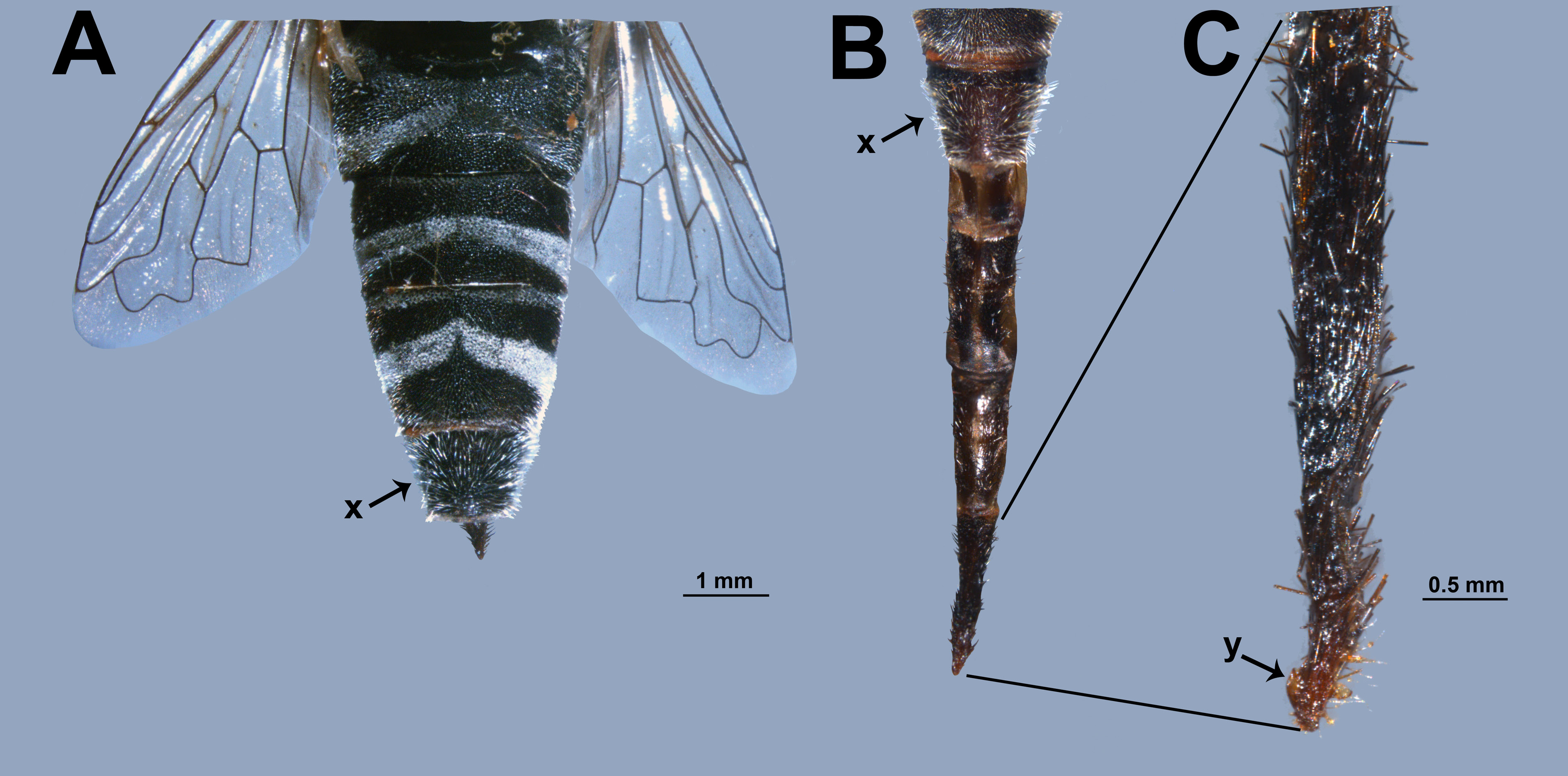
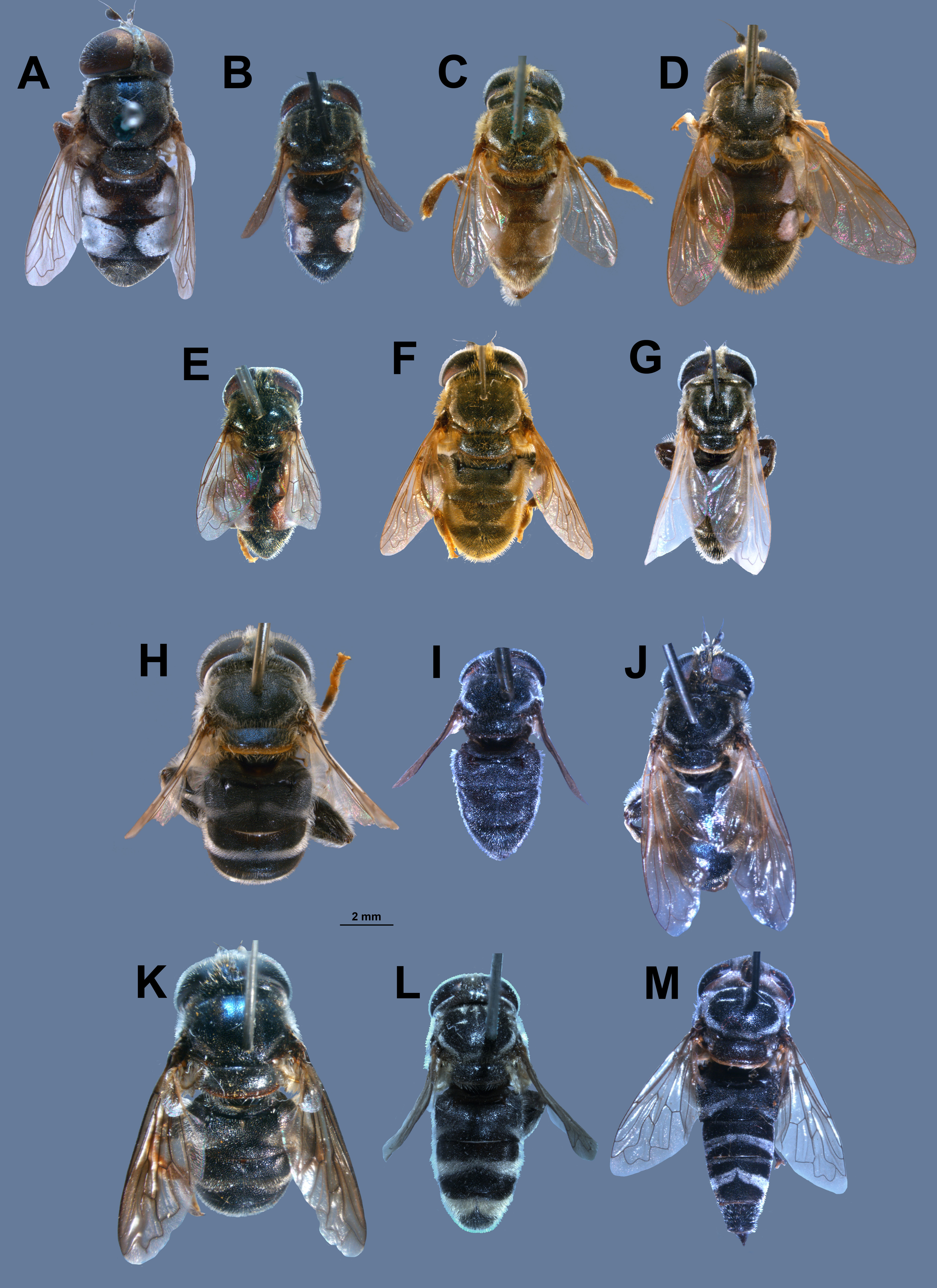
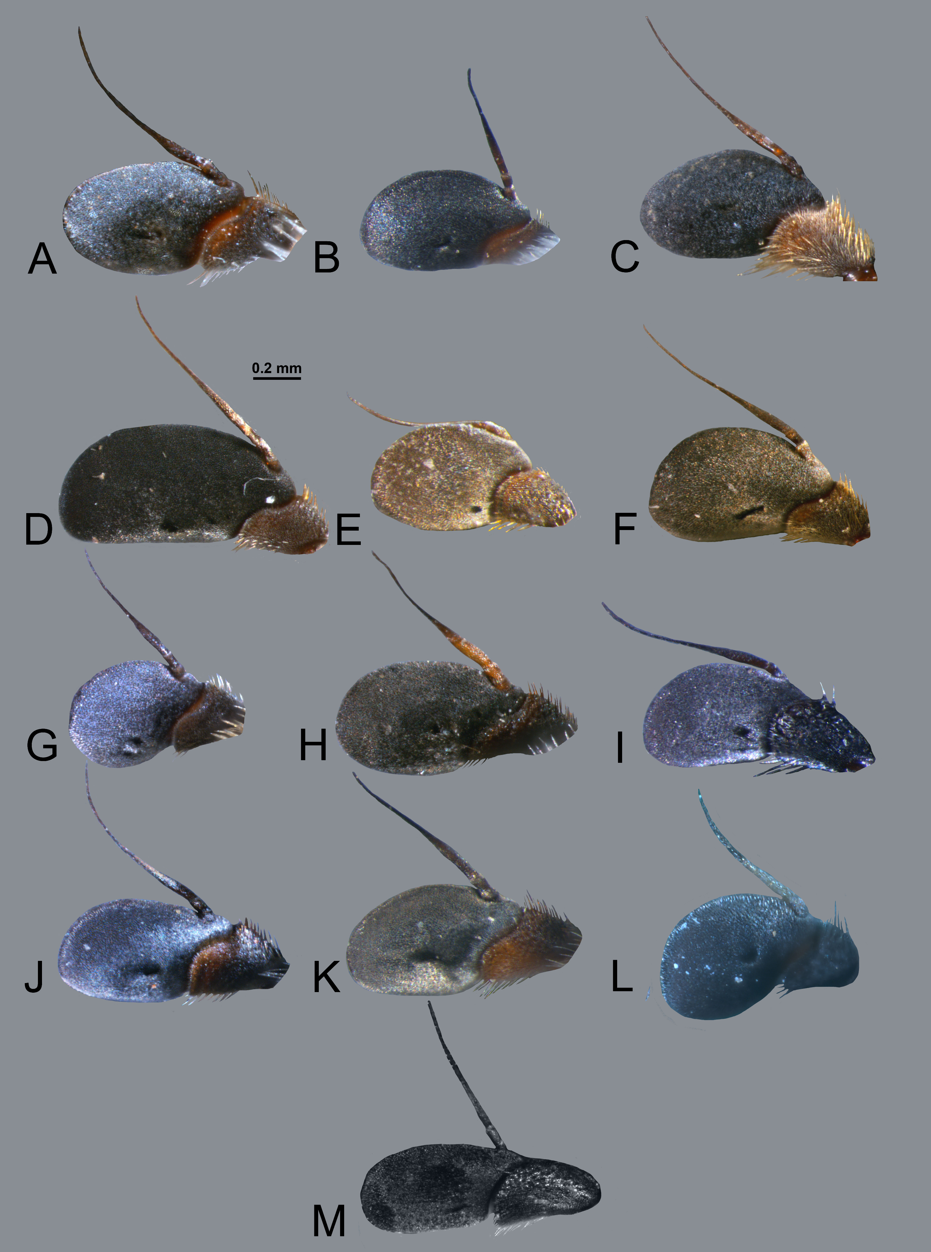
1. Katepisternum posteriorly setose over (almost) full width.Wing base extensively bare of microtrichia, esp. large parts of cells c, r 1, br, bm, and cup. The bare flat posterior margin of tergite 1 extensively microtrichiose. Thoracic setae not feathered but thickened and strongly glistening. ♂ terminalia with posterior surstyle lobe directed posteriorly, with a small hook at apex ( Fig. 10M View Fig :y). ♀ tergite 5 and sternite 5 with anteriorly directed setae ( Fig. 8 View Fig A–B), ovipositor with a subapical dorsal tooth ( Fig. 8C View Fig ) ………………………… nivalis group: M. nivalis ( Hull, 1964) comb. nov. ( Fig. 1M View Fig )
– Katepisternum either without setae between the dorsal and the ventral pile patches or with a few short setae below the dorsal patch. Wings either entirely microtrichiose or with small areas in cells br, bm, and/or cup bare of microtrichia.The bare flat posterior margin of tergite 1 (almost) bare of microtrichia. Thoracic setae serrate. ♂ terminalia with posterior surstyle lobe directed medially ( Fig. 13B, F View Fig :y). ♀ tergite 5 and sternite 5 with posteriorly directed setae, ovipositor without a tooth ……2
2. Posterior surstyle lobe straight, without hook ( Fig. 13E View Fig :y, F:y). Sternite 4 with inflated caudal lobes, each of which bears a small process on their median margin (usually visible only when the terminalia are extended), and with some part bare of microtrichia …………………………………………… sexfasciatus View in CoL group [not treated here at the species level]
– Posterior surstyle lobe forming a strong twisted hook( Fig.13A View Fig :y,B:y).Sternite 4with flat caudal lobes, without process at median margin, and entirely covered by microtrichia ……………3 argenteus group
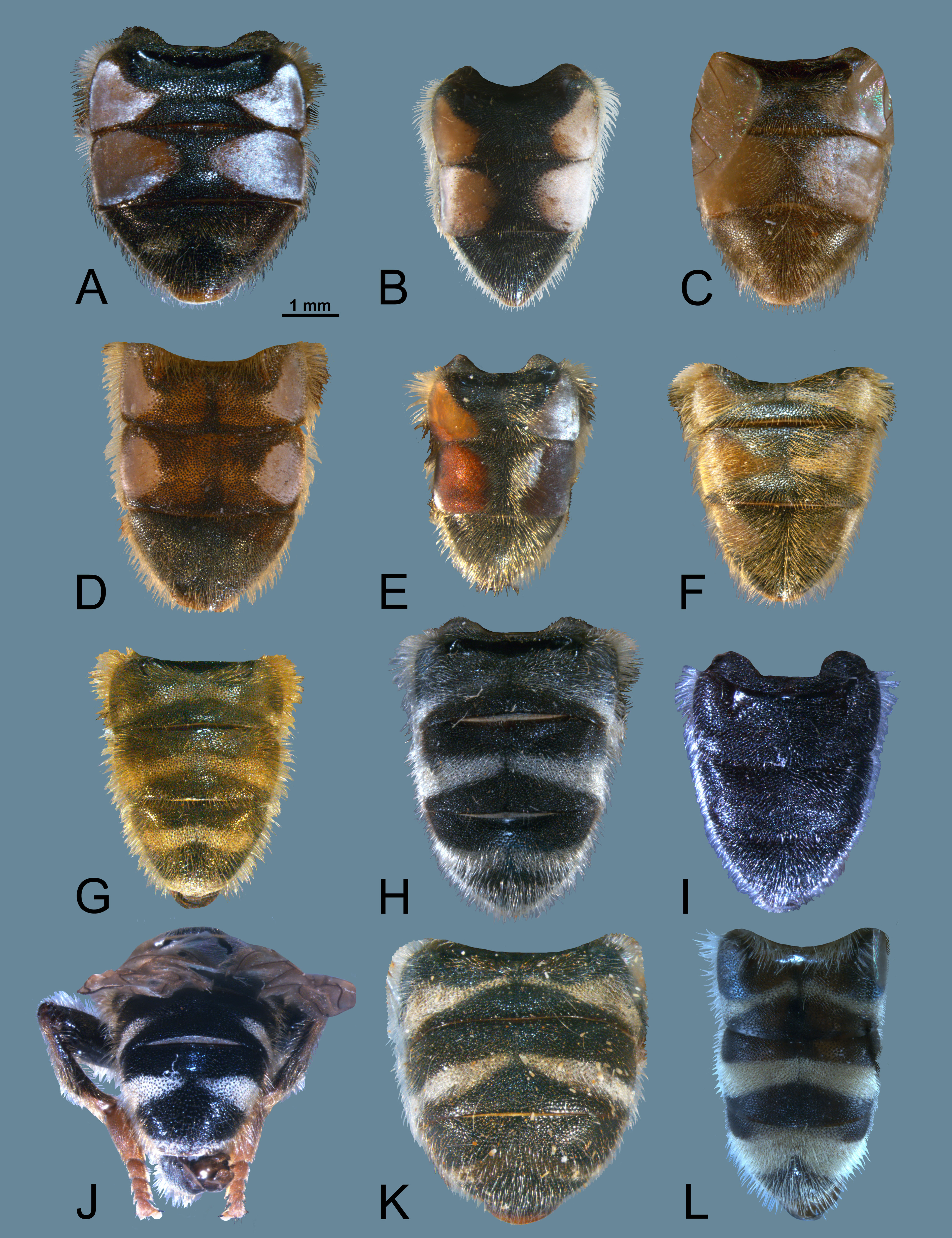
3. Tergites 2+3 with silver spots ( Fig. 7 View Fig A–F), i.e., areas densely covered by adpressed reflecting white microtrichia. These spots cover the lateral part of the tergite for more than three quarters the length of the tergite ……………………………………………………………………………4
– Tergites 2+3 without silver spots ( Fig. 7 View Fig G–K). Non-reflecting fasciae or spots of microtrichia are present, their maximum length less than half the length of the tergite, or all tergites without any markings ……………………………………………………………………………………………9
4. Costagial setae black. Ocellar triangle with a longitudinal groove. Mesofemur anteriorly black setose. Metatibia in proximal half dorsally with reduced microtrichia, not reaching scar. Tergite 4 with a pair of isolated submedian microtrichiose spots ( Fig. 7A View Fig ) ………………………………… …………………………………………………… M. argenteus ( Walker, 1852) comb. nov. ( Fig. 1A View Fig )
– Costagial setae yellow. Ocellar triangle without groove. Mesofemur without black setae. Metatibia in proximal half dorsally with a microtrichiose stripe that extends to the scar. Tergite 4 either without microtrichiose spots or present ( M. sexmaculatus View in CoL sp. nov.) anterolaterally……5
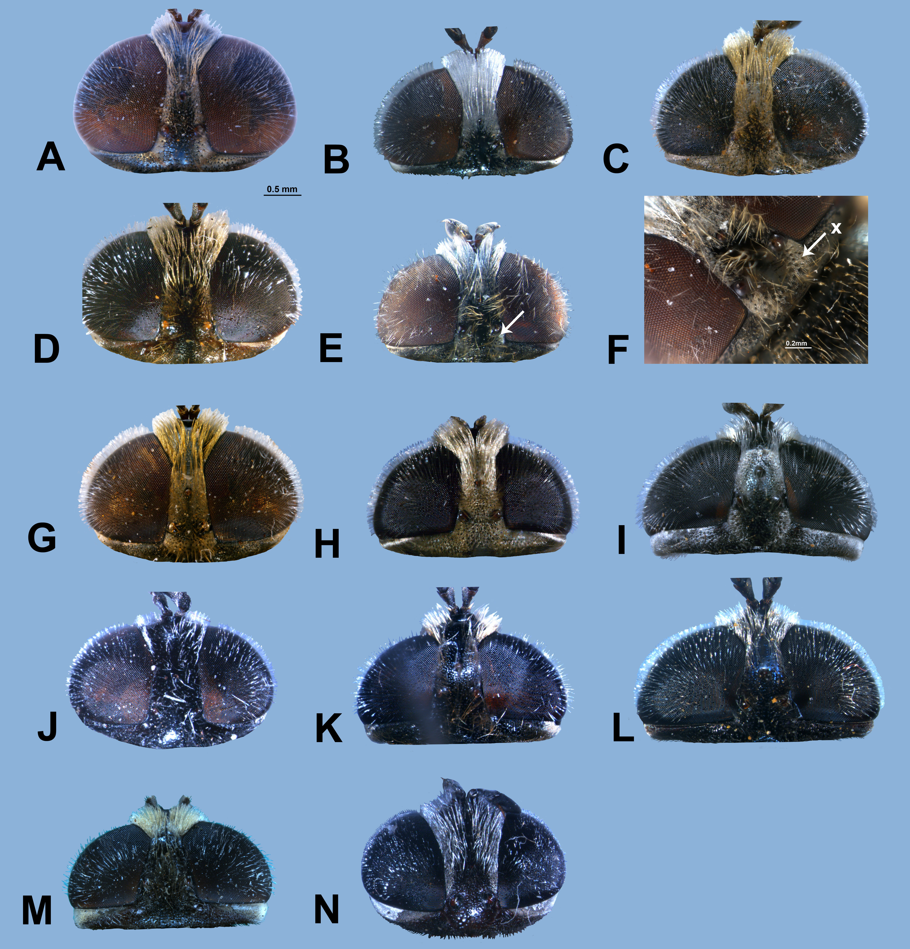
5. Tergite 4 with silver spots ( Fig. 7F View Fig ). Silver spots on tergite 3 completely and densely setose, setae directed towards the lateral margin, i.e., turned ca. 90° in relation to longitudinal axis. Vertex entirely microtrichiose except for a narrow stripe along the posterior margin ( Fig. 2G View Fig ) …… …………………………………………………………………… M. sexmaculatus View in CoL sp. nov. ( Fig. 1F View Fig )
– Tergite 4 without silver spots. Silver spots on tergite 3 for a large part without setae or (some M. magnicornis View in CoL sp. nov.) with scattered setae, setae turned to the side for ca 45°. At least ocellar triangle partly bare of microtrichia ………………………………………………………………6
6. Postpedicel elongated ( Fig. 3D View Fig ). Vertex almost bare of microtrichia ( Fig. 2D View Fig ). Silver spots on tergite 3 widely separated, their distance exceeds the length of the tergite, narrowly separated from anterior and posterior margins ( Fig. 7D View Fig ). Tergite 3 with extensive areas with black setae …………………………………………………………………… M. magnicornis View in CoL sp. nov. ( Fig. 1D View Fig )
– Postpedicel normal. Vertex in front of anterior ocellus entirely densely microtrichiose. Silver spots on tergite 3 narrowly separated, their distance less than half the length of the tergite, covering whole length of tergite. Tergite 3 with or without a few scattered black setae ……………………7
7. Distance between eyes 0.17 × width of head, ocellar triangle isosceles ( Fig. 2C View Fig ). Larger species, body length about 8 mm ………………………………… M. argentimaculatus View in CoL sp. nov. ( Fig. 1C View Fig )
– Distance between eyes larger, ocellar triangle equilateral (as in Fig. 2F View Fig ). Smaller species, body length 6.5–7.0 mm ………………………………………………………………8 (as in Fig. 1B, E View Fig )
8. Postocellar spot small, separated for about the distance between posterior ocelli ( Fig. 2B View Fig ). Median apex of the silver spot on tergite 2 near middle of tergite ( Fig. 7B View Fig ). Mesonotum with welldeveloped submedian and lateral (notopleuron, supraalar area) microtrichiose stripes ……… …………………………………………………………………… M. argentifrons View in CoL sp. nov. ( Fig. 1B View Fig )
– Postocellar spot large, almost confluent ( Fig. 2E, F View Fig ). Median apex of the silver spot on tergite 2 close to posterior margin of tergite ( Fig. 7E View Fig ). Mesonotum with traces only of the submedian and lateral microtrichiose stripes ( Fig. 4B View Fig ) …………………………………… M. natalensis View in CoL sp. nov. ( Fig. 1E View Fig )
9. Tergites without microtrichiose fasciae or spots ( Fig. 7I View Fig ). Distance between eyes big, frons and vertex with smooth surface (setae with simple alveoli, i.e., without tubercles), without microtrichia ( Fig. 2J View Fig ) …………………………………………… M. immaculatus View in CoL sp. nov. ( Fig. 1I View Fig )
– At least tergites 2+3 with microtrichiose fasciae or spots (as in Fig. 7K View Fig ). Distance between eyes narrower, frons and vertex with rough surface (setae with tuberculate alveoli), with microtrichiose pattern at least on vertex (as in Fig. 2I View Fig ) …………………………………………………………10
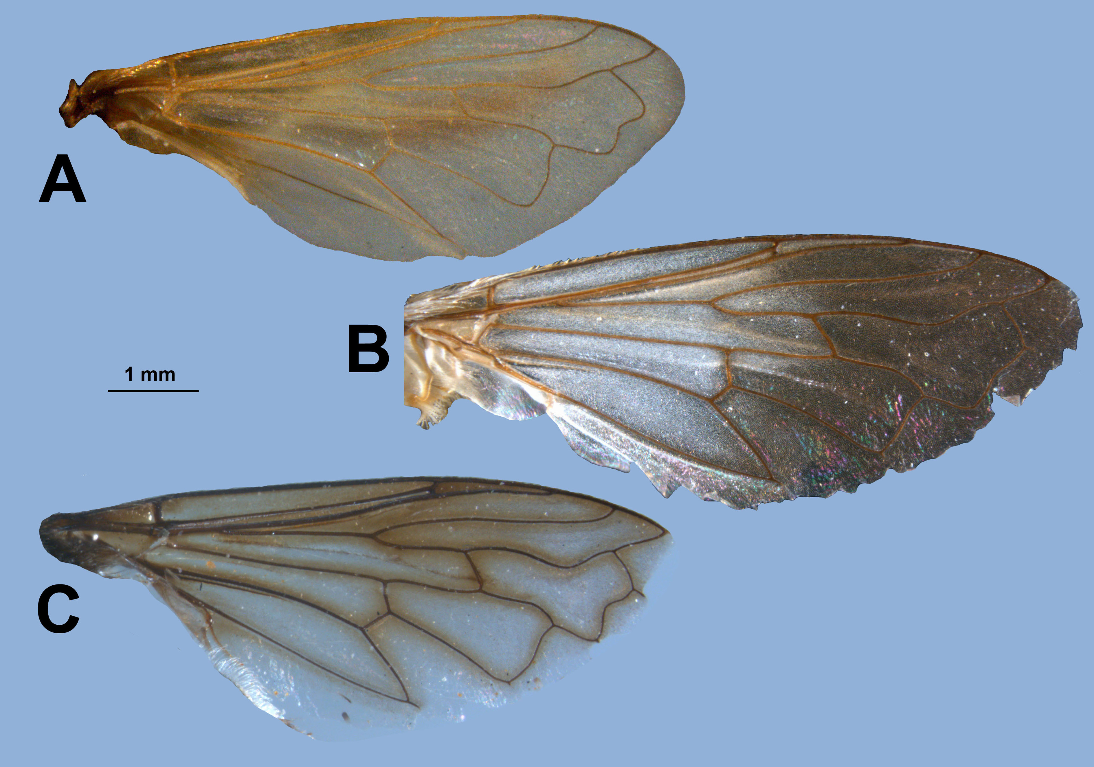
10. Postsutural pilosity of mesonotum black, with a intraalar microtrichiose stripe well separated from supraalar seta. Wing membrane brown along veins ( Fig. 5C View Fig ), posterior margin of alula bare of microtrichia. Tibiae 1–3 bright reddish, without dark rings ……… M. tabanoides View in CoL sp. nov. ( Fig. 1K View Fig )
– Mesonotal pilosity entirely light (white or yellow), with a supraalar microtrichiose stripe (as in Fig. 4C View Fig ). Wing membrane either clear or completely infuscated, alula entirely microtrichiose. Tibia 1 and 2 each with a dark ring, tibia 3 almost completely blackish. ……………………………………11
11. Tergites 2+3 with widely separated microtrichiose fasciae, tergite 4 with fasciae narrowly separated ( Fig. 7J View Fig ). Vertex almost bare of microtrichia (the very few microtrichia do not form a distinct pattern) ( Fig. 2K View Fig ). Wing membrane light brown …… M. ochreatus ( Hull, 1964) View in CoL comb. nov. ( Fig. 1J View Fig )
– Tergites 2–4 with fasciae ( Fig. 7G, H View Fig ). Vertex extensively densely microtrichiose ( Fig. 2H, I View Fig ). Wing membrane clear ……………………………………………………………………………………12
12. Wing with yellow veins, microtrichia equally dense and light (yellowish) on whole wing, appearing unicolorous ( Fig. 5A View Fig ). Frons, except for a stripe along the lunula, and vertex except for a narrow stripe at posterior edge entirely densely covered by yellowish microtrichia ( Fig. 2H View Fig ). Setae on all parts of the body golden. Tergites without black setae ……………… M. apiformis View in CoL sp. nov. ( Fig. 1G View Fig )
– Wing with brown veins, in proximal half densely covered by white microtrichia, in distal half with dark microtrichia, thereby wing appearing bicoloured ( Fig. 5B View Fig ). Nearly whole frons and a large posteromedian area on vertex without microtrichia ( Fig. 2I View Fig ). Tergites extensively black setose … …………………………………………………………………… M. cooksoni View in CoL sp. nov. ( Fig. 1H View Fig )


No known copyright restrictions apply. See Agosti, D., Egloff, W., 2009. Taxonomic information exchange and copyright: the Plazi approach. BMC Research Notes 2009, 2:53 for further explanation.
|
Kingdom |
|
|
Phylum |
|
|
Class |
|
|
Order |
|
|
Family |
|
|
SubFamily |
Eristalinae |
|
Tribe |
Merodontini |