
Ormiophasia Townsend, 1919
|
publication ID |
https://doi.org/ 10.11646/zootaxa.4643.1.1 |
|
publication LSID |
lsid:zoobank.org:pub:518ACC5F-A320-4EBD-B750-50006F40B054 |
|
persistent identifier |
https://treatment.plazi.org/id/03E98795-FF97-1175-FF53-F8CCFA71C7DF |
|
treatment provided by |
Felipe |
|
scientific name |
Ormiophasia Townsend, 1919 |
| status |
|
Ormiophasia Townsend, 1919 View in CoL
Ormiophasia Townsend, 1919: 164 View in CoL . Type species: Ormiophasia busckii Townsend, 1919 View in CoL , by original designation.
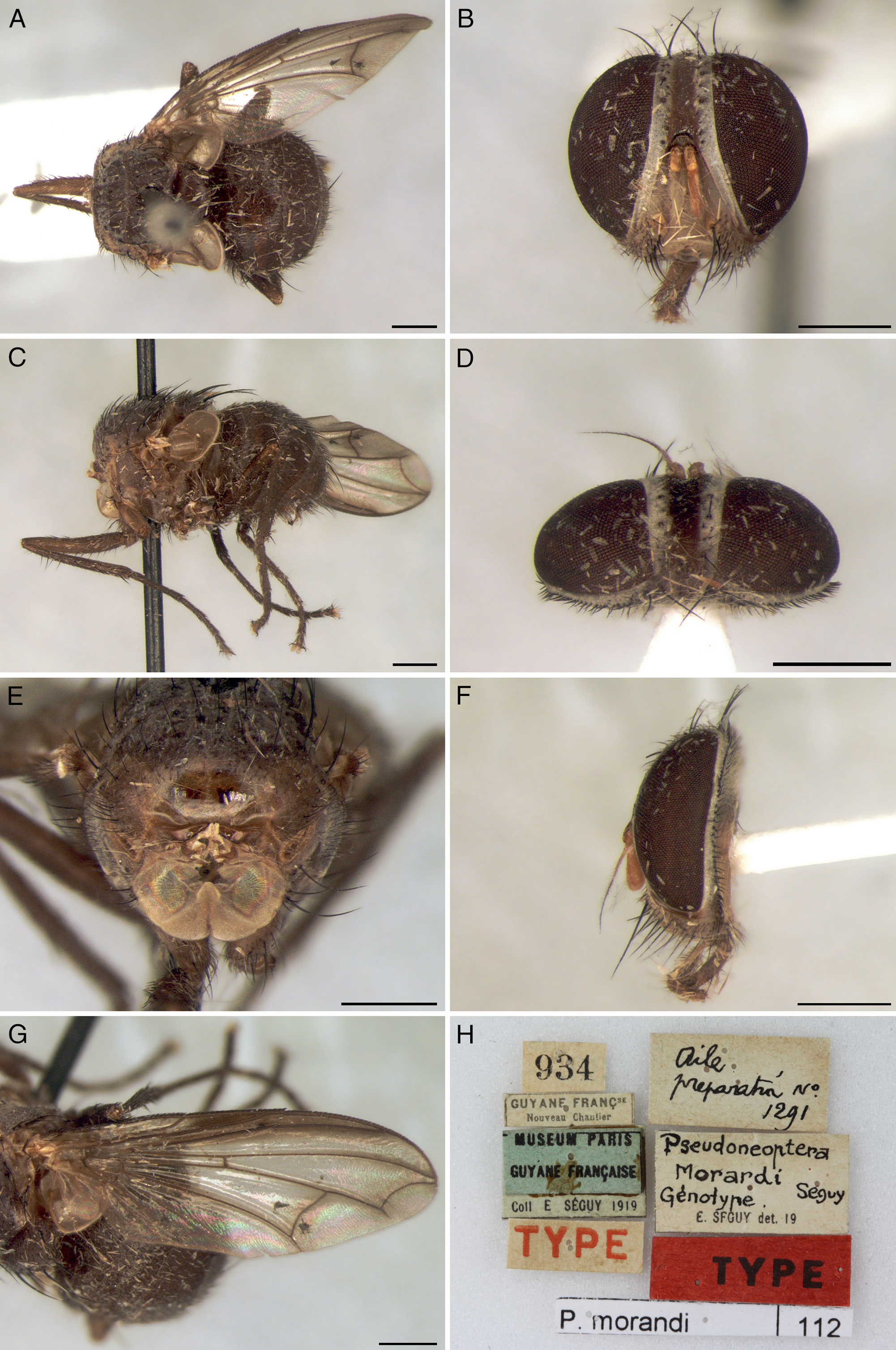
Pseudoneoptera Séguy, 1926b: 19 . Type species: Pseudoneoptera morardi Séguy, 1926b View in CoL , by monotypy; Séguy (1926b: 20, key to genera); Séguy (1927a: 423, key to genera; 424, catalog); Townsend (1931: 82, synonymy with Ormiophasia View in CoL ); Sabrosky (1953: 181, as Ormiophasia , catalog); Tavares (1964: 38, comments on synonymy).
Plagiatormia Séguy, 1926b: 19 (as Plagiotormia , incorrect original spelling). Type species: Plagiatormia obscura Séguy, 1926b , by monotypy; Séguy (1926b: 20, key to genera); Séguy (1927a: 423, as Plagiotormia , misspelling, key to genera; 424, catalog); Townsend (1931: 82, synonymy with Ormiophasia ); Sabrosky (1953: 181, as Ormiophasia , catalog); Tavares (1964: 38, comments on synonymy).
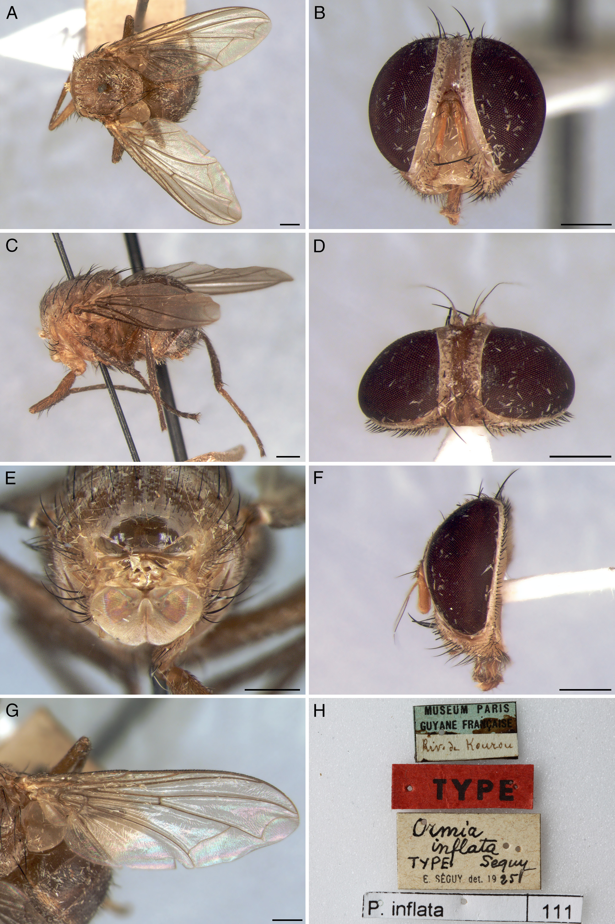
Pseudormia Séguy, 1927b: 262 . Type species: Pseudormia inflata Séguy, 1927b , by monotypy; Séguy (1925: 375, nomen nudum, comparison to Ormia View in CoL ; 376, key to genera); Séguy (1926a: 5, nomen nudum, comparison to Ormia View in CoL ; 9, as Peudormia, misspelling, key to genera); Séguy (1926b: 20, nomen nudum, key to genera); Séguy (1927a: 424, nomen nudum, key to genera, catalog); Townsend (1931: 82, synonymy with Ormiophasia View in CoL ); Sabrosky (1953: 181, as Ormiophasia View in CoL , catalog); Tavares (1964: 38, comments on synonymy).
Diversity and distribution. The ranges of the 16 species currently ascribed to Ormiophasia View in CoL cover an area from Southeast Mexico to northern Argentina.
References. Townsend (1919: 164, description of Ormiophasia View in CoL and of the type species O. busckii View in CoL ); Aldrich (1922: 5, comments on Ormia View in CoL , treated O rmiophasia as synonym of Ormia View in CoL ); Townsend (1927: 223, key to genera of Neotropical Muscoidea [sensu Townsend]); Malloch (1929: 279, as Ormia View in CoL , comments on validity of Ormiophasia View in CoL ); Townsend (1931: 82, synonymized Plagiatormia , Pseudormia and Pseudoneoptera with Ormiophasia View in CoL ); Townsend (1936: 100, comments on larval characters; 101, key to genera of Ormiini View in CoL , comments on synonymies of Plagiatormia , Pseudormia and Pseudoneoptera ); Townsend (1938: 236, redescription, description of larva); Townsend (1942: 325, illustration of larva); Sabrosky (1953: 171, key to genera of New World Ormiini View in CoL ; 172, key; 181, catalog, comments on synonymies of Plagiatormia , Pseudormia and Pseudoneoptera ); Thompson (1963: 455, comments on larvae being related to Euphasiopteryx and Ormia View in CoL ); Tavares (1964: 37–52, taxonomic revision, comments on synonymies of Plagiatormia , Pseudormia and Pseudoneoptera ); Tavares (1965a: 14, key to genera of Neotropical Ormiini View in CoL ); Guimarães (1971: 22; catalog); Tschorsnig (1985: 55, illustration of male ejaculatory apodeme; 97, description of male terminalia of Ormiini View in CoL ); Gramajo (1997: 96, record of Ormiophasia View in CoL , as Ormiaphasia, misspelling); Toma & Nihei (2006: 243, catalog of type material); Wood & Zumbado (2010: 1410, as Ormia Robineau-Desvoidy View in CoL ; treated Ormiophasia View in CoL as synonym of Ormia View in CoL ); Evenhuis et al. (2015: 197, catalog of Townsend’s genera); Nihei (2016: 914, catalog of Colombiam Tachinidae View in CoL ); O’Hara (2018: 55, catalog).
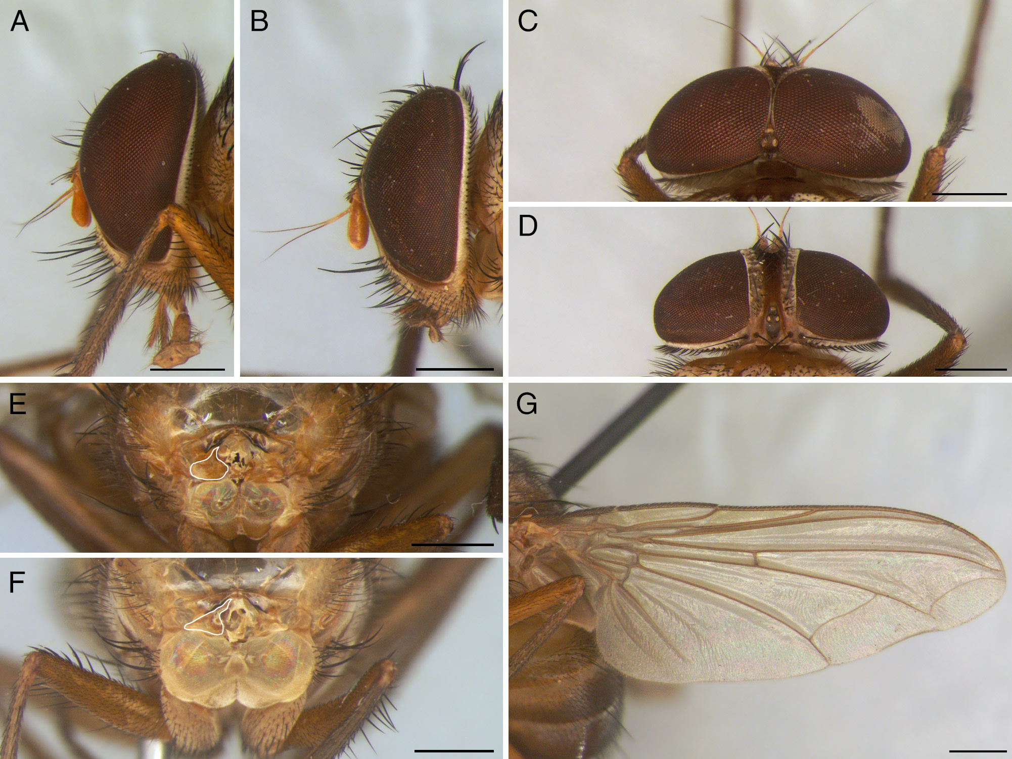
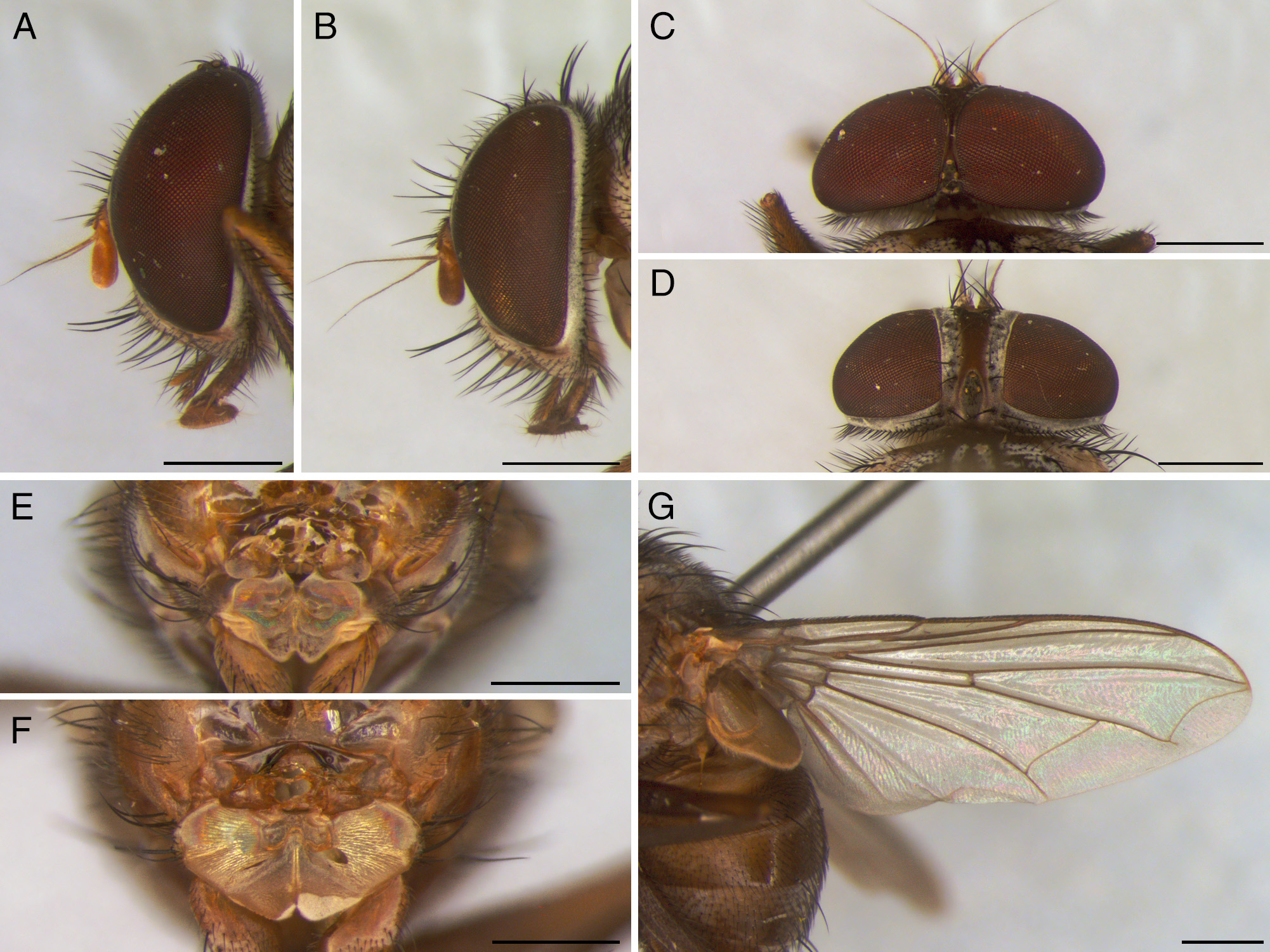
Diagnosis. Ormiophasia resembles Ormia , sharing many characters with this genus, such as: occiput extremely concave, especially in females; presternum with a pair of pits on anterior surface ( Fig. 1 View FIGURE 1 ); anterior surface of distiphallus membranous ( Figs 38–42 View FIGURE 38 View FIGURE 39 View FIGURE 40 View FIGURE 41 View FIGURE 42 ); hypoproct with a lateral patch of very short, spiniform setae ( Fig. 3B View FIGURE 3 ); and female tergite six with a row of strong, posteriorly curved setae. However, although many authors have considered Ormiophasia and Ormia as synonyms, Ormiophasia can be clearly distinguished by its darker body color, varying between brownish-yellow, brown and dark brown ( Figs 9–14 View FIGURE 9 View FIGURE 10 View FIGURE 11 View FIGURE 12 View FIGURE 13 View FIGURE 14 ) ( Ormia species generally have a pale yellow body color); ocelli well developed (vestigial or missing in Ormia ); one pair of presutural acrostichal setae (two to three pairs in Ormia ); tegula of same color as body ( Ormia usually with tegula black, contrasting with body color); male wing without callosities on veins C and R 2+3 ( Ormia usually with callosities on C and R 2+3) (see Tavares 1962, 1965a, 1965b); male terminalia with apex of cerci broad, at least 1/3 of total width of cerci ( Figs 38–42 View FIGURE 38 View FIGURE 39 View FIGURE 40 View FIGURE 41 View FIGURE 42 ) (in Ormia the cerci are tapered apically, less than 1/3 of total width of cerci) (see Tavares 1962, 1965a, 1965b); and female terminalia with hypoproct bare, without setulae ( Fig. 3B View FIGURE 3 , setulose in Ormia ). Additionally, Ormiophasia also differs from other Ormiini genera by having head hemispherical in profile, with genal length not exceeding 0.2 times length of head; face developed, at least 1.1 times width of facial ridge; facial ridge broad but not exceeding 2.9 times width of parafacial; and male terminalia with cerci completely fused.
Genus characterization. Both sexes. Head hemispherical in profile; oral axis shorter than antennal axis. Eye bare. Ocelli well developed. Antenna inserted in the middle of the eye, reaching halfway between lunule and lower facial margin; scape reduced, with row of marginal setulae; pedicel dorsally setulose, with one dorsal preapical seta; first flagellomere oblong; arista arising basally on dorsal surface, bare or weakly plumose, base pubescent and lightcolored, becoming tapered and darker distally. Fronto-orbital plate setulose to level of first anterior frontal seta. Parafacial bare. Facial ridge broad and subequal to facial length, setulose below vibrissa. Face slightly concave at vibrissal angle, with lower facial margin not visible in profile. Gena about 1/10 of head height, genal setae 4–5. Genal dilation setulose. Vibrissae strong and crossed, arising far above lower facial margin; supravibrissal setae 1–2, about 1/3 length of vibrissa; subvibrissal setae 3–4. Mouthparts well developed; clypeus globose; palpus developed, about 1.5 times length of prementum, clavate, covered with appressed setae from median area to apex; prementum setulose; labella padlike. Occiput covered with silver setulae, with row of short postocular setae; lower occipital area strongly concave.
Thorax densely covered with black setulae. Presternum bilobed ( Fig. 1 View FIGURE 1 ), each lobe with a pit on anterior surface. Basisternum bare, inflated; ventral surface grooved medially. Prosternal tympanal membrane corrugated, with corrugation equally distributed across entire membrane. Acrostichal setae 1+2–3; dorsocentral setae 2+3; intra-alar setae 1+2; supra-alar setae 1+2 (first postsutural seta weak, about 1/2 length of second postsutural seta). Postpronotal setae 3, with inner seta weaker. Notopleural setae 2, equal-sized. Postalar setae 2, equal-sized. Proepisternal setae 1, strong, upcurved, with 1–3 weaker setae near base. Katepisternal setae 2, divergent. Anepimeral seta 1. Katepimeron, katatergite, anatergite and postalar wall bare. Scutellar setae strong, convergent; basal pair subequal to subapical pair; lateral pair 2/3 length of subapical pair, closer to basal pair; apical pair absent; discal pair half length of subapical pair, more widely separated than subapical setae. Wing. Tegula setulose, with 1–2 inner marginal setae. Cells sc, c and r 1 with light yellow infuscation. Vein C ending just after vein M 1, at wing apex. Cell r 4+5 open; length of opening subequal to or shorter than crossvein r-m. Vein M 1 arched towards apex; bend of vein M angular or rounded, sometimes with a short stem. Legs. Fore coxa with an outer row of 3–4 anterior setae followed by 4–6 marginal setae. Fore tibia with 1 posterodorsal preapical seta, 2 posteroventral median setae, 1 preapical seta and 1 ventral preapical seta. Mid femur with 1 strong anteromedian seta and 2–3 strong posterodorsal preapical setae. Mid tibia with 1 strong anterodorsal postmedian seta, 1 apical seta, 1 posterodorsal apical seta, 2 posteromedian setae, 1 weak apical seta, 1 posteroventral apical seta, 1 weak ventral apical seta and 1 strong anteroventral apical seta. Hind femur with row of 3–4 posterodorsal basal setae and row of 11–12 weak posterodorsal setae from middle to apex. Hind tibia with 2 anterodorsal median setae, 1 preapical seta, 1 weak ventral apical seta, 2–3 weak anteroventral median setae and 1 weak apical seta. Pulvilli light yellow. Claws brown, with apex black.
Abdomen globose and widely connected with thorax, wider than long; densely covered with black setulae. Middorsal depression on syntergite 1+2 extending to hind margin of syntergite. Syntergite 1+2 and tergite 3 with one pair of marginal lateral setae. Tergite 4 with row of 8–10 marginal setae. Tergite 5 with row of 6–8 discal setae. Sternites visible, each with a weak row of marginal setae.
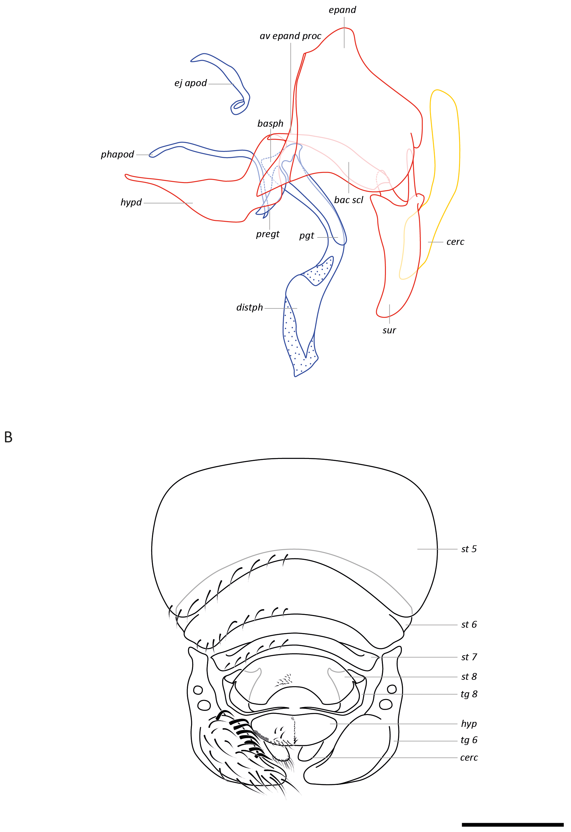
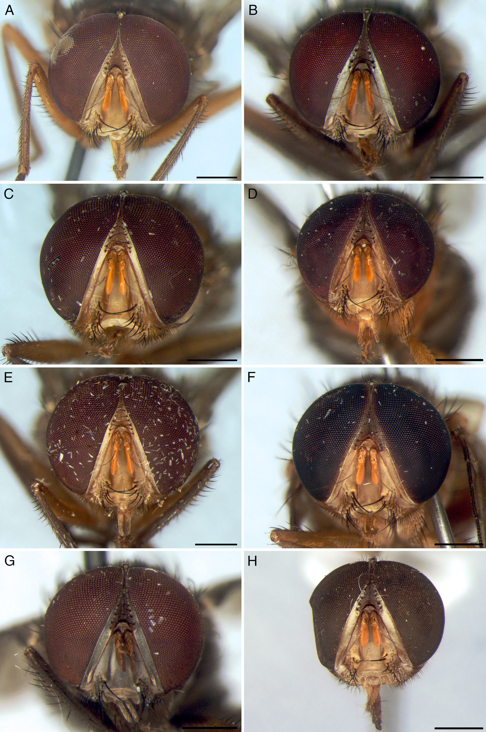
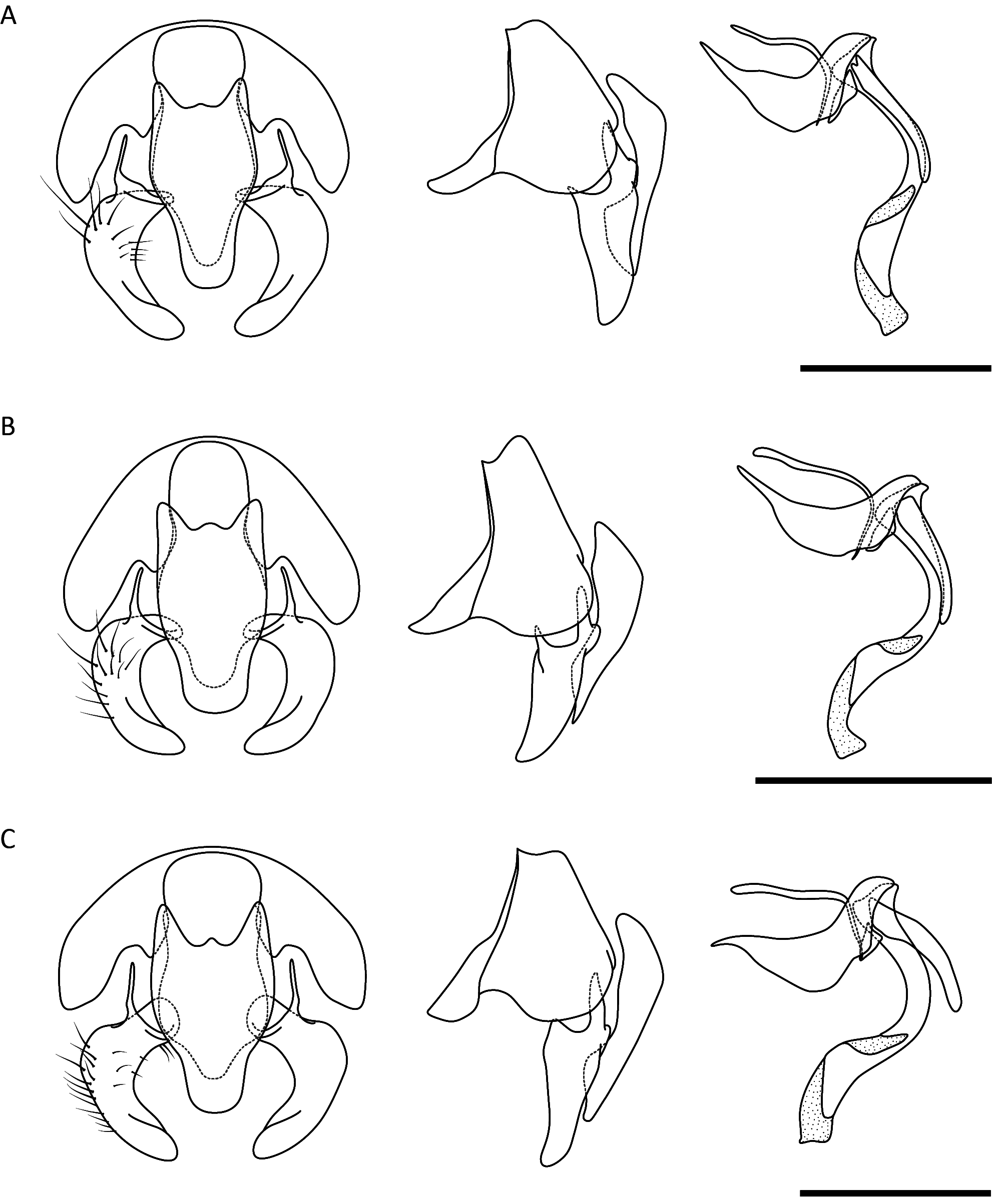
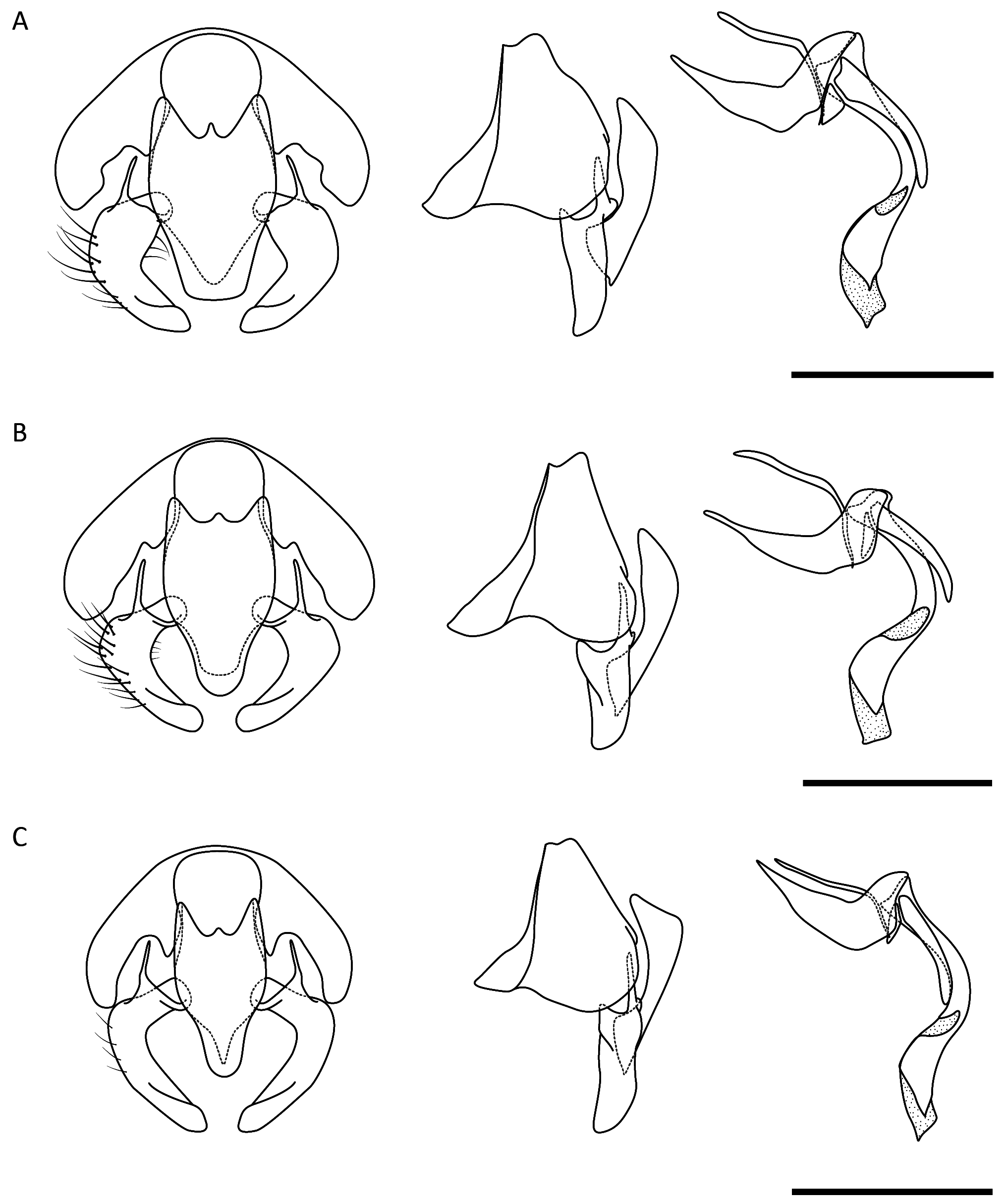
Male. Head holoptic ( Figs 5–6 View FIGURE 5 View FIGURE 6 ). Ocellar triangle constricted, tubercle-shaped. Dorsal ommatidia larger than peripheral ones. Thorax. Lateral cervical sclerite with external lobe oblong (e.g., Fig. 15E View FIGURE 15 ). Basisternum less inflated than in female. Prosternal tympanal membrane reduced. Posterior spiracle with posterior lappet shaped as an operculum. Terminalia ( Figs 3A View FIGURE 3 , 38–42 View FIGURE 38 View FIGURE 39 View FIGURE 40 View FIGURE 41 View FIGURE 42 ). Sternite 5 with posterior margin bilobed, setulose posteriorly. Epandrium with dorsal surface covered with strong, upcurved setae. Surstylus not fused with epandrium; strongly arcuate mediad; posterior surface with a slight distal, median ridge; articulation with bacilliform sclerite rounded; articulation with epandrium tapered and narrow; inner surface covered with microtrichia. Connection of bacilliform sclerite to hypandrium broad, becoming tapered towards surstylus. Anterior margin of hypandrium slightly longer than phallapodeme; concave; posterior area weakly sclerotized; hypandrial arms not fused with each other or with aedeagus. Basiphallus connected directly to distiphallus at a 90 o angle; distiphallus simple, smooth, with anterior surface grooved. Phallapodeme flat, slender. Ejaculatory apodeme narrow, with posterior apex slightly broader. Pregonites flat, bare, slightly concave, not fused with each other and not fused with hypandrium; anterior margin broad, reaching lower posterior margin of hypandrium. Postgonite long, bare, rod-like, with rounded apex, almost reaching connection between basiphallus and distiphallus. Cerci fused, with apex broad and at least 1/3 of total length of cerci; covered with setae.
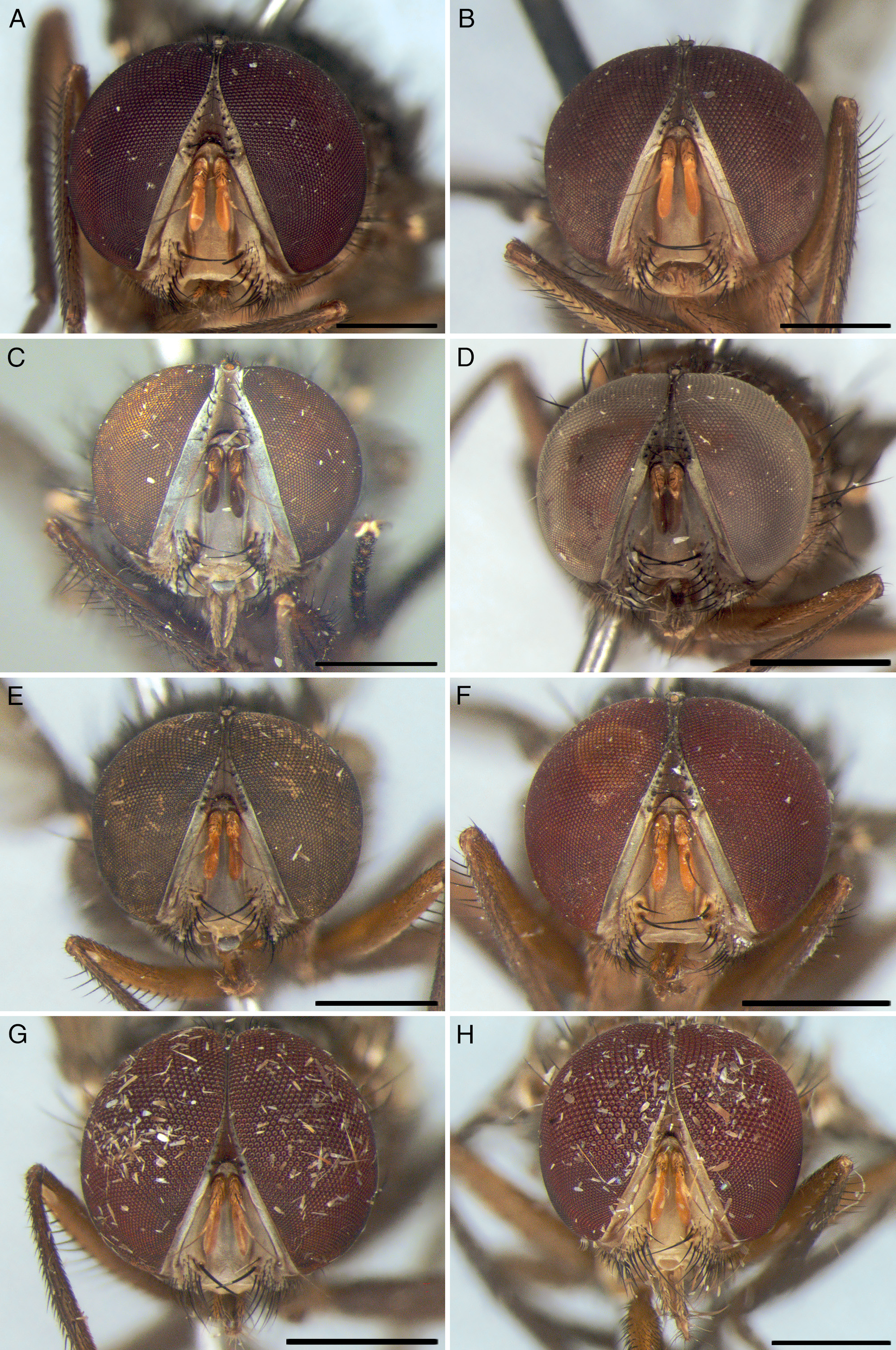
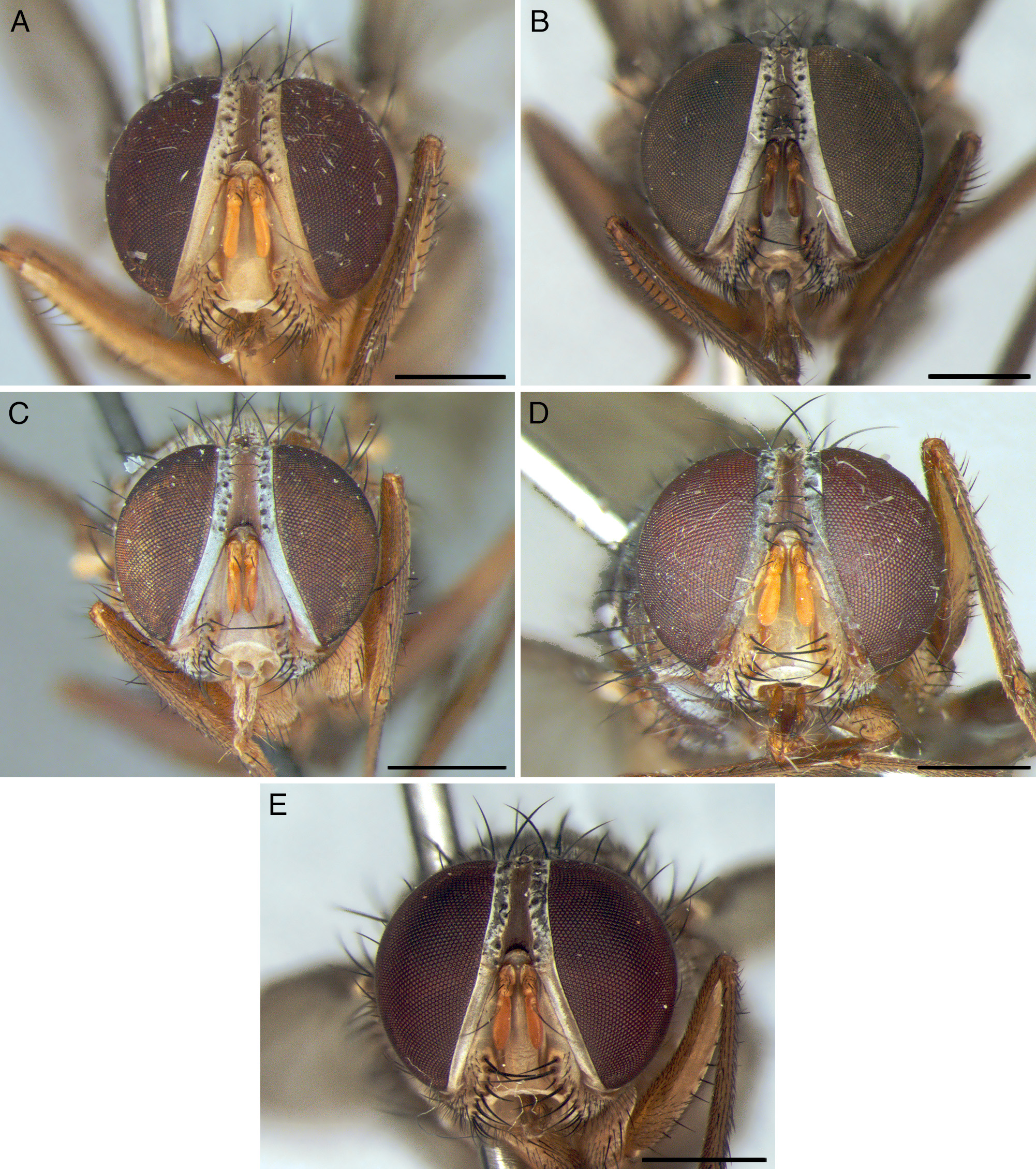
Female. Head dichoptic ( Figs 7–8 View FIGURE 7 View FIGURE 8 ). Ocellar triangle level with eyes, not protruding as a tubercle. Ommatidia all of the same size, not differentiated. One pair of inner vertical setae, strong and crossed, subequal to vibrissa. One pair of outer vertical setae, same size as inner vertical setae, divergent. One pair of upper orbital setae, divergent, half size of vertical setae. Two pairs of proclinate orbital setae. Thorax. Lateral cervical sclerite with external lobe extended and tapered at margin (e.g., Fig. 15F View FIGURE 15 ). Basisternum very inflated, much more than in male. Prosternal tympanal membrane broad. Posterior spiracle with posterior lappet stylus- or plume-shaped (except operculum-shaped in O. lanei ). Terminalia ( Fig. 3B View FIGURE 3 ). Tergite 6 broad, voluminous, setulose, separated into two equal-sized sclerites, bearing spiracles 6 and 7; inner margin with very strong, inward-curved setae. Tergite 7 absent. Tergite 8 developed, bare, separated into two equal-sized, concave sclerites. Epiproct (tergite 10), when present, represented by one pair of setae in membrane. Sternites 6, 7 and 8 each with a row of weak marginal setae. Hypoproct (sternite 10) bare; lateral surface with patch of very short, spiniform setae; ventral surface with longitudinal median row of short setae; posterior margin with row of slender, weak setae. Cerci free, not fused, with weak, slender marginal setae. Three equal-sized spermathechae, spherical, smooth.
Remarks. The first four species included in Ormiophasia were based mainly on female specimens, which probably caused most of the confusion about the validity of the genus. The male and female terminalia had not been studied and described at that time, but they include important diagnostic characters for the genus. Aldrich (1922) and Curran (1934) treated Ormiophasia as a junior synonym of Ormia , without justification. Only Malloch (1929) made some considerations based on characters such as the width of the female frontal vitta, which is narrow in Ormiophasia . Townsend (1919: 164) had already mentioned this character when describing Ormiophasia , and Sabrosky (1953: 172) also used it to differentiate Ormia and Ormiophasia in his key. Ormia punctata Robineau- Desvoidy, examined by Malloch, clearly has frontal vitta broader than in other Ormiophasia species, but there are also other species of Ormia with a narrower frontal vitta. Townsend did not know males of Ormiophasia for a long time after the original description ( Townsend 1927: 223, “ ♂ desconhecido [unknown]”), until he included descrip- tions of male terminalia in his Manual of Myiology ( Townsend 1936, 1938). These descriptions were superficial and incomplete, however, and relative to all Ormiini . In this context, the contributions and impact of the publications of Tavares (1962, 1964, 1965a, 1965b) on the taxonomy of New World Ormiini should be highlighted, since the male terminalia of a comprehensive number of species were described in detail for the first time. Furthermore, Tavares was the first to diagnose these genera based on male terminalia ( Tavares 1965a: 14). However, he described his species based only on males, without associating them with their respective females. The association of females with males and the dissections of female terminalia made in the present study have provided additional and valuable characters to differentiate Ormiophasia and Ormia .
Some comments about Tavares’s holotypes are also warranted. The right wing, antenna and male terminalia of his type material were mounted on slides and linked with their respective specimens by a code (see Fig. 19E View FIGURE 19 ). However, during the Brazilian military government (1964–1985), an event known as the “Massacre de Manguinhos” took place ( Jurberg 1993; Costa et al. 2008). This episode, which occurred on April 1st, 1970, greatly impacted the history of science in Instituto Oswaldo Cruz, Unidade Manguinhos, Rio de Janeiro, where Omar Tavares had worked and where his holotypes are deposited. The term “Massacre de Manguinhos” was coined by helmintologist Herman Lent ( Jurberg 1993), who worked for the same institute. The “Manguinhos” entomological collection is one of the most important historic scientific collections in Brazil. During the “Massacre de Manguinhos”, scientists were politically persecuted and the scientific collection was dismantled and transferred to another building inside the institute. The relocation of the Instituto Oswaldo Cruz collection resulted in the loss of much of the material.Although Tavares’s holotypes are still in good condition, their respective slides were lost, probably during this episode. Only the slides of the male terminalia of the paratypes, housed in MZSP and USNM, are available for study.
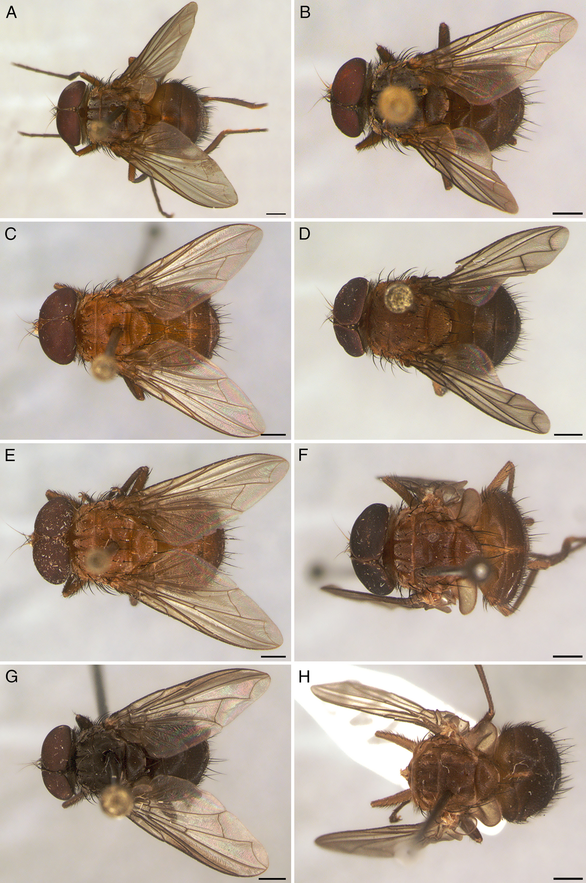
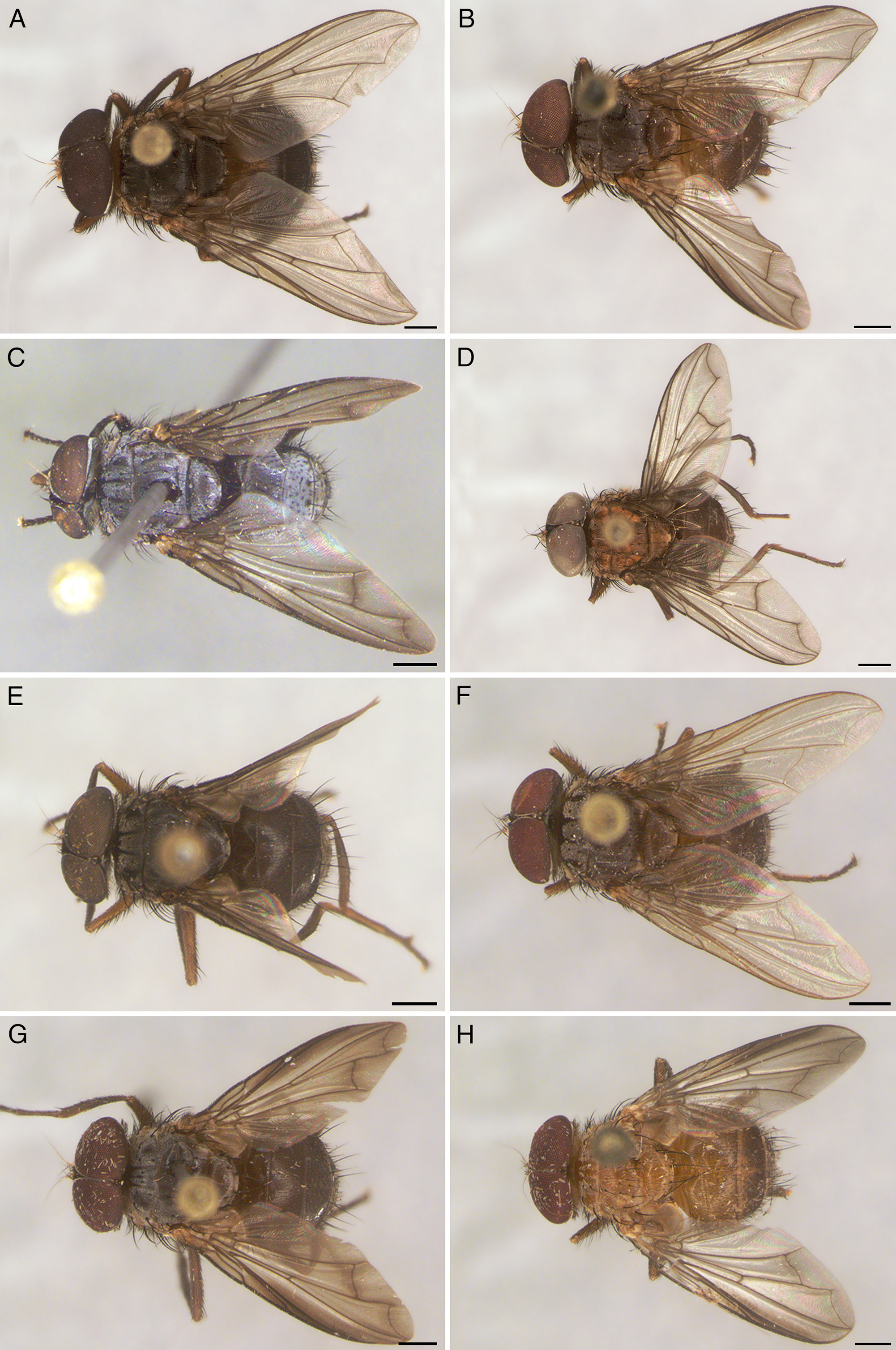
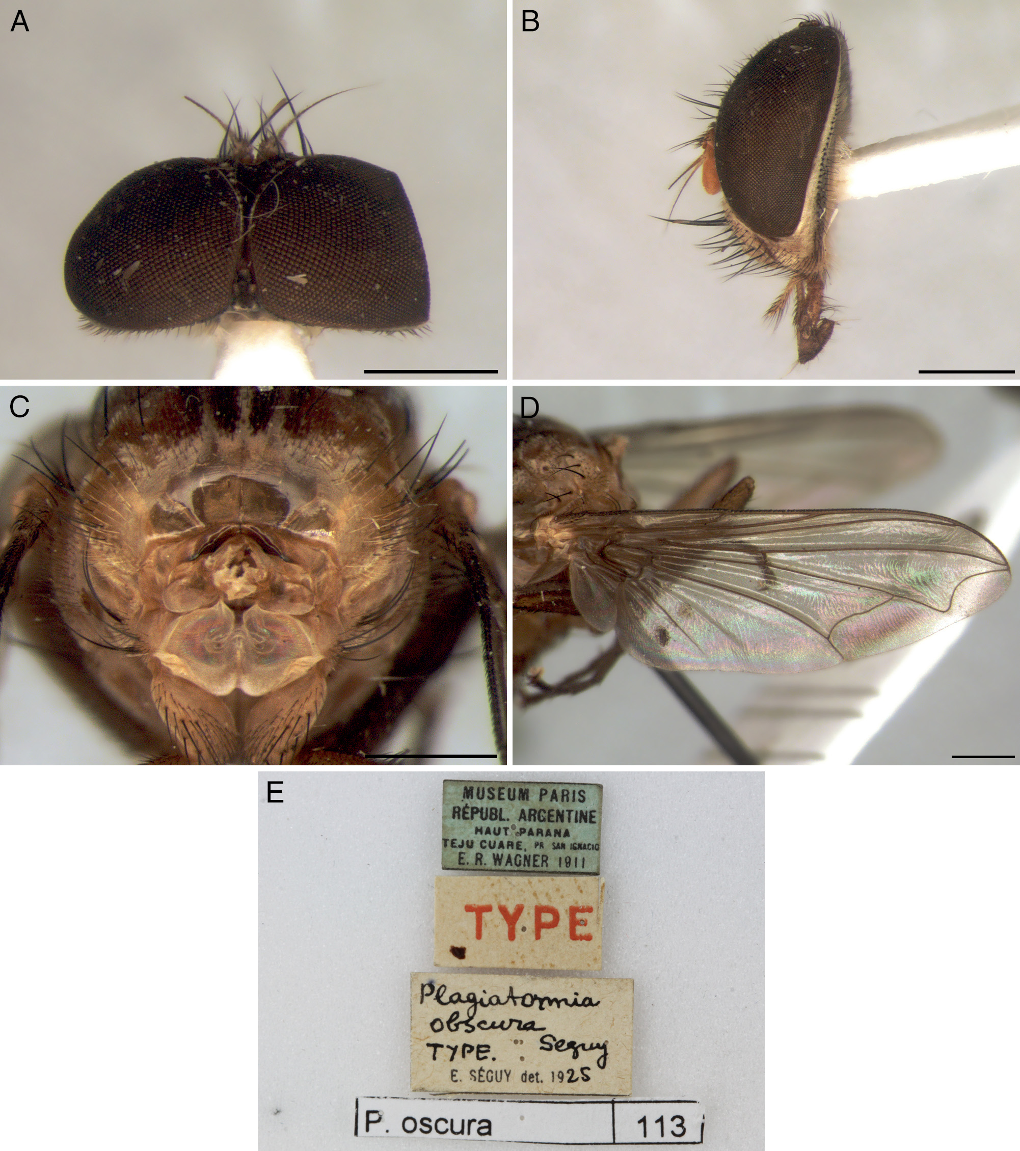
The type material of all nominal species of Ormiophasia was examined during this study. All of Séguy’s and Townsend’s holotypes are in good conditions of preservation ( Figs 16 View FIGURE 16 , 24 View FIGURE 24 , 29 View FIGURE 29 , 30 View FIGURE 30 ). The type specimens of Tavares ( Figs 18 View FIGURE 18 , 20 View FIGURE 20 , 22 View FIGURE 22 , 25 View FIGURE 25 , 27 View FIGURE 27 ) have damaged abdomens due to their dissection. The examination of type material allowed for a reliable identification of the material included in this study. Furthermore, as Sabrosky (1953: 173) remarked, Ormiophasia specimens exhibit a significant variation in body coloration, which ranges from brownish-yellow to almost black. The color and pruinosity of the head and thorax, and the male cerci and surstylus, were the most important characters supporting the description of the eight new species.
So far, there are no known host records for any Ormiophasia species.
Key to species of Ormiophasia View in CoL
Although Ormiophasia View in CoL (and Ormiini View in CoL ) species are sexually dimorphic, the key provided below aims to identify both male and female specimens (except for O. obscura , O. buoculus sp. nov. and O. townsendi View in CoL sp. nov., of which only males are known); when necessary, characters exclusive to one sex are included in couplets. The identification of female specimens, however, has to be done carefully, since the females of several species are very similar (e.g., O. costalimai View in CoL and O. inflata View in CoL or O. causeyi View in CoL and O. chapulini View in CoL sp. nov.). The male terminalia are the most reliable source of information allowing to confirm species’ identity.
Tavares (1964) provided the first key to Ormiophasia View in CoL species, but some characters used in his key fail when large series are examined, due to variation (e.g., of body color or of number of setae). Furthermore, there are some errors in the key, which are commented on in the Remarks under the respective species in the present paper. Some important characters used by Tavares are maintained here for some species (e.g., those of the male terminalia of O. lanei View in CoL ). Séguy (1926b) also provided a key to his monotypic genera Plagiatormia , Pseudoneoptera and Pseudormia , based on the length of the opening of cell r 4+5, the shape of the bend of vein M, the length of the antenna and the shape of the face. Although these characters can be seen in Séguy’s holotypes, they are variable in the additional material examined (except in O. obscura , so far known only from the holotype).
1 Basicosta broad, twice width of tegula ( Fig. 4B View FIGURE 4 ); female wing with strong brown infuscation around all veins except A 1 +CuA 2 (male wing with weak infuscation at the tip of vein R 2+3 and around veins M 1 and dm-cu, see Figs 33G View FIGURE 33 , 34F View FIGURE 34 ), and with veins R 2+3, R 4+5, M 1 and CuA 1 thickened ( Figs 33H View FIGURE 33 , 34G View FIGURE 34 ); thorax dark brown or brownish-yellow.......................... 2
- Basicosta subequal in width to tegula ( Fig. 4A View FIGURE 4 ); female and male wings hyaline ( Fig. 15G View FIGURE 15 ) or infuscated only around veins R 1, R 2+3, M 1 and dm-cu ( Fig. 28G View FIGURE 28 ), but without thickened veins; thorax brownish-yellow, brown or dark brown.............. 3
2 Thorax and abdomen dark brown ( Figs 10C View FIGURE 10 , 14B View FIGURE 14 ); male wing with section of vein M between crossvein dm-cu and M 1 slightly curved ( Fig. 33G View FIGURE 33 ); apex of male cerci long and about 2/5 length of cerci in posterior view ( Fig. 41A View FIGURE 41 ) (North Brazil, Costa Rica, French Guiana and Venezuela).......................................................... O. crassivena View in CoL sp. nov.
- Thorax brownish-yellow, contrasting with dark brown abdomen ( Figs 10D View FIGURE 10 , 14C View FIGURE 14 ); male wing with section of vein M between crossvein dm-cu and M 1 straight ( Fig. 34G View FIGURE 34 ); apex of male cerci short and about 1/3 length of cerci in posterior view ( Fig. 41B View FIGURE 41 ) (South and Southeast Brazil)......................................................... O. manguinhos View in CoL sp. nov.
3 Wing with strong brown infuscation around veins R 1 and R 2+3 ( Fig. 28G View FIGURE 28 ) and weak brown infuscation around veins M 1 and dm-cu; thorax and abdomen dark brown or brownish-yellow................................................... 4
- Wing hyaline ( O. cruzi View in CoL may have a very weak brown infuscation around veins M 1 and dm-cu, see Fig. 21G View FIGURE 21 ); thorax and abdomen dark brown, brown or brownish-yellow................................................................ 7
4 Thorax and abdomen dark brown; fronto-orbital plate and parafacial gray with silver pruinosity....................... 5
- Thorax and abdomen brown or brownish-yellow; fronto-orbital plate and parafacial brownish-yellow with yellow pruinosity 6
5 Male head with dorsal ommatidia larger than ocelli ( Fig. 6G View FIGURE 6 ); ocellar triangle very constricted, not visible in profile ( Venezuela)................................................................................ O. buoculus sp. nov .
- Male head with dorsal ommatidia not as large as above, about 0.6 times smaller than ocelli ( Fig. 5G View FIGURE 5 ); ocellar triangle constricted, visible as a tubercle in profile (North Brazil, Ecuador, French Guiana, Guyana, Panama and Peru)................................................................................................... O. morardi (Séguy) View in CoL
6 Male head with dorsal ommatidia larger than ocelli ( Fig. 6H View FIGURE 6 ); ocellar triangle very constricted, not visible in profile; thorax and abdomen brownish-yellow; surstylus slender, with outer posterior surface covered with weak setae in upper two-thirds ( Fig. 42C View FIGURE 42 ); apex of male cerci about 1/3 width of cerci in posterior view ( North Brazil)................. O. townsendi View in CoL sp. nov .
- Male head with dorsal ommatidia not as large as above, subequal to ocelli size ( Fig. 6B View FIGURE 6 ); ocellar triangle constricted, but visible as a tubercle in profile; thorax brown, abdomen brownish-yellow; surstylus stout, with outer posterior surface entirely covered with strong setae ( Fig. 40C View FIGURE 40 ); apex of male cerci about 1/2 width of cerci in posterior view ( Bolivia and Peru)................................................................................................... O. seguyi View in CoL sp. nov.
7 Fronto-orbital plate with silver pruinosity clearly contrasting with yellow pruinosity of lower parts of head (both sexes, but more obvious in females, see Fig. 7F View FIGURE 7 ); thorax and abdomen brown; surstylus slender in posterior view.................. 8
- Head pruinosity entirely silver or yellow, without contrast between fronto-orbital plate and rest of head; thorax and abdomen brown, dark brown or brownish-yellow; surstylus stout in posterior view......................................... 9
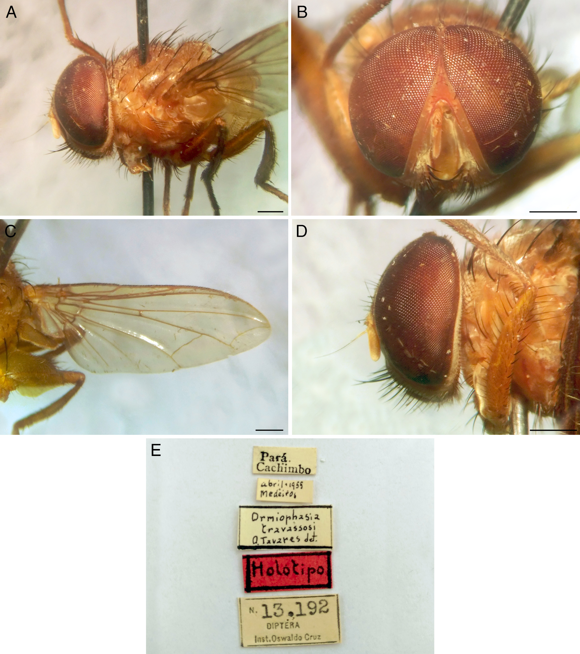
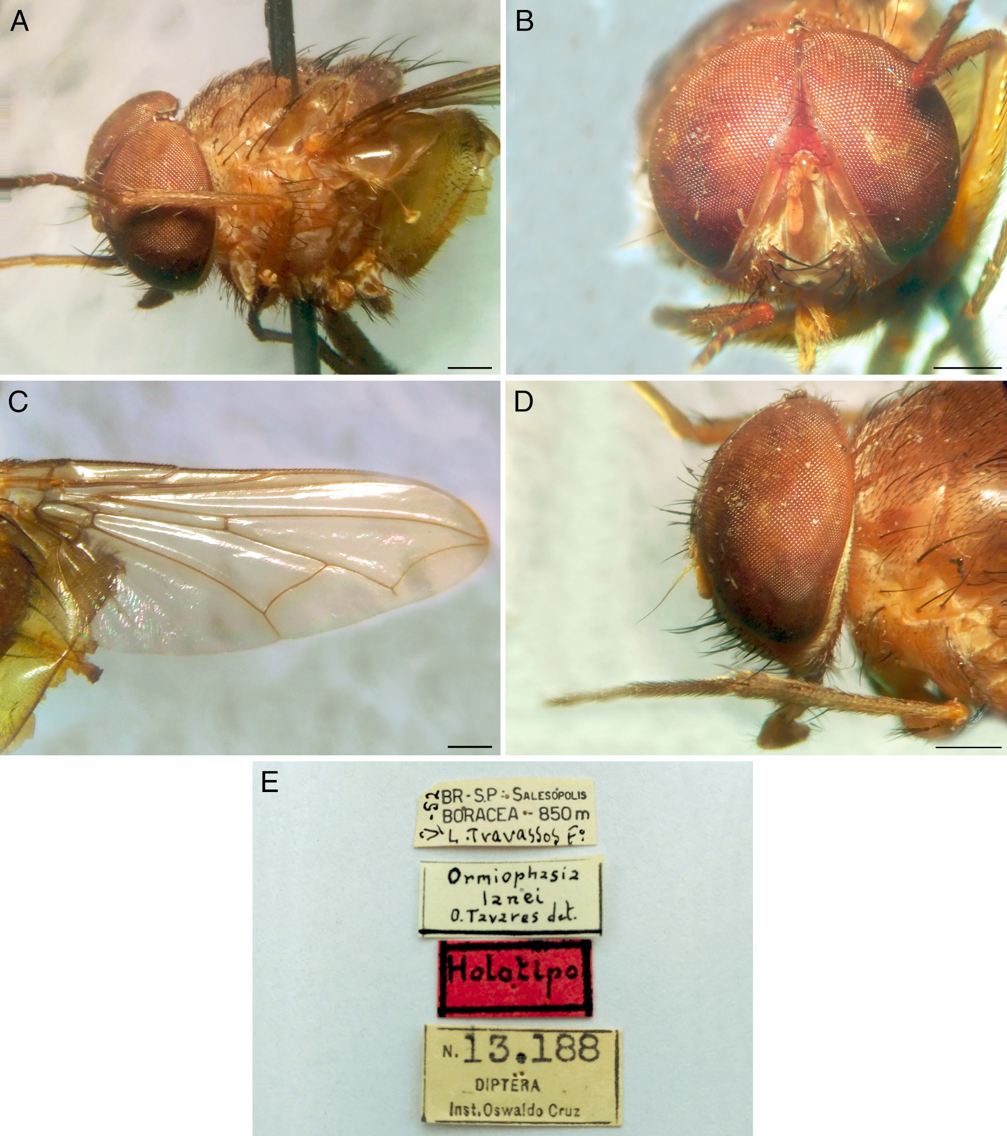
8 Clypeus darker than frontoclypeal membrane ( Figs 5F View FIGURE 5 , 7F View FIGURE 7 ); female posterior spiracle with posterior lappet shaped as an operculum; surstylus strongly inflected inward in posterior view, outer posterior surface covered with strong setae in upper two-thirds ( Fig. 39C View FIGURE 39 ); apex of male cerci subquadrate and abruptly constricted in posterior view (Southeast Brazil)... O. lanei Tavares View in CoL
- Clypeus of same color as frontoclypeal membrane ( Fig. 5D View FIGURE 5 ); female posterior spiracle with posterior lappet plume-shaped; surstylus not inflected inward as above, covered with weak setae in upper two-thirds ( Fig. 39A View FIGURE 39 ); apex of male cerci subquadrate and gradually constricted in posterior view (South and Southeast Brazil, Panama and Paraguay).......... O. cruzi Tavares View in CoL
9 Clypeus darker than frontoclypeal membrane ( Fig. 6E View FIGURE 6 ); thorax and abdomen dark brown........................... 10
- Clypeus of same color as frontoclypeal membrane ( Fig. 5A View FIGURE 5 ); thorax and abdomen brown or brownish-yellow ( O. guimaraesi View in CoL sp. nov. has a dark brown scutum, but the rest of the thorax is brown, see Figs 10A View FIGURE 10 , 12A View FIGURE 12 )........................... 12
10 Thorax and abdomen entirely dark brown ( Figs 10E View FIGURE 10 , 14D View FIGURE 14 ); anteroventral epandrial process extending well beyond ventral epandrial margin ( Fig. 41C View FIGURE 41 ); dorsal surface of epandrium with posterior margin at same level as anterior margin ( Fig. 41C View FIGURE 41 , lateral view); apex of male cerci about 1/2 width of cerci in posterior view, abruptly constricted ( Fig. 41C View FIGURE 41 ) ( Colombia, Costa Rica, Panama and Venezuela)................................................................. O. tavaresi View in CoL sp. nov.
- Thorax and abdomen dark brown, with postpronotal lobe brown ( Figs 9B View FIGURE 9 , 13B View FIGURE 13 ); anteroventral epandrial process continuous with ventral epandrial margin ( Fig. 42A View FIGURE 42 ); dorsal surface of epandrium with posterior margin higher than anterior margin ( Fig. 42A View FIGURE 42 , lateral view); apex of male cerci 1/3 ( Fig. 38B View FIGURE 38 ) or more than 1/2 width of cerci in posterior view ( Fig. 42A View FIGURE 42 ), gradually constricted.......................................................................................... 11
11 Arista weakly plumose ( Fig. 17 View FIGURE 17 A–B); apex of male cerci narrow, 1/3 width of cerci and rounded in posterior view ( Fig. 38B View FIGURE 38 ) (North Brazil, Guyana, Peru, Trinidad and Tobago and Venezuela)............................... O. causeyi Tavares View in CoL
- Arista bare ( Fig. 36 View FIGURE 36 A–B); apex of male cerci broad, more than 1/2 width of cerci and subquadrate in posterior view ( Fig. 42A View FIGURE 42 ) ( Costa Rica and Mexico)............................................................... O. chapulini View in CoL sp. nov.
12 Thorax with scutum dark brown and lateral surface brown ( Figs 10A View FIGURE 10 , 12A View FIGURE 12 ); apex of male cerci broad, subrectangular in posterior view ( Fig. 40B View FIGURE 40 ); surstylus stout in posterior view, with outer posterior surface densely covered with strong setae and inner posterior surface covered with short setae medially ( Colombia and Costa Rica)................... O. guimaraesi View in CoL sp. nov.
- Thorax entirely brown or brownish-yellow; apex of male cerci broad or narrow, rounded in posterior view; surstylus not as stout as above ( Figs 38A, 38C View FIGURE 38 , 39B View FIGURE 39 ), with inner posterior surface bare.............................................. 13
13 Thorax and abdomen entirely brown..................................................................... 14
- Thorax and abdomen entirely brownish-yellow............................................................. 15
14 Ocellar triangle bare, without setulae ( Fig. 15 View FIGURE 15 C–D); head yellow-pruinose ( Figs 5A View FIGURE 5 , 7A View FIGURE 7 ) ( Panama).... O. busckii Townsend View in CoL
- Ocellar triangle setulose ( Fig. 30A View FIGURE 30 ); head silver-pruinose ( Fig. 5H View FIGURE 5 ) ( Argentina)..................... O. obscura (Séguy)
15 Female head subtrapezoidal in frontal view, with gena about 0.15 times length of head ( Figs 7E View FIGURE 7 , 23B View FIGURE 23 ); apex of male cerci broad ( Fig. 39B View FIGURE 39 ), 3/5 width of cerci and rounded in posterior view, and gradually constricted ( North Brazil, French Guiana, Trinidad and Tobago and Venezuela)............................................................... O. inflata (Séguy) View in CoL
- Female head elliptic in frontal view, with gena about 0.12 times length of head ( Figs 7C View FIGURE 7 , 19B View FIGURE 19 ); apex of male cerci narrow ( Fig. 38C View FIGURE 38 ), 1/3 width of cerci and rounded in posterior view, and abruptly constricted (North Brazil, Colombia, Ecuador, Guyana, Peru, Suriname and Venezuela)......................................................... O. costalimai Tavares View in CoL


No known copyright restrictions apply. See Agosti, D., Egloff, W., 2009. Taxonomic information exchange and copyright: the Plazi approach. BMC Research Notes 2009, 2:53 for further explanation.
|
Kingdom |
|
|
Phylum |
|
|
Class |
|
|
Order |
|
|
Family |
Ormiophasia Townsend, 1919
| Gudin, Filipe M. & Nihei, Silvio S. 2019 |
Pseudormia Séguy, 1927b: 262
| Tavares, O. 1964: 38 |
| Sabrosky, C. W. 1953: 181 |
| Townsend, C. H. T. 1931: 82 |
| Seguy, E. 1927: 262 |
| Seguy, E. 1927: 424 |
| Seguy, E. 1926: 5 |
| Seguy, E. 1926: 20 |
| Seguy, E. 1925: 375 |
Ormiophasia
| Townsend, C. H. T. 1919: 164 |