
Phocoenidae, Boessenecker, 2013
|
publication ID |
https://doi.org/ 10.5252/g2013n4a5 |
|
DOI |
https://doi.org/10.5281/zenodo.4818521 |
|
persistent identifier |
https://treatment.plazi.org/id/03E7DD69-FF9C-860C-2794-786AFB003FB0 |
|
treatment provided by |
Felipe |
|
scientific name |
Phocoenidae |
| status |
|
Phocoenidae View in CoL unnamed genus 2
Odontoceti View in CoL incertae sedis – Barnes 1973: 37-42.
? Phocoenidae View in CoL – Barnes 1977: 331, table 5.
Phocoenidae View in CoL new genus – Racicot et al. 2007: 132 A.
REFERRED MATERIAL. — UCMP 219503, nearly complete skull collected by R.W. Boessenecker and C. Argento from UCMP locality V99854 View Materials ; and UCMP 219076, a pair of fused mandibles from UCMP locality V99859 View Materials .
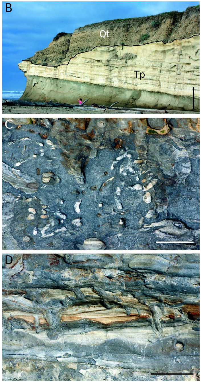
STRATIGRAPHIC OCCURRENCE. — Middle and upper parts of the San Gregorio section of the Purisima Formation, Early to Late Pliocene (c. 5-2.5 Ma; Zanclean-Gelasian equivalent; Fig. 2 View FIG ).
DESCRIPTION
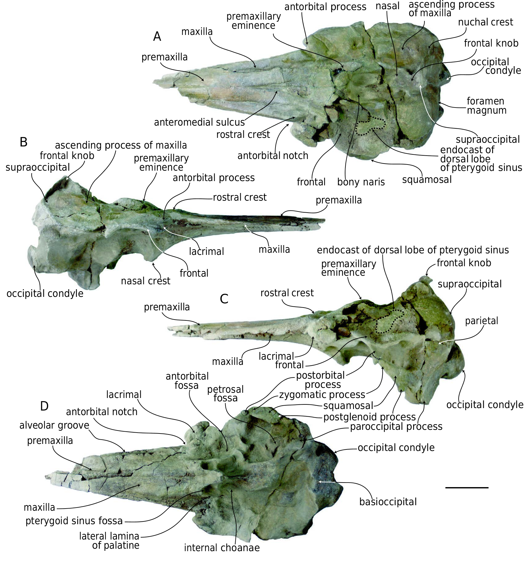
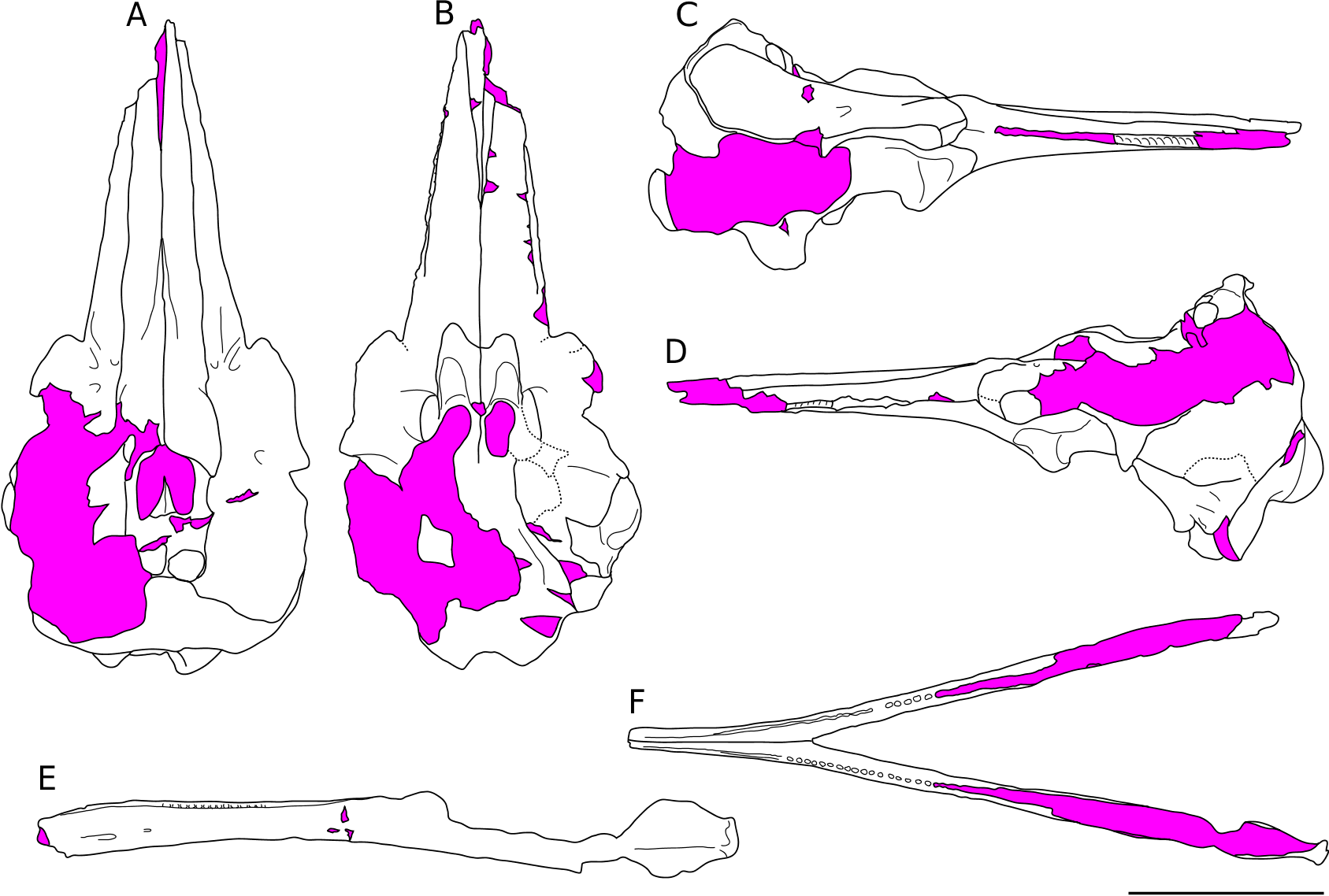
UCMP 219503 is a small ( Table 10 View TABLE ) partial skull missing part of the apex of the rostrum, most of the basicranium including both petrotympanics and the right squamosal, and the pterygoids ( Fig. 42 View FIG ). Ŋe dorsal surface is damaged and the left supraorbital and ascending processes of the maxilla are broken away, and the left premaxillary eminence is damaged. Ŋe rostrum is relatively long, triangular, dorsoventrally compressed, and the dorsal surface is slightly transversely convex. Ŋe rostral portion of the premaxilla is long and dorsally flat with nearly parallel medial and lateral margins. Posteriorly, the premaxilla is wider than the exposed rostral portion of the maxilla ( Fig. 42A View FIG ). Ŋe anteromedial sulcus is elongate and extends anteriorly to the midpoint of the rostrum, defining the acute prenarial triangle. Both premaxillary foramina are obscured by matrix, but are positioned anterior to the premaxillary eminence.Ŋe premaxillary eminence is convex and dorsally elevated; in dorsal aspect, the lateral margins of the eminences are slightly convex ( Fig. 42 View FIG A-C). Ŋe eminence is dorsally flat and overhangs laterally; the right eminence is transversely wider and slightly anteroposteriorly longer than the left. Ŋe ascending process of the premaxilla is reduced to a small conical process that slightly overlaps the maxilla posterior to the premaxillary eminence. Ŋe premaxilla-maxilla suture runs along the base of the eminences.
Ŋe rostral portion of the maxilla has a narrow dorsal exposure, and it tapers anteriorly ( Fig. 42A View FIG ). Posteriorly, the rostral portion of the maxilla is raised and faces dorsomedially. A distinct anterolaterally oriented rostral crest (sensu Mead & Fordyce 2009) is present medial to the antorbital notch on the dorsal surface of the maxilla, and anteriorly forms a small tubercle. Posteriorly adjacent to this crest is the anteriormost dorsal infraorbital foramen. Laterally adjacent to the rostral crest is a posteromedially oriented furrow that is confluent with the antorbital notch; medial to the rostral crest, an elongate shallow longitudinal trough is positioned along the medial margin of the maxilla. Ŋe antorbital process is large and knoblike, and composed of the maxilla, frontal, and the lacrimal ( Fig. 42A, B View FIG ). Posteromedially, the maxilla forms part of the margin of the bony naris; a slight sulcus runs parallel to this margin, which demarcates a narrow cylindrical ridge running posteriorly from the tip of the premaxilla and along the lateral side of the bony naris. Adjacent to the bony nares, the maxilla is dorsally concave where it forms a transversely narrow fossa for the inferior vestibule, whereas it is convex further posteriorly. Posteriorly, the dorsal surface of the maxilla is bordered by the frontal and posteromedially by the mesethmoid and nasals. Where the left ascending maxilla is broken away, a reniform, anterolaterally oriented matrix cast of the dorsal lobe of the pterygoid sinus occurs on the dorsal side of the frontal bone and lateral to the bony nares, and medial to the postorbital process of the frontal. In life, this sinus would have been situated between the frontal and ascending process of the maxilla. Ŋe palate is smooth and flat anteriorly, and transversely convex posteriorly ( Fig. 42D View FIG ). Adjacent to the palatine, the maxilla is transversely concave. Poorly preserved alveolar grooves are present; well-preserved alveoli are only present on the right maxilla, along the middle of the preserved portion of the rostrum.
Ŋe right nasal is damaged, and the left nasal is large, inflated, and sub-spherical; both nasals are positioned on the anterior surface of the frontal knob ( Fig. 42A, C View FIG ). Much of the left frontal is visible where the left supraorbital and ascending processes of the maxilla have broken away. Ŋe lateral margin of the frontal (and facial region of the skull) forms a slight corner in dorsal view at the position of the postorbital process. Ŋe ventrally projecting postorbital process, better preserved on the left side, is narrow and triangular in lateral aspect. Posteriorly, the suture between the frontal and the supraoccipital along the nuchal crest is difficult to detect and appears fused. Ŋe left nuchal crest is eroded away; the right nuchal crest is tall, anteroposteriorly thin, highest adjacent to the frontal knob, and decreases in height laterally ( Fig. 42A, B View FIG ). Posterior to the vertex, the nuchal crest forms a posteriorly directed and sharp crest. Ŋere is a deep fossa in the right side of the frontal knob, where it meets the nuchal crest; this feature is damaged on the left side. Ŋe frontal knob is tall and dorsally prominent; the nasal-frontal sutures are clear, but it is unclear whether the interparietal is exposed or not. Ŋe frontoparietal suture in the region of the temporal fossa is indistinct. Ŋe parietal is gently convex and exposed within the temporal fossa. Ŋe temporal fossa is proportionally long but dorsoventrally low in height, triangular, and is delineated dorsally by the frontal, anteriorly by the postorbital process, ventrally by the zygomatic process, and posteriorly by the temporal crest.
Ŋere are large (and presumably natural) fenestrae in the supraoccipital dorsolateral to the occipital condyles; these appear to have been enlarged due to breakage. A median furrow rises from the foramen magnum to the vertex. A small, posteriorly overhanging crest is present on the posterior side of the frontal knob. Ŋe occipital condyles are convex, D-shaped, and not defined by a prominent edge or neck. Ŋe foramen magnum is oval shaped, and narrower ventrally. Ŋe jugular notch is a slight incision between the paroccipital process of the exoccipital and the basioccipital crest. Only the posteriormost portion of the flat basioccipital and left basioccipital crest are preserved; the basisphenoid is lost.
Ŋe zygomatic process of the squamosal is triangular in lateral view, anterodorsally directed, is dorsally flat, and bears a wide and shallow mandibular fossa ventrally; a shallow tympanosquamosal recess is present medially. Ŋe postglenoid process forms a distinct posterior margin of the fossa, and the postglenoid notch extends dorsal to the postglenoid process, distinguishing it from the rest of the squamosal. Ŋe postglenoid process is knob-like. Ŋe supramastoid crest is dorsally concave and sharp.
Part of the vomerine crest is exposed in ventral view ( Fig. 42D View FIG ); it merges dorsally with the trans-
versely thin nasal septum; anteriorly, the nasal septum converges with the well-developed, conical, and ventrally-directed ventral tubercle. Dorsally, there is a well-developed median ridge along the mesethmoid in the region of the bony nares. Both palatines are poorly preserved, but the lateral laminae are transversely thin and nearly parallel, and delineate transversely narrow fossae for the hamular lobes of the pterygoid sinus anterior to the choanae. Ŋe maxillo-palatine suture is anterolaterally convex.
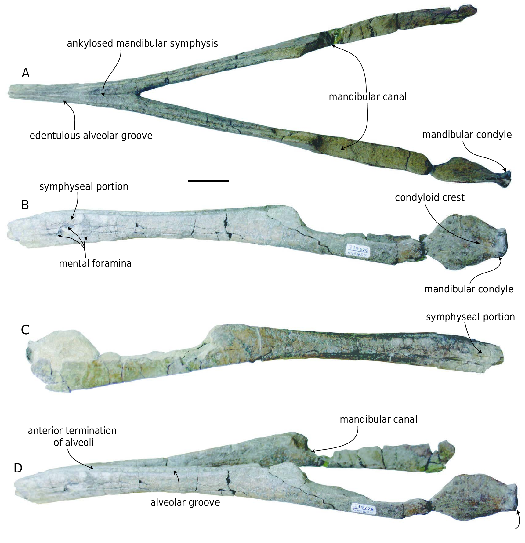
Ŋe mandibles (UCMP 219076) are fused at the symphysis ( Figs43-45 View FIG View FIG View FIG ; Table 13 View TABLE ). Ŋe anterior extremity of the symphyseal portion is missing, as is the right mandibular condyle, and the dorsal margin of both coronoid crests ( Fig.43D View FIG ). Anterior to the left condyle, the ventral margin is damaged. Ŋe mandibular condyle is oval-shaped and faces posteriorly. Ŋe medial surface of the condyle is hollowed out as part of the large (but incompletely preserved) mandibular foramen. Ŋe medial part of the condyle is a dorsoventrally oriented, bladelike process. Ŋe condyloid crest occurs as a slight ridge on the lateral face. On the better preserved left side, approximately 25 alveoli are preserved ( Fig. 43A, D View FIG ), although this is a minimum number as many interalveolar septa are missing. Ŋe toothrows are slightly laterally concave in dorsal aspect, and in lateral aspect the toothrow is slightly concave dorsally. In dorsal aspect, the toothrows are widely divergent posterior to the symphysis, but at the elongate symphysis, the toothrows are near parallel. Ŋe alveoli terminate within the alveolar groove 2 cm anterior to the posterior end of the symphyseal portion. From that level, the alveolar groove transforms into a thin, but deep sulcus; the left and right sulci are separated medially along the symphyseal portion by a high, transversely narrow median bony ridge( Fig. 43A, D View FIG ). Ŋe ventral margin of the ramus is ventrally concave in lateral aspect; the horizontal ramus is dorsoventrally shallowest just posterior to the mandibular symphysis, and it deepens posteriorly and anteriorly toward the symphysis. Ŋe symphyseal portion of the mandible is elongate and transversely narrow with flat lateral surfaces. It tapers dorsoventrally toward the anterior tip in lateral view, and is slightly downturned ( Figs 43B, C View FIG ; 44E View FIG ; 45D View FIG ). Several mental foramina occur on the lateral surface of the symphyseal portion. Dorsally, a flat triangular surface is present between the alveolar grooves along the posterior portion of the symphysis; a thin median groove is visible here, and a deep, wide groove is present on the posteroventral margin of the symphysis. Ŋe rami diverge at with an angle of 24°.
REMARKS AND COMPARISONS
Ŋe skull exhibits several phocoenid synapomorphies, including the presence of a premaxillary eminence, ascending process of the premaxilla that terminates anterior to the nasal, a frontal knob, and a welldeveloped dorsal lobe of the pterygoid sinus fossa ( Figs 42-45 View FIG View FIG View FIG View FIG ; Barnes 1985a; Muizon 1988a; Lambert 2008a; Murakami et al. 2012a, b). Ŋe skull and mandible differ from an undescribed broad-headed phocoenid from the San Diego Formation (UCMP 38340) in exhibiting a more elongate and attenuate rostrum with a distinct rostral crest medial to the antorbital notch, a much narrower antorbital notch and less prominent antorbital process, and mandibles that are fused at the symphysis and edentulous anteriorly. Ŋese two specimens from the Purisima Formation share several bizarre features with an undescribed phocoenid from the San Diego Formation preliminarily introduced by Racicot et al. (2007) including a fused, transversely flattened and edentulous symphyseal region of the mandible, a large, hypertrophied premaxillary eminence, and a knoblike rostral crest on the maxilla ( Figs 42-45 View FIG View FIG View FIG View FIG ). Ŋese features distinguish the Purisima and San Diego Formation material from all other fossil and
mandibular condyle
modern phocoenids. UCMP 219503 further differs from extant phocoenids in having a premaxilla wider than the maxilla at the base of the rostrum, having asymmetrical premaxillae, and a transversely convex palate. Ŋese specimens (UCMP 219503, 219076) appear to represent a taxon congeneric or conspecific with the undescribed porpoise from the San Diego Formation Racicot et al. (2007). More complete material of this taxon from the San Diego Formation includes a partial skeleton with skull and mandibles, partial postcranial skeleton, additional mandibles, partial crania, and petrosals (SDNHM 22452, 23194, 24710, 64742, 65276, 83724). Only a preliminary description of the Purisima Formation specimens is presented here owing to the ongoing work by Racicot and colleagues. Ŋe bizarre symphyseal portion of the mandible of this taxon is unique among phocoenids, although it is important to note that Lomacetus ginsburgi Muizon, 1986 exhibits a mandible with an elongate (but unfused) mandibular symphysis ( Muizon 1988b; Fig.43B, C View FIG ). Furthermore, Muizon (1988a) identified strong prognathism of the mandibles as a uniting feature of Phocoenoides dalli True, 1885 , Phocoena dioptrica Lahille, 1912 , Salumiphocoena stocktoni (Wilson, 1973), and Piscolithax Muizon, 1983 . Additional material referable to this taxon from the Santa Cruz section of the Purisima Formation includes partial rostra (UCMP 219483, 219582) and several isolated petrosals (UCMP 137472, 219484), based on comparison with the material from the San Diego Formation. Isolated petrosals of this taxon (UCMP 88582 and 88583) from the San Diego Formation were identified and figured by Barnes (1973) as Odontoceti incertae sedis.

No known copyright restrictions apply. See Agosti, D., Egloff, W., 2009. Taxonomic information exchange and copyright: the Plazi approach. BMC Research Notes 2009, 2:53 for further explanation.
|
Kingdom |
|
|
Phylum |
|
|
Class |
|
|
Order |
|
|
SubOrder |
Odontoceti |
|
Family |
Phocoenidae
| Boessenecker, Robert W. 2013 |
Phocoenidae
| RACICOT R. & DEMERE T. A. & ROWE T. 2007: 132 |
Phocoenidae
| BARNES L. G. 1977: 331 |
Odontoceti
| BARNES L. G. 1973: 37 |