
Trapania safracornia, Fahey, Shireen J., 2004
|
publication ID |
https://doi.org/10.5281/zenodo.157364 |
|
DOI |
https://doi.org/10.5281/zenodo.5629148 |
|
persistent identifier |
https://treatment.plazi.org/id/03E787D2-FFB0-FFCE-FE98-FA016D90FD30 |
|
treatment provided by |
Plazi |
|
scientific name |
Trapania safracornia |
| status |
sp. nov. |
Trapania safracornia View in CoL sp. nov.
( Figures 1–4 View FIGURE 1 View FIGURE 2 View FIGURE 3 View FIGURE 4 )
Type material. Holotype: CASIZ 156067. North side, Rottnest Island, Western Australia, Australia, 32.00S, 115.30E, collected by G. Gunness, 12 April 2001, 28 m.
Paratype: CASIZ 162641. From type locality.
Additional specimens not collected: Two specimens, from type locality, photographed by G. Gunness, February 2004.
Distribution. This species is known only from the type locality.
Etymology. The specific name safracornia is taken from the Arabic safra meaning yellow and the Latin cornus meaning horn. The name refers to the yellow processes on this new species.
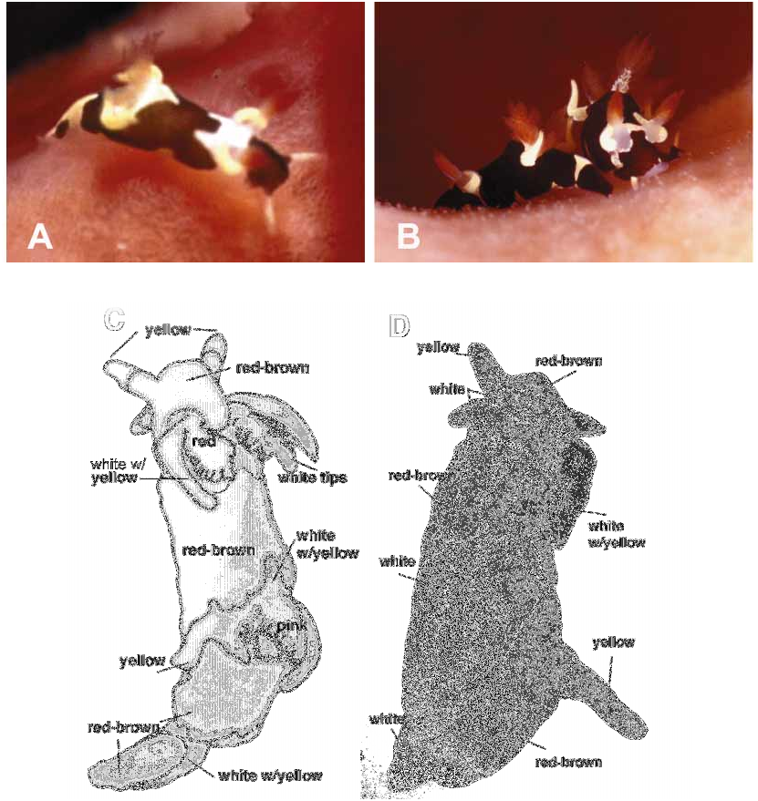
External Morphology. Living animals 7 and 8 mm in length. Body shape soft, elongate; plump midsection ( Figs 1 View FIGURE 1 A, B). No distinct mantle edge. Elongate processes next to each rhinophore and on each side of the gill, curving upwards, towards posterior of animal. Gill and rhinophores not retractile, no rhinophore or gill pockets. Gill composed of three bi or tripinnate branchial leaves. Anus situated within circle formed by gill branches. Long rhinophores with 9–10 lamellae on posterior side. Two long tapered oral tentacles, anterior edge of foot extends to elongate tapered corners and curved leading edge ( Fig 1 View FIGURE 1 C). Genital aperture located on right side of body in anterior third.
Brown background color of living animal with symmetrical white patches between rhinophores, at gill, tail tip and posterior third of dorsum ( Fig 1 View FIGURE 1 C). White patch at gill saddleshaped and patch at rhinophores extends only posteriorly in two points. No white spots in addition to large white patches on dorsum. Irregularly shaped overlay of yellow pigment on white patch at posterior third of dorsum and on tail tip. Yellow pigment covers white ground color of elongate processes next to gill and rhinophores. White ring around rhinophoral apertures. Translucent rhinophore stalks with red club and white tips. Translucent gill branches with pinkishtan apex. No distinguishing color on axes. Ventral side with long white patch extending length of animal, three symmetrical extensions of white color up body sides ( Fig 1 View FIGURE 1 D). White oral tentacles, foot corner extensions. Redbrown anterior of foot.
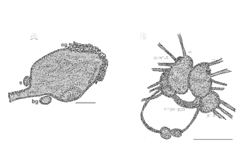
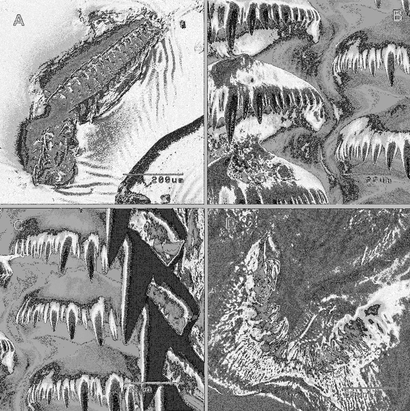
Internal Anatomy. Oval buccal bulb ( Fig 2 View FIGURE 2 A), small buccal pump and not as prominent as compared to other Trapania species ( Rudman 1987). Tiny seedshaped oral glands around the mouth. Radular formula 21 x 1.0. 1 in both specimens (CASIZ156067, 162641). Teeth increase in size from oldest to newest ( Fig 3 View FIGURE 3 A). Rachidian teeth absent. One long cusp on outer edge of each tooth with multiple large denticles (10–14) that vary in size, smallest located near inner edge ( Fig 3 View FIGURE 3 B). Largest of main denticles always second or third from main cusp. In between largest denticles 1–2 minute denticles, up to eight per tooth. Largest cusp at outer edge of tooth with one small broadly triangular denticle at base of outer edge ( Fig 3 View FIGURE 3 C). Jaw, a thickened plate with two rows of pointed rodlets around open edge ( Fig 3 View FIGURE 3 D).
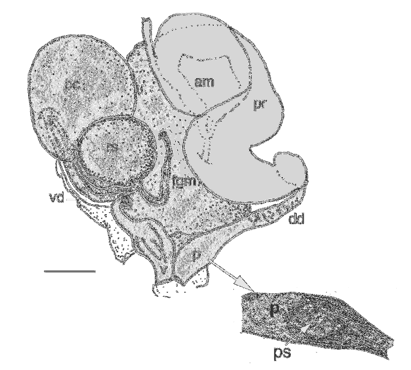
Ampulla large, nearly round ( Fig 4 View FIGURE 4 ), branches into oviduct and prostate. Hermaphroditic duct enters ampulla terminally. Thin oviduct enters large female gland mass. Thick tubular prostate folds once after exiting ampulla, narrows into short, muscular deferent duct. Deferent duct widens into elongate penis. Sparse, small spines in distal portion of penis. Vaginal duct twice as long as deferent duct, lacks spines. Vaginal duct enters large, round bursa copulatrix at proximal end. Distal end of vaginal duct widens into bulbous vagina, which has longitudinal folds. From bursa copulatrix, separate oviduct connects to large, round receptaculum seminis. Bursa copulatrix approximately two times as large as receptaculum seminis. Short uterine duct from receptaculum seminis to female gland mass.
Central nervous system with fused cerebral and pleural ganglia ( Fig. 2 View FIGURE 2 B). Eyes sessile on cerebral ganglia. Two equal sized pedal ganglia behind and slightly below cerebropleural complex, joined by short commissure. Three prominent nerves leading from pedal ganglia and four nerves including the rhinophoral nerve, originate from cerebropleural ganglia. Two buccal ganglia positioned ventral to esophagus.
No known copyright restrictions apply. See Agosti, D., Egloff, W., 2009. Taxonomic information exchange and copyright: the Plazi approach. BMC Research Notes 2009, 2:53 for further explanation.
|
Kingdom |
|
|
Phylum |
|
|
Class |
|
|
Order |
|
|
Family |
|
|
Genus |