
Diplura lineata ( Lucas, 1857 )
|
publication ID |
https://doi.org/ 10.5852/ejt.2016.210 |
|
publication LSID |
lsid:zoobank.org:pub:E239C584-B41B-4AC7-89E1-E3D8A2EB921A |
|
DOI |
https://doi.org/10.5281/zenodo.5541658 |
|
persistent identifier |
https://treatment.plazi.org/id/03E7878E-437B-FFB7-BE72-E770FAC3FE75 |
|
treatment provided by |
Plazi |
|
scientific name |
Diplura lineata ( Lucas, 1857 ) |
| status |
|
Diplura lineata ( Lucas, 1857) View in CoL
Figs 14–26 View Figs 14 – 25 View Fig. 26
Mygale lineata Lucas, 1857: 14 , fig. 1a–c (♂).
Thalerothele fasciata Bertkau, 1880: 24 , pl. 1, f. 6 (♀) syn. nov.! Harmonicon nigridorsi Mello-Leitão, 1924: 186 (♀) syn. nov.!
Diplura fasciata View in CoL – Simon 1889: 183.
Thalerothele fasciata – Simon 1903: 963. — Mello-Leitão 1923: 101, f. 10. Thalerothele lineata – Mello-Leitão 1926: 309.
Harmonicon nigridorsi – Mello-Leitão 1926: 315, fig. 4. — Bücherl et al. 1971: 122, figs 15–16 (♂). Paraharmonicon nigridorsi – Mello-Leitão 1926: 316 (tentative superfluous name only).
Type material
Mygale lineata : BRAZIL, Rio de Janeiro: Rio de Janeiro, [no date], near Rio [“environs de Rio”, probably Tijuca ] (♂ holotype, MNHN, not located) .
Thalerothele fasciata : BRAZIL, Rio de Janeiro, Tijuca [no date], van Beneden leg. (♀ holotype, IRSNB, not located) .
Harmonicon nigridorsi : BRAZIL, Rio de Janeiro, [no date], W.S. Bristowe leg. (♀ holotype + ♂ probably mixed later, MNRJ 17 , MLPC 847 , examined) .
Material Examined
BRASIL: 2♂♂, 1 ♀, 1 juv., Rio de Janeiro, Casimiro de Abreu: Barra de São João, Morro de São João , 21–24 Mar. 2003, Expedition Arachné ( MNRJ 4322 ) ; 1 ♀, 24 Mar. 2003, Expedition Arachné ( MNRJ 4335 ) ; Mangaratiba: 1 ♂, 1 ♀, Ilha de Itacuruçá, Águas Lindas , 24–25 Mar. 2007, R.L.C. Baptista, C.S. Costa & A.R. Oliveira leg. ( UFRJ 0332 ) ; 1 juv., Reserva Ecológica Rio das Pedras , 11– 12 Nov. 2004, A.P.L. Giupponi leg. ( MNRJ 4312 ) ; 1 juv., Nova Iguaçu: Parque Municipal de Nova Iguaçu , 21 Jul. 2004, C. Lima leg. ( MNRJ) ; 1 ♂, PM Nova Iguaçu, ( MNRJ 4311 ) ; 1 ♀, Rio de Janeiro, Parque Estadual da Pedra Branca , ( MNRJ 4310 ) ; 1 ♀, Camorim, Açude , 31 Mar. 2014, P. Castanheira leg. ( MNRJ 6820 ) ; 1 ♂, Serra do Mendanha, 485 m, Pitfall, 12 Dec. 2008, J.A.L. Pontes leg. ( MNRJ 18434 ) ; 1 ♀, Floresta da Tijuca , 20 Apr. 1986, R.L.C. Baptista leg. ( MNRJ 3112 [CRB T005]) ; 1 ♀, 1 juv., Floresta da Tijuca , 400 m, 27 Nov. 1987, A.P.L. Giupponi leg. ( MNRJ 3410 ) ; 1 ♀, Floresta da Tijuca , 7 Jan. 1998, A.P.L. Giupponi leg. ( MNRJ 1854 ) ; 1 ♀, Floresta da Tijuca , 400 m, 3 Mar. 2001, A.P.L. Giupponi, D.R. Pedroso & D.F. Almeida leg. ( MNRJ 1856 ) ; 1 ♂, Parque Nacional da Tijuca , 25 May 2001, A.P.L. Giupponi, D.R. Pedroso & R.L.C. Baptista leg. ( MNRJ 3568 ) ; 1 ♀, Parque Nacional da Tijuca , 10 Jan. 2005, D.R. Pedroso & R.L.C. Baptista leg. ( MNRJ 4302 [ AER]) ; 1 ♀, Parque Nacional da Tijuca , 25 Aug. 2004, D.R. Pedroso & A.P.L. Giupponi leg. ( MNRJ 4308 ) ; 1 ♀, Parque Nacional da Tijuca , Archer , 22 Jan. 2005, D.R. Pedroso & A.P.L. Giupponi leg. ( MNRJ 4309 ) ; 1 juv., Parque Nacional da Tijuca , Gávea , 18 Jan. 2005, D.R. Pedroso leg. ( MNRJ 4307 [AER]) ; 2 ♀♀, Parque Nacional da Tijuca , Gávea , 18 Jan. 2005, D.R. Pedroso leg. ( MNRJ 4304 ) ; 2 ♀♀, Parque Nacional da Tijuca , Pai Ricardo , 21 Jan. 2005 D.R. Pedroso leg. ( MNRJ 4305 [AER]) ; 1 ♀, Parque Nacional da Tijuca , Pai Ricardo , 21 Jan. 2005, D.R. Pedroso leg. ( MNRJ 4306 [AER]) ; 1 ♀, Parque Nacional da Tijuca, Pai Ricardo , 21 Jan. 2005, D.R. Pedroso leg. ( MNRJ [AER]) ; 1 ♀, Parque Nacional da Tijuca, Sumaré , 22 Jan. 2005, D.R. Pedroso leg. ( MNRJ 4303 [AER]) .
Diagnosis
Both sexes of this species have a characteristic color pattern, similar to the Amazonian D. sanguinea (F.O. Pickard-Cambridge, 1896) . The abdomen of both species have a dark brown dorsum bearing beige broad transversal stripes. In D. lineata , the stripes are short and broad, with irregular outline, covering only the side margins of the dorsum. At the sides, there are several small beige spots among and under the stripes (sometimes fused with them). On the other hand, D. sanguinea have longer and a bit thinner stripes, without connecting beige spots at the sides. The copulatory bulb is very similar in both species, but the spermatic duct is very constricted at the basis of the embolus, becoming almost filiform afterwards, in D. lineata ( Fig. 20 View Figs 14 – 25 ), while in D. sanguinea the constriction is small, and the basis of embolus harbors a large duct, which tapers regularly towards the apex. The spermathecae in D. lineata ( Fig. 25 View Figs 14 – 25 ) have a thick stem and three distal lobules, while D. sanguinea have a thin stem and six to seven distal lobules.
Description
Male (MNRJ 4311)
Total length 18.5. Carapace: 7.4 long, 6.0 wide, chelicerae 2.7. Abdomen: 7.6 long, 3.9 wide. Spinnerets: PMS 0.9 long; PLS, total length 10.0, basal article 3.3, middle 3.3, distal 3.3, respectively. Legs: see Table 3 View Table 3 . Carapace: length/width 1.2; flat, cephalic area slightly raised, thoracic furrows shallow and wide. Fovea: short, deep, slightly recurved. Carapace covered with short, thin setae, interspersed with some longer and thicker setae; border with abundant long and thick setae pointing outwards, increasing in number towards posterior angles. Clypeus narrow and small, but clearly visible, not totally hidden by the eye tubercle, frontal margin bearing 6 thick, long, erect setae. Eye tubercle: 0.5 long, 1.1 wide, one thick, long seta on tubercle anterior margin, area between posterior eyes covered with thin setae and bearing 3 thicker, longer setae. AME 0.3, almost spherical, but a bit longer than wide, set apart by half its diameter.ALE elliptical, much longer than wide, just a bit longer than the AME diameter. PME small, with flattened lens, longer than wide, its length around 0.6× AME diameter. PLE elliptical, much longer than wide, its length a bit less than 0.9× AME diameter. PME and PLE closely spaced by around 0.2× the PME length. Anterior eye row slightly recurved, posterior eye row recurved. Eye rows with similar width. Chelicerae: formed by 11 teeth on promargin on both left and right chelicera. Plectrum with 4 thick, long setae. Labium: length/width 0.8, no cuspules. Labio-sternal groove deep, with elongated sigilla. Sternal groove deep, with elongated sigilla. Sternum slightly longer than wide. Posterior angle in a blunt point, not separating coxae IV. Sigilla: three pairs, elliptical, increasing in size from anterior to posterior, all near margin. Palp ( Fig. 17 View Figs 14 – 25 ): relatively short, femur: d1–3–0, pl0–0–1, rl0–0–1, tibia pl 0–2–0, v0. Tibia: 3.1 long, 1.0 wide, short, incrassated, thinner at the basis and apex. Maxillae: length/ width 1.5. Cuspules: 15 spread over ventral inner heel. Lyra: located at the ventral side of the maxilla, formed by 6 modified thick setae, the basalmost much thinner and shorter than the others, which slightly increase in size from basis to apex of the lyra, all setae weakly curved at apical portion, with rounded apex. Legs ( Figs 15–16 View Figs 14 – 25 ): Leg formula 4123. Legs covered with short, thin, horizontal black setae and with some longer, thicker, erect black setae. All tarsi with thin scopula, throughout the length of the article. Tarsi I–III with scopula almost undivided, with only some isolated thicker setae at the middle line of the basal third of the ventral face. Tarsi IV with scopula partially divided, with thicker setae covering the basal half of the ventral face. Metatarsi I–II almost without scopula, with only a few setae near the apex. Metatarsi III–IV without scopula. All tarsi provided with numerous small cracks covering almost all the ventral and lateral faces, except by the basis and tip of the article. Tibia I around 4.3× longer than wide. Retrolateral distal spur placed at the ventral corner of tibia I, with a wide-base, bearing at its apex a pointed and almost erect megaspine, with similar length of the spur. Metatarsus I relatively long and a little sinuous on ventral view, with a protruding retrolateral tubercle, placed ventrally on its basal third, faced towards the apex of the article. Ventral side with an apical spine and two spines on the median third, the basalmost located in advance to the tubercle. Fringe formed by many spiniform setae and spines (clasper) covering the median portion of the prolateral side of metatarsus I ( Fig. 16 View Figs 14 – 25 ). Spines: leg I: femur d1–2–1, pld0–2–1, rld1–1–1 left, rld0–1–2 right; patella 0 left, 0–0–1 right; tibia p0–1–1, v0–1–1ap (apophysis); metatarsus p0–1–0, v0–2–1ap; leg II: femur d1–2–1 left, d1–2–0 right, pld0– 2–1, rld0–2–0 left, rld0–2–1 right; patella 0–1–1 left, 0–0–1 right; tibia p0–1–1, v1–1 –2ap; metatarsus pl0–2–0, v1–2 –2; leg III: femur d1–2–0, pld0–2–1, rld0–2–1 left, rld1–1–1 right; patella pld0–1–0 left, pld0–1–1 right, rld0–1–0; tibia d1–1–0 left, d0–1–0 right, p0–1–0 left, p1–1–0 right, r0–1–1 left, r1–2–0 right, v2–2 –2ap; metatarsus d3–1–1 left, d2–3–2 right, p0–2–0 left, p1–1–0 right, r0–1–0; v1–3 –3ap; leg IV: femur d1–2–0, pld0–2–1, rld1–2–1 left, rld0–2–1 right; patella 0–1–0 left, 0–0–1 right, tibia d0–1–0, p0–1–1 left, p0–2–0 right, r2–1–1, v2–2 –2; metatarsus d2–2–2, p1–1–1, r1–1–0, v2–3 –3ap. Claws: ITC without teeth. Teeth at STC: all claws with a wide and high spur at the basis in both sides leg I: inner and outer rows 9–10; leg II: inner row 9, outer row 8–9; leg III: inner row 6–7, outer row 6; leg IV: inner and outer rows 6–7. Bulb ( Figs 18–21 View Figs 14 – 25 ): globose, slightly wider than long, with embolus moderately long, around 2× the bulb size. Bulb with an abrupt curve near the base of the embolus and a strong constriction on ventral view. Embolus with a relatively thin base, gradually tapering towards the apex, either on prolateral or retrolateral view. Embolus on ventral view slightly curved at its beginning, straight through most of its length, with the apex bended retrolaterally. Also on ventral view, a strong bulge near the base of the embolus and spermatic duct wide, abruptly tapering near the base of the embolus, becoming thinner, almost filiform.
Female (MNRJ 6820) ( Fig 14 View Figs 14 – 25 )
Total length 15.3. Carapace: 6.7 long, 5.4 wide, chelicerae 2.6. Abdomen: 8.6 long, 4.9 wide. Spinnerets: PMS 1.1 long; PLS, total length 6.9, basal article 2.5, middle 2.2, distal 2.2; respectively. Legs: see Table 4 View Table 4 . Females are very similar to males except by its bigger size and the following characteristics: carapace length/width 1.2. Clypeus narrow, around ½ AME diameter, frontal margin with 7 setae. Eye tubercle with 4 thick setae on anterior margin, area between posterior eyes bearing 5 thick setae. 13 teeth on promargin of chelicera. Maxilla with 19 (left) and 16 (right) cuspules. Lyra ( Fig. 22-24 View Figs 14 – 25 ) with 8 setae. All tarsi with thin scopula, throughout the length of the article. Tarsi I–II with scopula divided by two series of thicker setae at the middle line of the ventral face, but tarsi III–IV with many setae arranged in several rows covering most of the ventral face throughout the article. Metatarsi I–II with undivided thin scopula, covering both sides and the ventral face. Metatarsi III–IV without scopula. All tarsi provided with few small cracks covering only the median area of the ventral and lateral faces. Spines: leg I: femur d2–1–1 left, d1–2–0 right; patella 0; tibia p0–0–1, r0–0–1 left, r0 right, v0–0–2; metatarsus v0–3–2ap; leg II: femur d1–2–0, pld0–0–1; patella 0; tibia p0–0–1, v0–0–2ap; metatarsus v1–2 –2ap; leg III: femur d1–0–0 left, d0 right, pld0–0–1, rld0–0–1 left, rld0–0–2 right; patella r0–0–1 left, r0–1–0 right, pld0– 0–1 right; tibia p1–2–0 left, p0–1–0 right, r1–1–1left, r1–2–0 right, v0–0–2ap; metatarsus p1–3–1 left, p2–3–2 right, r1–2–1, v1–3 –3ap left, v1–5 –3ap right; leg IV: femur d1–2–0, rld0–0–2, patella p0–0–1, r0–1–0, tibia p0–2–0, r1–1–1, v0–1–2ap; metatarsus p1–1–0, r1–1–0, v1–2 –1ap. Spermathecae ( Fig. 25 View Figs 14 – 25 ) separated by about 50% of its length, with a thick stem and similar width up to the distal lobules. Stem strongly bent forward at the distal third, bearing one large internal lobe just before the curvature and two apical lobules, all with similar size.
Color pattern
Both sexes with carapace reddish brown, thoracic furrows and cephalic area slightly darker, sometimes all the carapace dark reddish brown. Eye area black. Chelicera reddish brown; labium, sternum and leg coxae reddish orange brown with darker sigillae. Legs orange brown. Abdomen dark brown, bearing short and large light brown transversal stripes with irregular outlines, placed only over the flanks, many light brown spots between and underneath the stripes. Venter pale brown.
Variation
Total length may vary from 16.2 to 18.5 in males and from 15.3 to 26 in females. Cheliceral teeth on promargin varying from 8 to 11 in both males and females. The maxillary cuspules may vary between 12 and 15 in males and 14 and 19 in females. In females, the lyra is composed by 6 to 8 setae.
Synonymy and notes
Lucas 1857 described Mygale lineata based on a male specimen from the surroundings of the city of Rio de Janeiro. The collecting locality was probably Tijuca Forest, as many foreigners had houses or farms in the area and collecting trips to other localities were not encouraged by Brazilian government. Considering the common practice in that time and the illustration of the habitus ( Lucas 1857: fig. 1), the male holotype was probably dried and pinned. The small abdomen seems shrunk and folded at both sides of the dorsum, what gives an impression of a median light longitudinal stripe. The holotype may be lost, as it was not found at the MNHN collection during a visit by the first author and it was not located by the MNHN curator afterward.
Bertkau (1880) described Thalerothele fasciata based on a female from Tijuca. The holotype of this species is probably lost, like most Bertkau’s type material (e.g., Levi 1969: 71, 1991: 203, 210; Höfer & Brescovit 2000: 332). The description given by Bertkau (1880: 24) for Thalerothele fasciata , including an illustration of the female habitus ( Bertkau 1880: fig. 6), is extensive. However, it does not give details of the genitalia, as Bertkau considered that it was probably an immature female ( Bertkau 1880: 25). On the other hand, the illustration depicts the typical color pattern of D. lineata . The 14.5 mm body length of the immature female holotype is just a little less than the 15.3 to 23.5 mm found in D. lineata fully grown females. There are 8 teeth on the promargin of the chelicera, what is similar to the 8 to 11 teeth in D. lineata . Compared to the range of 14–19 cuspules in D. lineata , the smaller number (6–7) of T. fasciata holotype is probably due to its small age, as immature D. lineata have less cuspules on maxilla and there is a well-known increase in number of cuspules with age and size in many Mygalomorphae.
Mello-Leitão (1924: 186) established Harmonicon nigridorsi (now Diplura nigridorsi ) for one female holotype from the city of Rio de Janeiro. Furthermore, the redundant description of the same species ( Mello-Leitão 1926: 10) also mentioned only the female holotype (“typo”). Both descriptions of Harmonicon nigridorsi ( Mello-Leitão 1924, 1926) are short, without mention to genitalia or any clear diagnostic trait in relation to D. lineata . When examining the type vial (MNRJ 17, MLPC 847), we found a male from the same species, besides the female holotype. Bücherl et al. (1971: 119) considered the male “tipo” (holotype) and the female as a “síntipo” (paratype), in spite of clear indication by Mello- Leitão that the holotype was a female, without mention to any additional specimen. The female holotype is badly preserved and darkened, with chelicerae, legs and most of the pedipalps separated from the body. Only the coxa (maxilla) and trochanter of the right pedipalp are still connected to the holotype body, and the maxilla of the left pedipalp is missing. Dissection of the genital area revealed an small genitalia, with lobes not completely developed, indicating that the female was not old, but show the pattern found in D. lineata ( Fig. 25 View Figs 14 – 25 ). The color pattern is barely visible, probably due to former events of desiccation. The original description mentioned a large dark median band on the back of abdomen, with sinuous margins, delimited by light stripes at each side. Also, there were light spots scattered over the sides. To date, the shrunk abdomen still displays the light brown transversal stripes with irregular outlines and some light brown spots underneath the stripes also found in typical D. lineata . The male (total length 19.5) was clearly erroneously included in the holotype vial and seems to have been collected later, as it is in a better state than the female, although also shrunk and darkened. The chelicerae, coxae and trochanter II–IV and left femur IV are still attached to the body. The severely shrunk abdomen still shows the light brown stripe as in the female. The copulatory bulb, lyra with 6 setae, tibia I retrolateral spur and metatarsus I tubercle are also similar to D. lineata . The illustration of the copulatory bulb by Bücherl et al. (1971: fig. 15) is misleading, as it depicts a very elongated and wide embolus, compared to smaller and thinner embolus found in the vial, similar to D. lineata ( Figs 17–21 View Figs 14 – 25 ). On the other hand, the tibia and metatarsus I in fig. 16 are accurate and similar to D. lineata ( Fig. 16 View Figs 14 – 25 ).
In relation to the original description, there are two numbers for the total length of the holotype of D. nigridorsi : 28 mm ( Mello-Leitão 1924) and 23 mm ( Mello-Leitão 1926). Probablly both numbers are wrong, but 23 mm falls in the range observed for females of D. lineata (15.3–26) and is closer to the current shrunk size of the holotype (13.5). Mello-Leitão cited only 8 cheliceral teeth, but there are also 3 small teeth he may have overlooked. He also cited 12–14 cuspules on the maxilla of the holotype. The inner corner of the right maxilla has 14 cuspules, but there is a scar on that area, what may indicates that the original number of cuspules was higher. Anyway, the number of teeth and cuspules both fall within the range of D. lineata , but there is a lot of variation in the number of those structures throughout species of Diplura . There are 5 setae in the lyra of the holotype of D. nigridorsi , just one less than the minimal number found in D. lineata . This is obviously just intraspecific variation, as the female is a young mature specimen. Finally, only one species of Diplura has been found in the dozens of collection trips to forested areas in Rio de Janeiro city. Following the reasons above, the three described species are considered synonyms, and D. lineata prevails as the senior synonym.
Habitat
Specimens of D. lineata have been found under fallen logs in humid areas of the Atlantic forest. The spiders do not make funnel-webs, but apply silk to the tunnels or log cavities. Sometimes they build small entrance silk tubes or connect some tubes in a small silk network inside log cavities.
Distribution ( Fig. 26 View Fig. 26 )
Known only from the state of Rio de Janeiro, Brazil, restricted to the municipalities of Casimiro de Abreu, Nova Iguaçu, Rio de Janeiro and Mangaratiba. The records for Venezuela and Colombia ( Simon 1889: 188) and for the state of Santa Catarina, Brazil ( Mello-Leitão 1923: 101–102) are probably erroneous. The specimens cited by Simon may belong to D. sanguinea (F.O. Pickard-Cambridge, 1896) or a related species, as we have examined specimens from Colombia belonging to that species-group.
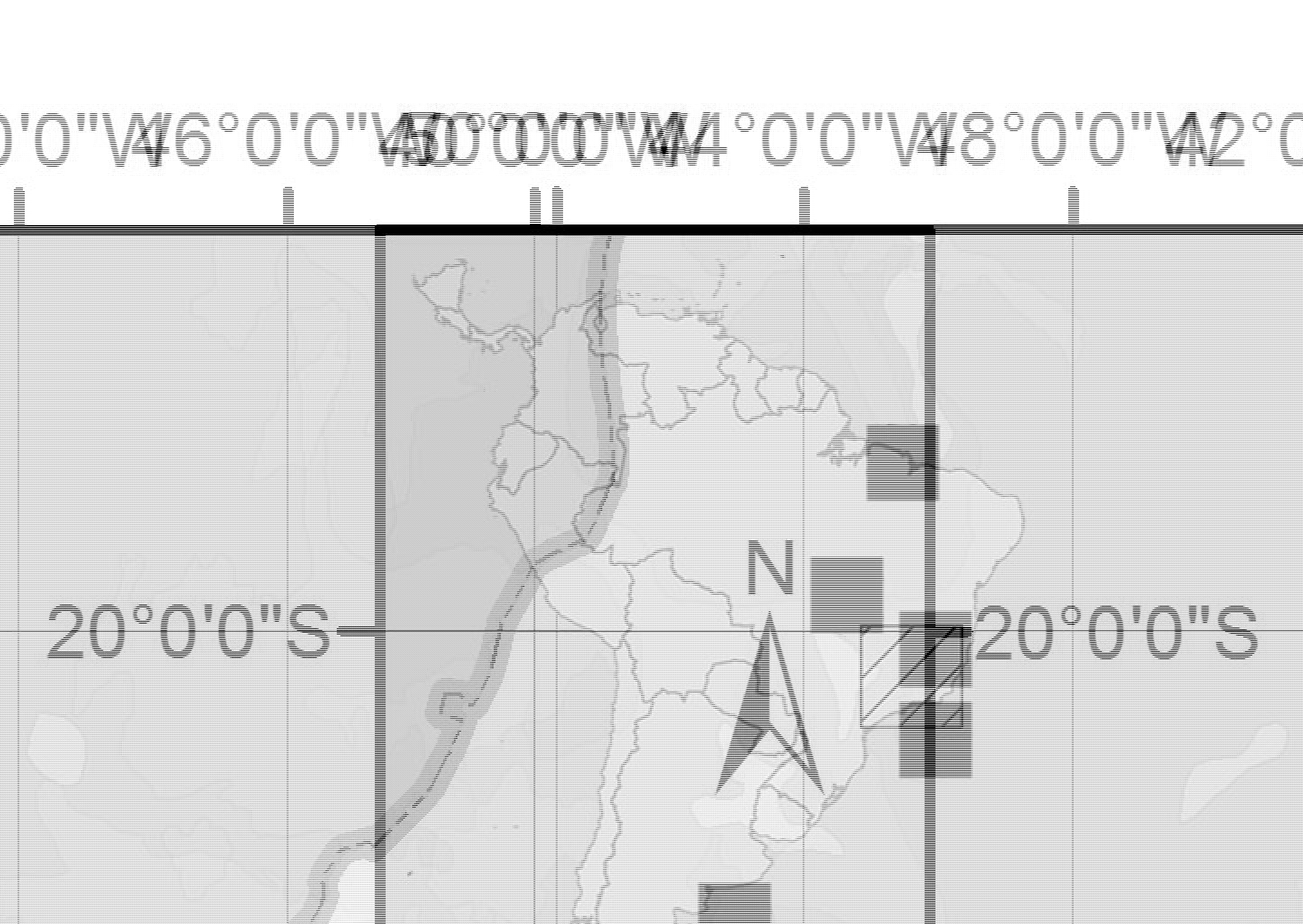
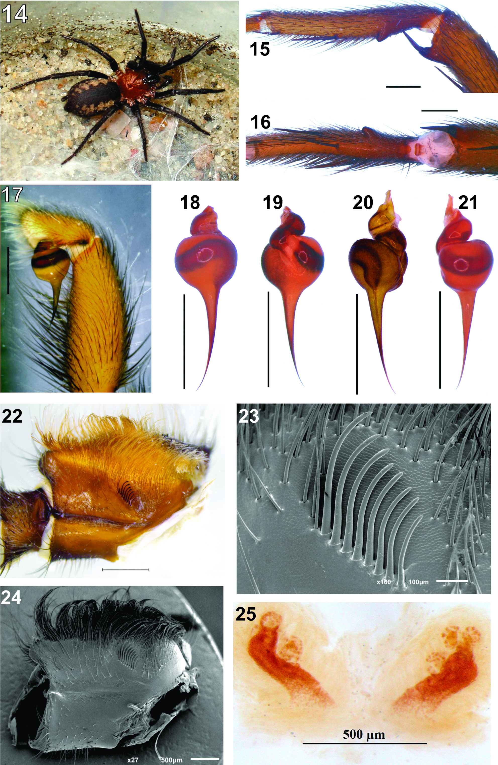
Table 3. Diplura lineata (Lucas, 1857), ♂. Length of left leg articles (dorsal view).
| Leg I | Leg II | Leg III | Leg IV | |
|---|---|---|---|---|
| Fe | 6.7 | 6.4 | 6.0 | 7.6 |
| Pa | 4.0 | 3.3 | 2.8 | 3.2 |
| Ti | 5.6 | 5.3 | 4.7 | 6.1 |
| Mt | 5.3 | 5.9 | 6.6 | 9.1 |
| Ta | 5.0 | 4.6 | 4.0 | 5.0 |
| Total | 26.6 | 25.5 | 24.1 | 31.1 |
Table 4. Diplura lineata (Lucas, 1857), ♀. Length of left leg articles (dorsal view).
| Leg I | Leg II | Leg III | Leg IV | |
|---|---|---|---|---|
| Fe | 7.5 | 7.0 | 6.6 | 7.4 |
| Pa | 4.6 | 4.4 | 3.7 | 4.1 |
| Ti | 5.5 | 5.2 | 4.6 | 5.9 |
| Mt | 5.5 | 5.5 | 5.9 | 6.9 |
| Ta | 4.1 | 4.0 | 3.6 | 3.5 |
| Total | 27.2 | 26.1 | 24.4 | 27.8 |
| MNHN |
France, Paris, Museum National d'Histoire Naturelle |
| IRSNB |
Belgium, Brussels, Institut Royal des Sciences Naturelles de Belgique |
| MNRJ |
Brazil, Rio de Janeiro, Sao Cristovao, Universidade do Rio Janeiro, Museu Nacional |
No known copyright restrictions apply. See Agosti, D., Egloff, W., 2009. Taxonomic information exchange and copyright: the Plazi approach. BMC Research Notes 2009, 2:53 for further explanation.
|
Kingdom |
|
|
Phylum |
|
|
Class |
|
|
Order |
|
|
Family |
|
|
Genus |
Diplura lineata ( Lucas, 1857 )
| Denis Rafael Pedroso, Pedro de Souza Castanheira & Renner Luiz Cerqueira Baptista 2016 |
Harmonicon nigridorsi Mello-Leitão, 1924 : 186
| Mello-Leitao 1924: 186 |
Thalerothele fasciata
| Bertkau 1880: 24 |
Mygale lineata
| Lucas 1857: 14 |