
Microplana monacensis ( Heinzel, 1929 )
|
publication ID |
https://doi.org/ 10.1080/00222933.2010.536267 |
|
DOI |
https://doi.org/10.5281/zenodo.10536969 |
|
persistent identifier |
https://treatment.plazi.org/id/03E687FA-FFEE-BC07-FE4F-FAC6FDF4FCB3 |
|
treatment provided by |
Felipe |
|
scientific name |
Microplana monacensis ( Heinzel, 1929 ) |
| status |
|
Microplana monacensis ( Heinzel, 1929) View in CoL
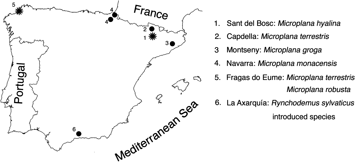
( Figures 1 View Figure 1 , 4 View Figure 4 , Table 1)
Material examined
Holotype. NHMW 2849 View Materials , transverse sections of the anterior part on two slides, transverse sections of the middle part of the body on two slides, sagittal sections of the posterior region on three slides.
Other material. ZMA V. Pl 6856.1, Etxalar, close to an irrigation channel, Navarra ( Northern Spain ), 12 May 2006, sagittal sections on six slides. CRBA-3777a-b, Etxalar , close to an irrigation channel, Navarra ( Northern Spain ), 12 May 2006, sagittal sections on two slides. CRBA-3778, Irurita , in the shore of the river Artesiaga , Navarra ( Northern Spain ), 14 May 2006, sagittal sections on one slide .
Comparative discussion
This species was described from Monaco on the basis of only one specimen ( Heinzel 1929). Minelli (1977a) considered M. monacensis to be a junior synonym of M. scharffi . However, Jones et al. (2008), after the re-analysis of the type specimen, pointed out the validity of the species. The species, whose external appearance has not previously been described, is characterized by a well-developed penis bulb into which the vasa deferentia enter separately and, subsequently, unite to form the ejaculatory duct, which is surrounded by a very thick layer of circular muscle fibres. The narrow bursal canal is lined with a ciliated, nucleated epithelium. After it has received the separate openings of the oviducts, the canal curves dorsally and communicates with the copulatory bursa, which communicates through a short genito-intestinal duct with the gut.
We have collected specimens of M. monacensis from two localities in Navarra, Etxalar and Irurita ( Table 1, Figure 1 View Figure 1 ). In elongated state the living, sexually mature specimens measured 20 mm in length, with a width of about 2 mm ( Figure 4A View Figure 4 ). The dorsal surface is orange and the anterior end is dark brown. The very wide creeping sole occupies about two-thirds of the body width. The internal anatomy of these specimens has been compared with that of the type material housed in the Naturhistorisches Museum Wien in Vienna.
We consider these specimens to belong to M. monacensis , albeit that they exhibit a few differences when compared with the type material from Monaco. First, in the anteroventral part of the body of the Iberian specimens (from about half way along the pharynx to the anterior tip) there is a thick and conspicuous zone of tissue absent in the holotype. It consists of mesenchyme interspersed with many cavities. The zone is located in the lateral regions of the body and lies between the ventral epidermis and the mesenchymal layer of longitudinal muscles ( Figure 4B View Figure 4 ). We believe that this zone of tissue corresponds to what has been described in the older literature for other species as the spongy structure of parts of the ventral mesenchyma (“Bindegewebslücken”; cf. Von Graff 1912 –17: 2762). Second, the vasa deferentia open separately into a small bulbar cavity ( Figure 4C View Figure 4 ) surrounded by two distinct layers of circular muscle fibres of different orientation. This cavity is not depicted in the reconstruction provided by Heinzel (1929). Third, the gonopore lies under the middle of the ventral wall of the male atrium, whereas it is placed in the common atrium in the animal from Monaco. Fourth, the oviducts open close to the anterior section of the bursal canal. In the type specimen they open in the central section of the bursal canal. Fifth, in specimens ZMA V.Pl. 68.56.1 and CRBA-3777a-b the copulatory bursa extends laterally (as an elongated tube) and eventually seems to open to the exterior, but the poor state of the tissue prevents a proper observation of this feature. This presumed “opening”, not observed in the type specimen, occurs on the dorsolateral side in one animal but ventrally in the other ( Figure 4D View Figure 4 ).
We consider that our specimens belong to M. monacensis despite the observed anatomical differences. However, because of the lack of material from the type locality we cannot rule out the possibility that our specimens belong to a different species. We will be able to clarify this point only if new material from the type locality becomes available.
The new records considerably extend the distribution range of the species, as it was previously only known from Monaco.

| ZMA |
Universiteit van Amsterdam, Zoologisch Museum |
| V |
Royal British Columbia Museum - Herbarium |
No known copyright restrictions apply. See Agosti, D., Egloff, W., 2009. Taxonomic information exchange and copyright: the Plazi approach. BMC Research Notes 2009, 2:53 for further explanation.