
Rineloricaria langei, Ingenito & Ghazzi & Duboc & Abilhoa, 2008
|
publication ID |
https://doi.org/ 10.1590/S1679-62252008000300009 |
|
persistent identifier |
https://treatment.plazi.org/id/03E687BD-FF99-FFC3-FC56-8DEB9821F81B |
|
treatment provided by |
Carolina |
|
scientific name |
Rineloricaria langei |
| status |
sp. nov. |
Rineloricaria langei View in CoL , new species
Rineloricaria sp. ; Ingenito et al., 2004: key, picture and comments (in part, only MHNCI 8937). [Picture shows a specimen from MHNCI 8937, but legend erroneously indicates MHNCI 9133].
Holotype. MCP 42506, male, 114.9 mm SL, Brazil, Paraná, Quatro Barras, rio Iraí, at Fazenda Moura-Daz , downstream Reservoir of rio Iraí – SANEPAR, 49°05’26”W 25°29’23”S, Jan 2003, V. Abilhoa. GoogleMaps
Paratypes. MCP 23459, 4 View Materials (1 male, 3 females), 71.7-116.5 mm SL . MHNCI 8937 View Materials , 10 View Materials (1 male, 9 females), 51.7-111.9 mm SL . MNRJ 31157 View Materials , 5 View Materials (females, 1 c&s), 81.1-106.2 mm SL. All specimens collected with the holotype .
Diagnosis. Rineloricaria langei can be distinguished from all species of the genus except R. catamarcensis , R. longicauda , R. maquinensis , R. misionera , R. nigricauda , R. quadrensis , and R. steindachneri by its narrow body (cleithral width 13.0-15.7% of SL; vs. 17.9-22.4% in R. aequalicuspis , 20.0-22.2% in R. anhaguapitan , 17.8-19.2% in R. anitae , 18.0- 21.8% in R. baliola , 16.0-19.1% in R. cadeae , 18.3-22.6% in R. capitonia , 17.7% in R. felipponei , 21.7-22.0% in R. jaraguensis , 17.1-20.6% in R. kronei , 19.1-20.6% in R. latirostris , 16.8-19.3% in R. malabarbai , 18.3-24.0% in R. microlepidogaster , 17.9% in R. pareiacantha , 18.7-19.9% in R. pentamaculata , 18.3-23.6% in R. reisi , 17.6-19.6% in R. tropeira , 16.0-18.1% in R. sanga , 17.6-19.9% in R. setepovos , 16.6-19.9% in R. stellata , 15.8-21.4% in R. strigilata , 18.2% in R. thrissoceps , and 16.2-21.6% in R. zaina ). Rineloricaria langei differs from R. catamarcensis , R. nigricauda and R. steindachneri by having the dorsal caudal ray not prolonged
L. F. S. Ingenito, M. S. Ghazzi, L. F. Duboc & V. Abilhoa 357
358 Two new species of Rineloricaria from the rio Iguaçu basin as a filament (vs. dorsal caudal ray produced as a short filament). It is distinguished from R. quadrensis by having larger interorbital width (19.7-24.0% vs. 18.5-19.3% of HL) and longer dorsal-spine length (18.3-22.2% vs. 17.4-18.6% of SL); differs from R. longicauda by elliptical naked area at tip of snout, not reaching most anterior pore of the infraorbital sensory canal (vs. elongate naked area at tip of snout reaching the most anterior pore of the infraorbital sensory canal). In addition, R. langei can be easily distinguished from R. maquinensis (with abdomen almost naked) and R. misionera (with pectoral girdle not covered by plates) by abdomen and pectoral girdle completely covered by plates.
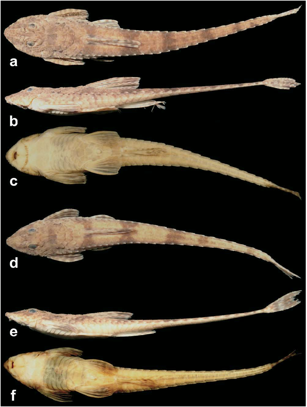
Description. Morphometric data in Table 1. Head and body deeply depressed. Head narrow in females and immature males, and broad in mature males. Body narrow. Body depth greatest at dorsal-fin origin. Head triangular in dorsal view. Dorsal profile of head convex from snout tip to anterior margin of parietosupraocciptal, thereafter slightly concave to posterior margin of first predorsal plate. Predorsal region straight. Trunk almost straight and gradually depressed from dorsal-fin origin to caudal-fin base. Ventral profile of body nearly straight from snout tip to anal-fin origin, then becoming more depressed towards caudal-fin base. Caudal peduncle depressed forming lateral keels with 12(1), 13(9), 14(8*), or 15 (2) coalesced plates from 29(6*), 30(13) or 31(1) total lateral plates in middle series. Four lateral series of plates, mid-dorsal series absent.Abdomen and ventral region of pectoral girdle completely covered by plates, even in specimens smaller than 85.5 mm SL; plates almost reaching transverse line crossing through anterior margin of branchial openings. Ventral region of scapular girdle covered by several irregularly arranged platelets smaller than abdominal plates. One (2), two (1), three (16*) or four (1) series of abdominal plates, usually regularly distributed in adults. Abdominal plates of irregular shapes and sizes. Pre-anal plate present and anteriorly rounded by three polygonal plates. Lateral abdominal plates four (1), five (9*), six (9) or seven (1). Dorsal-fin base with five plate rows. Anal-fin base with two (8) or three (12*) plate rows. Two (9) or three (11*) rows of plates between urogenital pore and anal fin. Plate counts on opposite sides of body usually different, except at dorsal- and anal-fin bases.
Top of head and parieto-supraocciptal wrinkled, with welldeveloped ridges. Ridges of parieto-supraocciptal diverging posteriorly in adult specimens and nearly parallel in young. Predorsal ridges parallel. Plates of first three mid-dorsal series with evident ridges. Upper edge of orbit raised. Postorbital notch shallow, short and wide, not surpassing one third of orbital diameter.
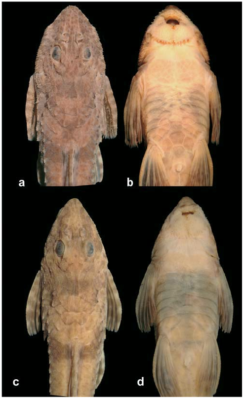
Head and body covered by small odontodes, somewhat more developed on top of head. Mature males with hypertrophied odontodes distributed only on lateral margins of head and dorsum of pectoral fin ( Fig. 2 View Fig ). Snout tip with small elliptical naked area, not reaching most anterior pore of infraorbital ramus of sensory canal. Naked area of snout separated from upper lip by two to four series of inconspicuous odontodes. Lips well developed and covered by papillae; only one irregular row of papillae at anterior most area of upper lip. Two rows of inconspicuous papillae separating upper and lower lips. Maxillary barbel thin and shorter than orbital diameter. Notch present in lower lip. Teeth bicuspid with lateral cusp smaller than medial. Premaxilla with five (3), six (8), seven (7) or eight (2*) teeth. Dentary with five (2), six (6), seven (6) or eight (6*) teeth. Total number of vertebrae 33. Four ribs attached to vertebrae 7 to 10.
Pectoral-fin rays seven (i,6); fin margin reaching pelvicfin origin when adpressed. Pelvic-fin rays six (i,5). Dorsal-fin rays eight (i,7); first ray shorter than head length; its origin located dorsal of pelvic-fin base. Anal-fin rays six (i,5). Caudal-fin rays 12 (i,10,i); its distal margin slightly concave; dorsal principal rays longer than ventral rays; in some specimens dorsal unbranched ray slightly elongated, extending distally less than one-third of orbital diameter and not prolonged as filament.
Color in alcohol. Ground color of dorsal surface of adults dark brown; specimens smaller than 85.5 mm SL light brown with small dark spots or vermiculated lines, mainly on snout. Dorsum of head with inconspicuous and scattered dark brown blotches, sometimes forming two weak longitudinal stripes extending from snout tip to anterior region of orbit, passing medially through nares. Longitudinal stripes usually join with greater number of chromatophores laterally and between eyes forming darker region on top of head. Five (4) or six (16*) dark brown transverse bars across body; first on dorsal-fin origin, second on distal margin of dorsal-fin rays, third immediately after vertical line passing through tip of anal-fin rays, fourth and fifth on caudal peduncle, and sixth at end of caudal peduncle, very close to base of caudal fin. Fourth and fifth transverse bars sometimes slightly wider. Fourth and fifth or fifth and sixth bars sometimes join into wider bar (forming five bars instead of six). Some pores of laterosensory system with dark chromatophores, more evident at anterior half of side of body. Color of sensory pores of head indistinct from background. Ventral surface of body pale yellowish. Some specimens with scattered chromatophores on median region of ventral surface of caudal peduncle, at anal-fin origin, and at base of pectoral and pelvic fins.
Fins yellowish with interradial skin hyaline and small darkbrown blotches on its rays. Pectoral fin with interradial skin dark brown, in most specimens its blotches usually expand from rays to interradial skin, mainly on branched portion. Pelvic fin hyaline in most of proximal and medial regions, becoming darker at its branched portion. Dorsal fin with blotches from distal region of first two to four fin rays expanded and joined on interradial skin, forming singular vertical elongated blotch with wider upper region.Anal fin hyaline, with blotches on its distal region. Blotches from distal margin of caudal fin very expanded and joined over interradial skin, forming one or two wide bars occupying most of area. Caudal fin with darkish base.
L. F. S. Ingenito, M. S. Ghazzi, L. F. Duboc & V. Abilhoa 359
the specimens. That unnatural coloration was caused by an artifact during process of fixation and/or preservation in alcohol.
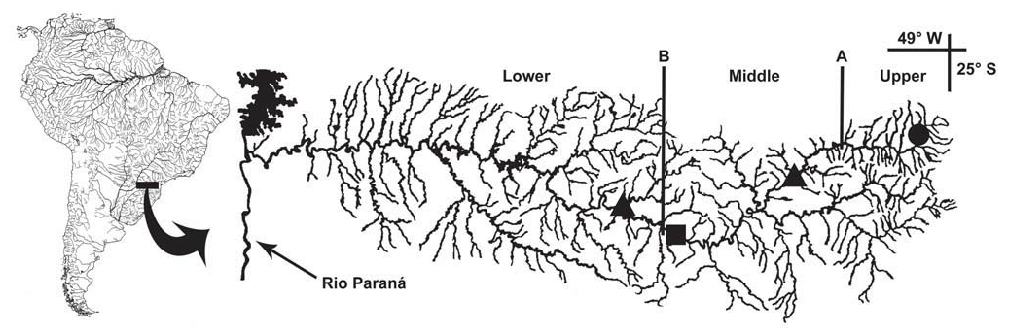
Distribution and habitat. Rineloricaria langei is known from the rio Iraí (a headwater river of upper rio Iguaçu drainage), at the metropolitan region of Curitiba, Paraná State ( Fig. 6 View Fig ). This species inhabits small streams with light to moderate streamflow, over a substrate of sand, some rocks and vegetal debris.
Etymology. The specific name langei is given in honor of Professor Rudolf Bruno Lange, one of the first curators of the zoological collections of Museu de História Natural Capão da Imbuia (MHNCI) during the forties of the Twentieth Century, in which the ichthyological collection is included.
Remarks. The paratypes MHNCI 8937 and MNRJ 31157 exhibit an artificial green color, easily seen on ventral surface of
| MCP |
Pontificia Universidade Catolica do Rio Grande do Sul |
| V |
Royal British Columbia Museum - Herbarium |
No known copyright restrictions apply. See Agosti, D., Egloff, W., 2009. Taxonomic information exchange and copyright: the Plazi approach. BMC Research Notes 2009, 2:53 for further explanation.
|
Kingdom |
|
|
Phylum |
|
|
Class |
|
|
Order |
|
|
Family |
|
|
Genus |