
Paranaiara inajae, Kihara, Terue Cristina & Huys, Rony, 2009
|
publication ID |
https://doi.org/ 10.5281/zenodo.190348 |
|
DOI |
https://doi.org/10.5281/zenodo.6214350 |
|
persistent identifier |
https://treatment.plazi.org/id/03E5D87E-FFB6-FFF0-D09B-8B01FA8AFCC5 |
|
treatment provided by |
Plazi |
|
scientific name |
Paranaiara inajae |
| status |
sp. nov. |
Paranaiara inajae sp. nov.
( Figs 1 View FIGURE 1 –11)
Type locality. Brazil, São Paulo State, Ubatuba (23º37.2’ S, 45º01.2’ W), 41 m depth; for additional environmental parameters see Table 1 (station 18V).
Type material. Holotype female in ethanol (reg. no MZUSP 19054) from station 18V, March 1989. Undissected paratypes (in ethanol) deposited in MZUSP (reg nos 19055–19062) include 1 female from station 5V, March 1989; 1 female from station 7V, March 1989; 1 female and 2 males from station 16V, March 1989; 1 female from station 18V, March 1989; 1 female and 1 male from station 26V, March 1989; 1 female from station 5I, August 1989; 1 female and 2 males from station 18I, August 1989; 1 female from station 26I, August 1989. Additional undissected paratypes (in ethanol) deposited in NHM include 1 female from station 7V, March 1989 (reg. no 2008.3651); 2 males from station 16V, March 1989 (reg. nos 2008.3652–3653); 1 female from station 18V, March 1989 (reg. no 2008.3654); 2 females from station 26V, March 1989 (reg. nos 2008.3655–3656); 1 female from station 27V, March 1989 (reg. no 2008.3657); 2 females from station 18I, August 1989 (reg. nos 2008.3658–3659); and 1 female from station 27I, August 1989 (reg. no 2008.3660). Dissected paratypes in the collection of C.E.F. da Rocha (Departamento de Zoologia, Instituto de Biociências, Universidade de São Paulo): 1 female from station 5I, August 1989; 3 females from station 26I, August 1989; 4 males from station 27I, August 1989; and 2 females from station 28I, August 1989. All material collected by T. Corbisier.
recorded during the interdisciplinary project “Rational use of the coastal ecosystem from the Brazilian tropical region:
São Paulo State” conducted by the Biological Oceanography Department – Oceanographic Institute, University of São
Paulo. Stations were sampled across the inner continental shelf of São Paulo State between São Sebastião Island and
Ubatumirim inlet, Ubatuba during March (V stations) and August 1989 (I stations). Lat. = latitude, Long. = longitude;
Temp. = temperature; MZ = grain size; GS = sorting; C org = organic carbon. Sediment type according to Shepard’s (1954)
classification.
Station Lat. Long. Depth Temp. MZ GS Sand Silt Clay C org CaCO3 Sediment
type (S) (W) (m) (º C) (Ø) (φ) (%) (%) (%) (%) (%)
Paranaiara inajae () , scanning electron micrographs: (A) surface ornamentation of pedigerous somite, showing minute denticles and reticulation near posterior margin, dorsal; (B) rostrum, dorsal; (C) antennulary segments 4–7, anterodorsal; (D) antennulary segments 4–5, anterior; (E) antennulary segment 6, anterior. Scale bars: 2 μm (E); 5 μm (A, D), 10 μm (B, C).
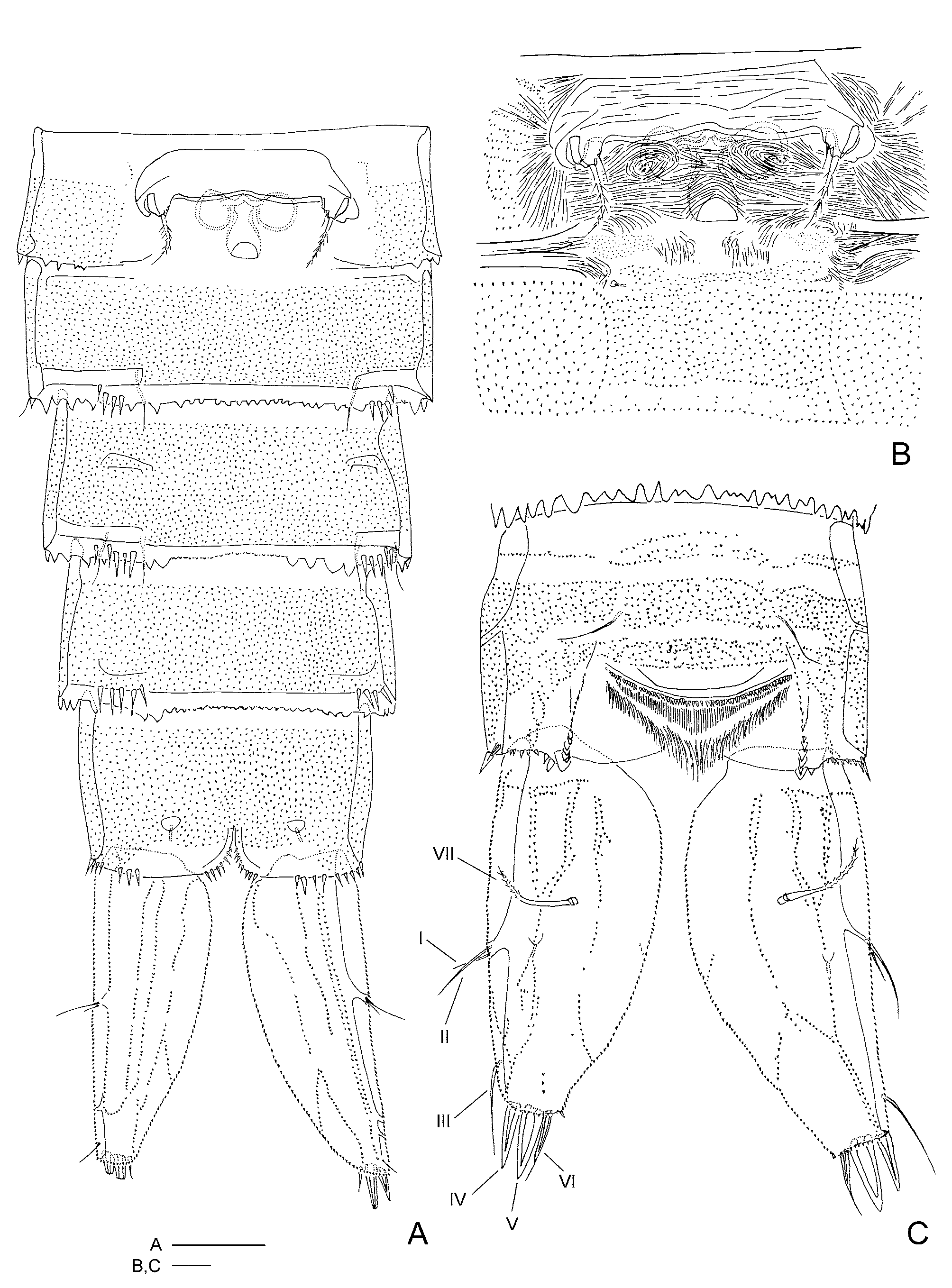
Description. FEMALE ( Figs 1–7 View FIGURE 1 View FIGURE 2 View FIGURE 3 View FIGURE 4 View FIGURE 5 View FIGURE 6 View FIGURE 7 ). Total body length 725–1014 µm (N = 14; mean = 849 µm). Largest width measured at posterior margin of cephalic shield: 192 µm. Urosome slightly narrower than prosome ( Fig. 1 View FIGURE 1 A–B).
Cephalic shield with serrulate posterior margin; pleural area well developed and rounded, with sensilla as illustrated in figures 1A–B; without areolation and without minute spinules as found on free body somites.
Pedigerous somites ( Fig. 1 View FIGURE 1 A–B) covered with minute spinules and a pattern of sensilla and pores as illustrated; hyaline frill not developed; pleurotergites well developed, rounded, ventral portion without minute spinules; posterior margins serrate and with fine reticulation as shown in figure 1C.
Urosomites ( Figs 1 View FIGURE 1 A–B; 2A) with surface ornamentation consisting of minute spinules dorsally and ventrally. Hyaline frill not developed but posterior margin distinctly serrate dorsally and ventrolaterally. Posterior margin of urosomites 2–4 crenulate midventrally and with a few lateroventral spinules.
Genital double-somite ( Figs 1 View FIGURE 1 A–B; 2A) with original segmentation marked by transverse serrate surface ridge dorsally and dorsolaterally and a short surface suture ventrolaterally; completely fused ventrally. Genital field ( Fig. 2 View FIGURE 2 B) with pattern of dense surface striations; copulatory pore large, located in midventral depression, without surrounding spinules. Gonopores fused medially forming a single genital slit covered on both sides by opercula derived from the sixth legs. P6 with a small protuberance bearing 1 pinnate outer seta and 1 minute inner seta.
Anal somite ( Fig. 2 View FIGURE 2 A, C) with well developed rounded, denticulate anal operculum flanked by a row of small spinous processes. Anal opening with a fringe of fine setules and bordered by small spinules midventrally. Surface ornamentation consisting of a pair of sensilla dorsally and a pair of pores ventrally; posterior margin with few spinules ventrally and dorsally.
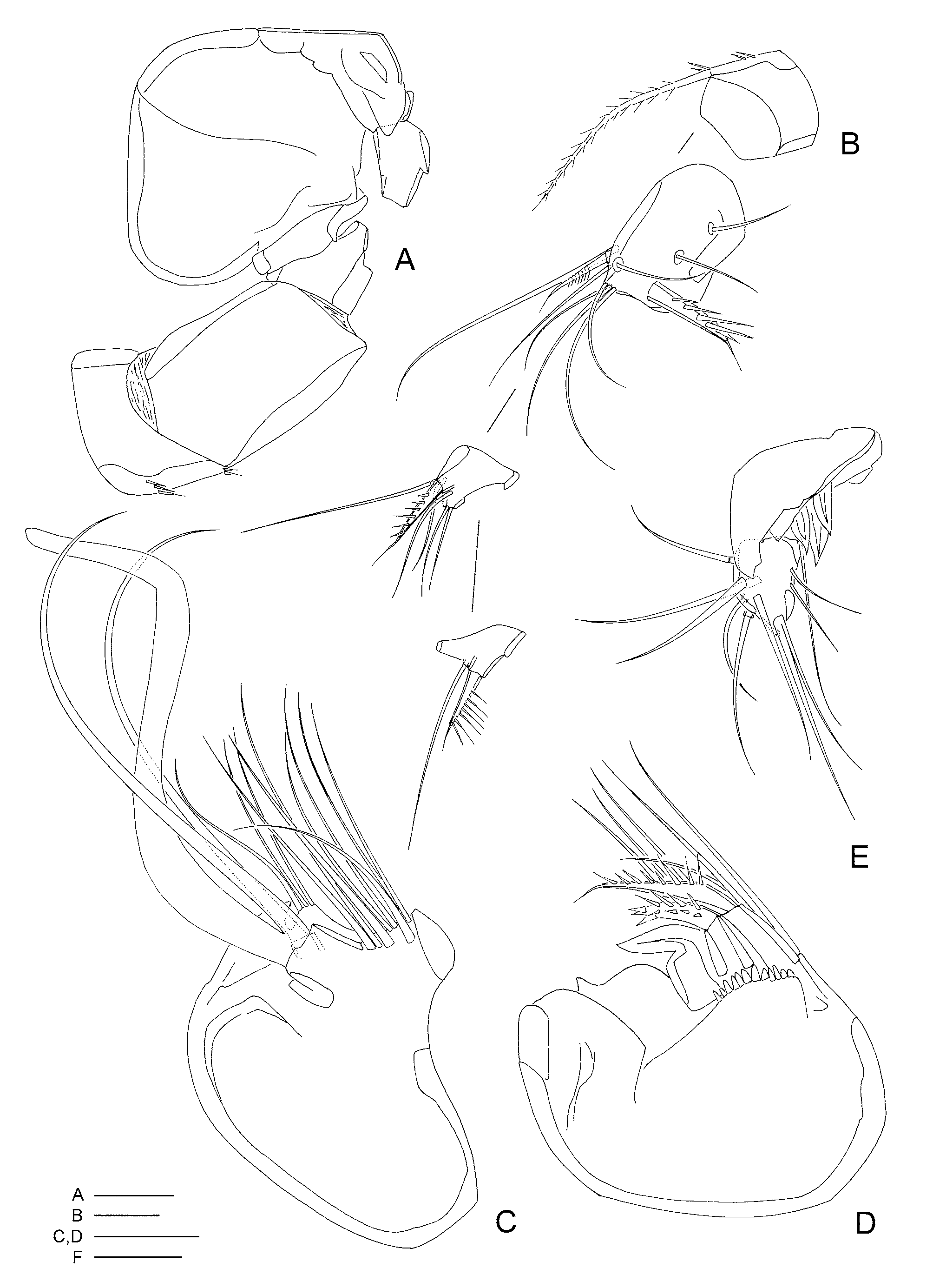
Caudal rami ( Figs 2 View FIGURE 2 C; 3A–B) lamelliform and elongate, about 3 times as long as wide; with straight outer margin and convex inner margin. Each ramus with a dorsal pore medially, 1 tube-pore laterally and 7 setae: seta I naked, shortest and closely set to naked seta II; seta III naked and positioned ventrolaterally; setae IV and V represented by rudimentary, conical, smooth spines, not fused basally; seta VI naked; seta VII biarticulate at its base and sparsely pinnate. Surface ornamentation of each ramus consisting of rows of minute denticles as shown in figures 2C, 3A–B. Posterior margin denticulate and with a ventral semi-circular extension covering bases of setae IV–VI.
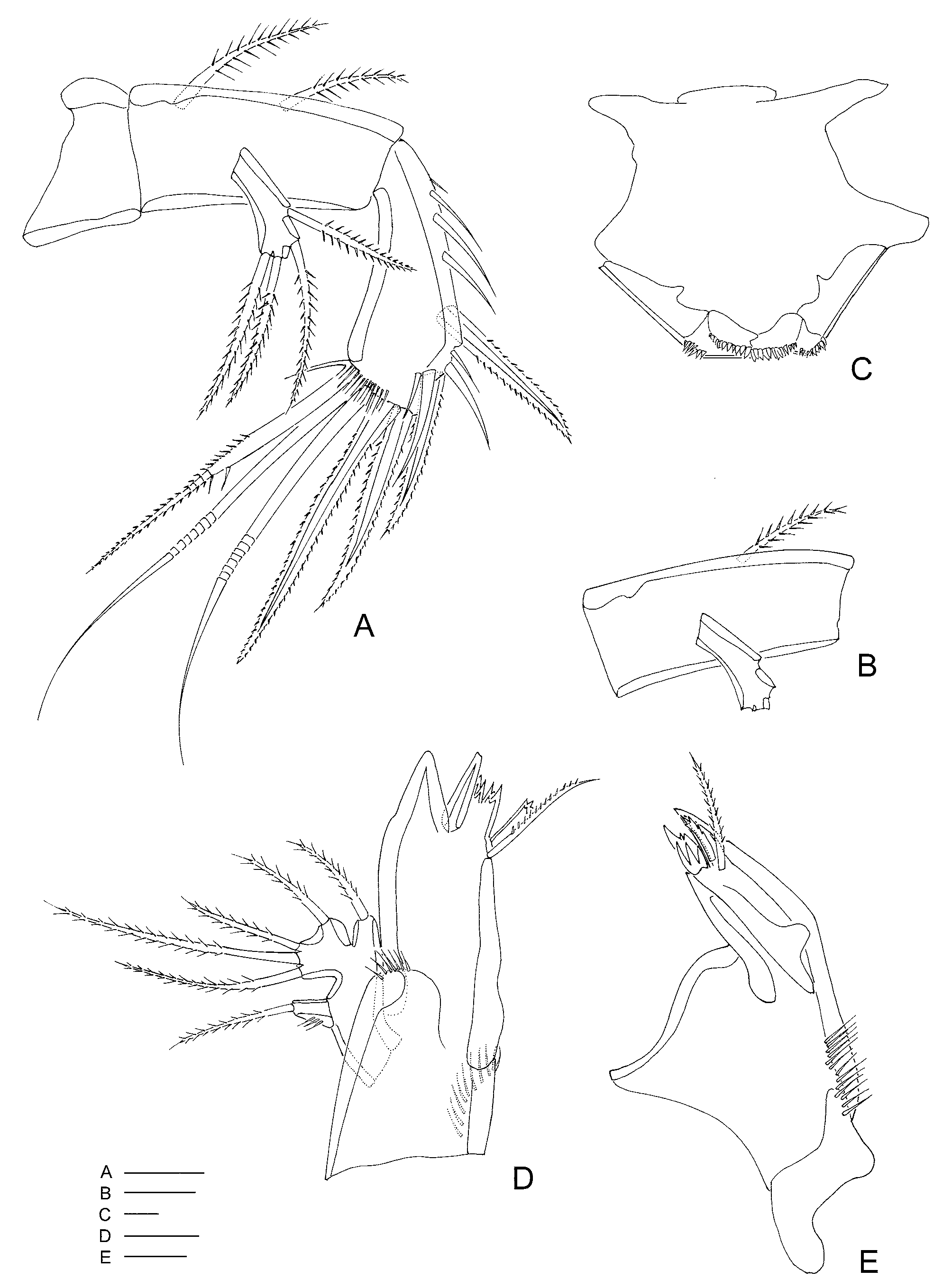
Rostrum ( Fig. 3 View FIGURE 3 C) triangular, prominent and tapering abruptly towards apex; completely defined at the base; with pair of tiny sensilla and a middorsal tube-pore near the apex; dorsal and ventral surface without ornamentation.
Antennule ( Fig. 3 View FIGURE 3 D) short, 6-segmented, segment 3 the longest. Segment 1 with spinular rows around anterior margin and at base of seta. Segment 3 with aesthetasc fused basally to a seta and arising from a distinct pedestal. Armature formula: 1-[1 pinnate], 2-[7 pinnate + 1 pinnate spine (+ 2 elements missing in holotype, indicated by dorsal scars in Fig. 3 View FIGURE 3 D)], 3-[5 + 2 pinnate spines + (1 + ae)], 4-[1 pinnate], 5-[1 + 1 pinnate + 1 pinnate spine], 6-[7 + 1 pinnate spine + acrothek]. Acrothek consisting of 1 slender and 1 strong pinnate seta. Pinnate spines on segments 5 and 6 very large and with coarse spinules.
Antenna ( Fig. 4 View FIGURE 4 A) 3-segmented comprising coxa, allobasis and and free distal segment of endopod. Coxa small, without ornamentation. Basis and proximal endopod segment completely fused, forming an elongate allobasis bearing 2 abexopodal pinnate setae. Exopod 3 times longer than wide, with 2 pinnate setae laterally and 2 pinnate setae apically. Free endopod segment as long as allobasis, abexopodal margin with a row of long spinules and distal margin with a row of fine spinules; lateral armature consisting of 2 pinnate spines; distal armature consisting of 2 pinnate spines and 3 geniculate setae, outermost one being pinnate and fused basally to a short seta.
Labrum ( Fig. 4 View FIGURE 4 C) well developed, with spinular ornamentation along distal margin. Paragnaths not observed.
Mandible ( Fig. 4 View FIGURE 4 D–E) with well developed gnathobase bearing several multicuspidate teeth around distal margin and 1 pinnate seta at dorsal corner. Palp small, biramous but only the exopod is defined at its base. Basis with 1 pinnate seta. Exopod 1-segmented, small, with 1 plumose seta apically and a few spinules along outer margin. Endopod fused to basis, with 1 lateral and 3 distal plumose setae.
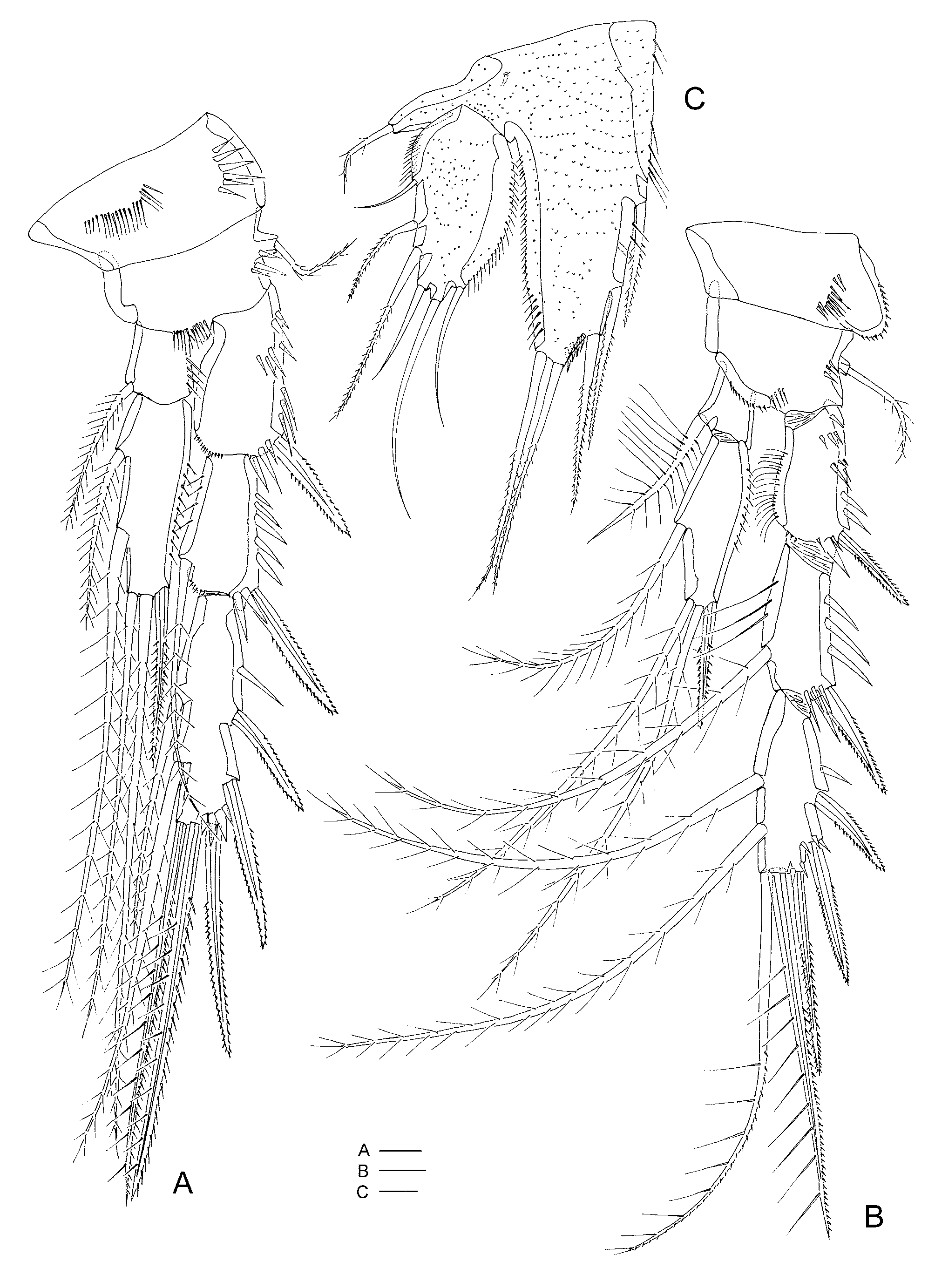
Maxillule ( Fig. 5 View FIGURE 5 A–B) with large praecoxa bearing a few short distal spinules around outer margin; arthrite strongly developed, with 2 naked setae on anterior surface and 7 spines/setae around distal margin, innermost one fused at its base ( Fig. 5 View FIGURE 5 B). Coxa with cylindrical endite bearing 1 pinnate seta. Basis with 2 endites, represented by 2 plumose and 3 pinnate setae, respectively; with a row of spinules on posterior surface. Endopod incorporated with basis, represented by 2 plumose setae. Exopod 1-segmented, with 2 sparsely plumose setae.
Maxilla ( Fig. 5 View FIGURE 5 C) with 3 endites on syncoxa; praecoxal endite small and cylindrical, with one pinnate seta; proximal coxal endite with one multicuspidate spine fused to endite, and 2 pinnate setae; distal coxal endite with 2 pinnate spines and 1 naked seta. Allobasis drawn out into strong, slightly curved claw. Accessory armature consisting of 2 naked setae. Endopod represented by 3 naked setae.
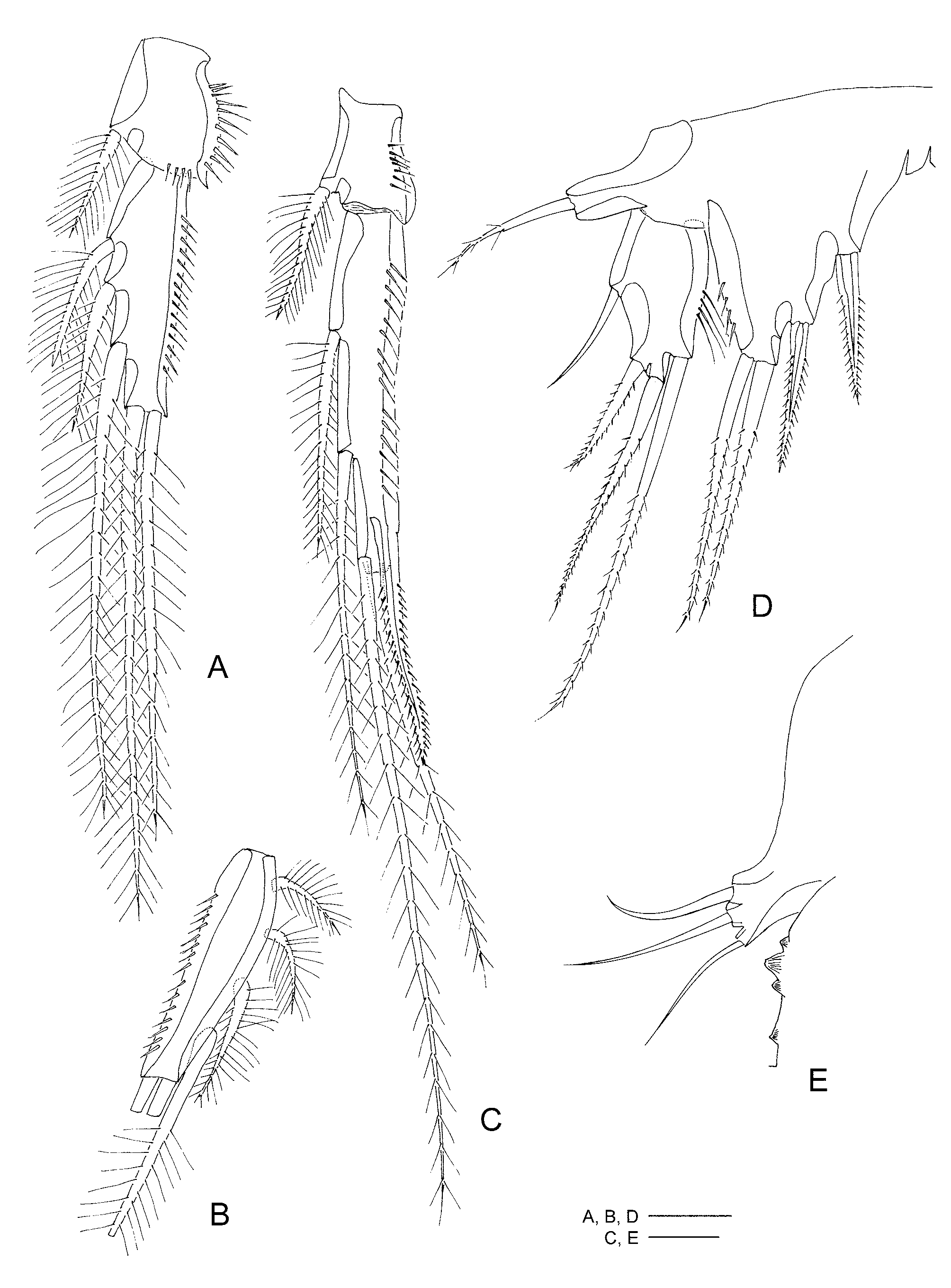
Maxilliped ( Fig. 5 View FIGURE 5 D) without armature or ornamentation on syncoxa. Basis with a few spinules near outer distal corner. Endopod drawn out into a long pinnate claw; accessory armature consisting of a long naked seta. Swimming legs P1–P4 ( Figs 6 View FIGURE 6 A–B; 7A–B) with 3-segmented exopods and 2-segmented endopods.
P1 ( Fig. 6 View FIGURE 6 A) with a well developed coxa bearing strong spinules along outer margin, minute denticles near outer distal corner and fine setules on anterior surface. Basis with setules along inner margin; anterior surface with a pore and 4 spinular rows as figured; armature consisting of strong, pinnate outer and inner spines. Exopodal segments with strong spinules along outer margin and outer distal corner; exp-2 and -3 also with setules along inner margin; exp-1 with 1 stout pinnate outer spine; exp-2 with pinnate outer spine and plumose inner seta (extending beyond distal margin of exp-3); exp-3 with 3 pinnate outer spines and 2 geniculate distal setae. Endopod 0.85 times as long as exopod; segments with setules along inner margin and spinules along outer margin as figured; enp-1 slightly shorter than enp-2, unarmed. Enp-2 with 1 plumose and 1 strong naked setae apically, and 1 plumose inner seta.
P2–P4 ( Figs 6 View FIGURE 6 B; 7A–B). Coxa and basis with spinular rows along outer margin. Coxa with 1 (P4) or 2 (P2–P3) rows of setules/spinules on anterior surface. Basis with spinules near insertion of endopod near base of outer spine/seta; anterior surface with setular row in P2; with outer bipinnate spine (P2) or sparsely plumose seta (P3–P4). Exopodal segments with strong spinules along outer margin and outer distal corner; exp-1 and -2 also with fine setules along inner margin. Endopodal segments with spinules along outer margin (except P4 enp-1); P2 enp-2 twice as long as enp-1, endopod reaching to proximal third of exp-3; P3 enp-2 3 times as long as enp-1, endopod reaching to distal margin of exp-2; P4 enp-2 2.3 times longer than enp-1, endopod reaching to middle of exp-2. Spine and setal formula as for genus.
P5 ( Fig. 7 View FIGURE 7 C) baseoendopod with a short outer setophore bearing a short, plumose basal seta; with one pore near proximal margin. Endopodal lobe elongate, extending beyond distal margin of exopod, with 3 pinnate setae along inner margin and 2 pinnate setae apically; outer margin with spinules; inner margin with spinules, setules and 2 tube-pores as figured. Exopod oval, tapering distally, inner and proximal outer margins with setules; with 1 naked and 2 pinnate setae along outer margin, 2 naked setae around apex and 1 naked seta along inner margin.
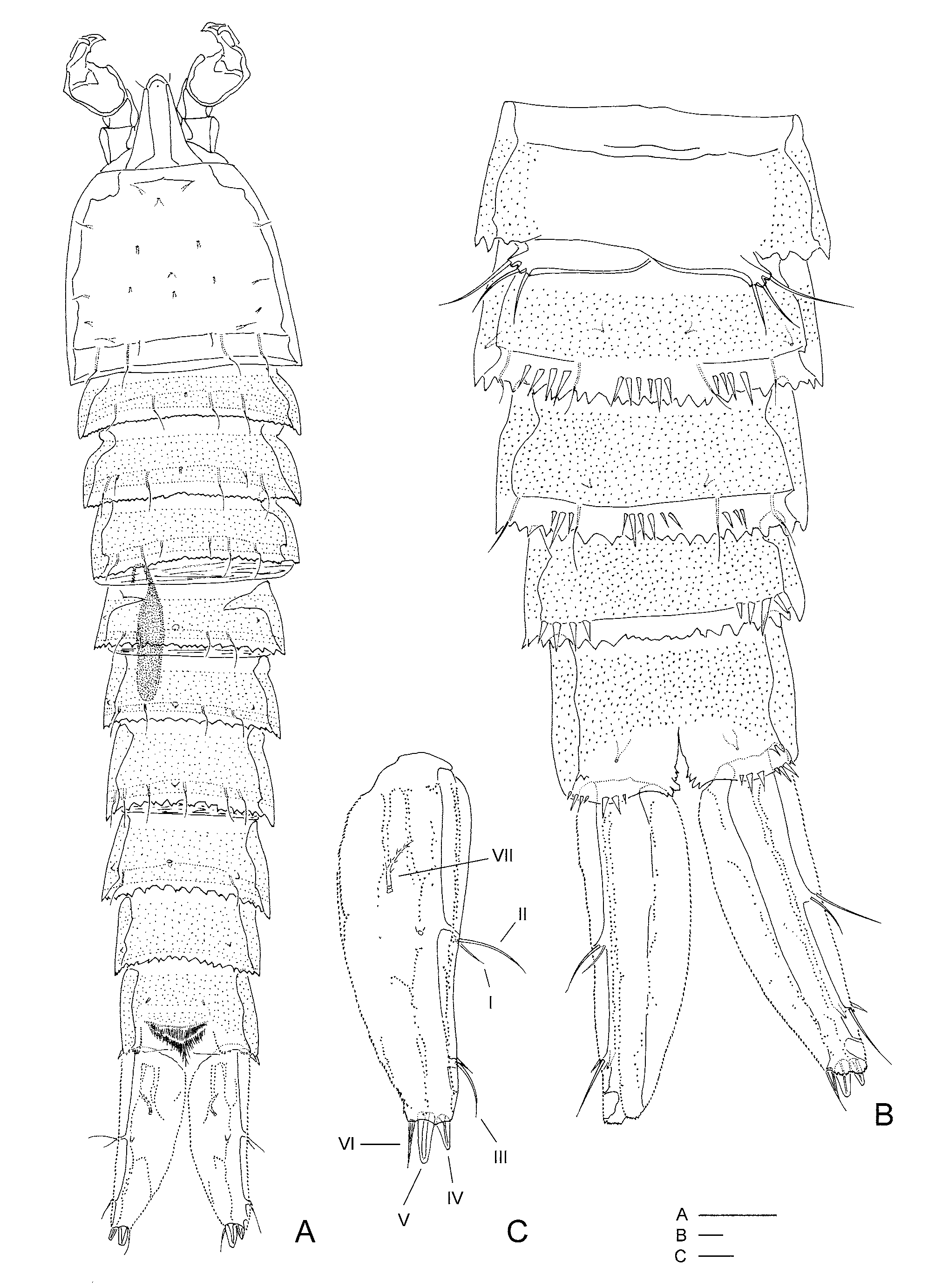
MALE ( Figs 8 View FIGURE 8 –11). Body more slender than in female. Body length 652–913 µm (N = 10; mean = 808 µm). Largest width measured at distal margin of P3-bearing somite: 128 µm. Urosome narrower than prosome ( Fig. 8 View FIGURE 8 A). Rostrum distinct at base as in female (Fig. 11B). Cephalic shield with smooth posterior margin; ornamentation consisting of sensilla and pores as figured. Pedigerous somites covered with small denticles (Fig. 11A); with serrate posterior margin and delicate reticulation (Fig. 11A). Surface ornamentation of urosome ( Fig. 8 View FIGURE 8 A–B) consisting of patches of minute spinules, sensilla and pores. Posterior margins with weaker serrations than in female; with ventral spinule rows. Caudal rami ( Fig. 8 View FIGURE 8 B–C) more slender than in female, with additional tube-pore and spinules near base of seta III.
Antennule ( Figs 9 View FIGURE 9 A–E; 11C–E) 7-segmented. Subchirocer with geniculation between segments 5 and 6. Segment 1 with 2 rows of spinules along anterior margin. Segment 4 represented by small sclerite ( Fig. 9 View FIGURE 9 B). Segment 5 largest and swollen, with partial surface suture ( Figs 9 View FIGURE 9 C–D; 11C–D). Segment 6 forming dorsal spinous process overlying anterior part of segment 7 ( Fig. 9 View FIGURE 9 E). Armature formula: 1-[1 pinnate], 2-[9 + 1 pinnate +1 pinnate spine], 3-[5 + 1 pinnate spine], 4-[1 + 1 pinnate spine], 5-[18 + 2 pinnate spines + 1 fused spine + (1 + ae)], 6-[1 + 3 modified elements], 7-[8 + acrothek]. Pinnate spines on segments 2–4 with coarse spinules (Fig. 11C–D). Modified elements on segment 6 fused at base and transversally elongate (Fig. 11E). Acrothek consisting of 2 short, basally fused, naked setae.
P2 endopod ( Fig. 10 View FIGURE 10 A) 2-segmented; both apical setae of enp-2 distinctly shorter than in female.
P3 endopod ( Fig. 10 View FIGURE 10 C) 2-segmented and modified. Enp-2 more slender and longer than in female; outer margin with a subdistal, straight, bipinnate apophysis (homologous with outer spine of enp-2 of female). Inner seta of enp-1 and both distal inner and outer distal seta of enp-2 shorter than in female.
Fifth pair of legs ( Fig. 10 View FIGURE 10 D) fused medially; defined at base. Baseoendopod with a short outer setophore bearing a plumose basal seta. Endopodal lobe triangular, much shorter than in female, not reaching distal margin of exopod; with 2 inner pinnate spines and 2 apical pinnate setae; with a row of small spinules along outer margin. Exopod about twice as long as maximum width; ornamented with a few fine setules along inner margin; armature consisting of 1 long pinnate inner seta, 1 pinnate apical seta and 2 outer elements (proximal one naked, distal one pinnate).
Sixth pair of legs ( Figs 8 View FIGURE 8 B; 10E) asymmetrical, represented on both sides by a small plate; right plate distinct from the somite but left one is fused with it. Outer distal corner produced into a cylindrical process bearing 3 naked setae.
Variability. One female paratype showed only the distal abexopodal seta (derived from the proximal endopod segment) on the allobasis of the left antenna ( Fig. 4 View FIGURE 4 B). One male paratype displayed three short plumose setae along the inner margin of the left P2 enp-2 ( Fig. 10 View FIGURE 10 B).
Etymology. The specific name inajae is dedicated to the senior author’s mother, Inajá Batista Kihara.


| MZUSP |
Museu de Zoologia da Universidade de Sao Paulo |
No known copyright restrictions apply. See Agosti, D., Egloff, W., 2009. Taxonomic information exchange and copyright: the Plazi approach. BMC Research Notes 2009, 2:53 for further explanation.
|
Kingdom |
|
|
Phylum |
|
|
Class |
|
|
Order |
|
|
Family |
|
|
Genus |