
Protemnodon anak Owen, 1874
|
publication ID |
https://doi.org/ 10.11646/megataxa.11.1.1 |
|
DOI |
https://doi.org/10.5281/zenodo.10993689 |
|
persistent identifier |
https://treatment.plazi.org/id/03E587FD-FFE5-D50F-FCA2-726CFB01FE09 |
|
treatment provided by |
Felipe |
|
scientific name |
Protemnodon anak Owen, 1874 |
| status |
|
Macropus anak (Owen, 1859) : Quart. J. Geol. Soc. Lond. 15, p. 185 (nomen nudum).
Protemnodon anak Owen, 1873 : Proc. Roy. Soc. Lond. 21, p. 128 (nomen nudum).
Protemnodon anak Owen, 1874 : Phil. Trans. Roy. Soc. 164, p. 277. See also Owen (1877) pp. 428–430, pl. 85, figs 1–4 & 7–14); Etheridge & Jack (1892), p. 677; Raven (1929), p. 255; Simpson (1930), p. 76; Tate & Archbold (1937), p. 410; Tate (1948), p. 297; Troughton (1957), p. 187; Stirton (1963), p. 137, fig. 13a; Bartholomai (1973), pp. 318–330, pl. 9–12; Hope (1973), pp. 167, 182; Dawson (1985), p. 66, table 1; Helgen et al. (2006), p. 303, appendix 2; Jones et al. (2021), p. 36; Janis et al. (2023), figs 2–4, SI table 1.
Protemnodon og Owen, 1874: Phil. Trans. Roy. Soc. 164, p. 277, pl. 25, figs 5–6. See also Owen (1877), p. 430, pl. 85, figs 5–6; Etheridge & Jack (1892), p. 678; Palmer (1904), p. 883; Simpson (1930), p. 76; Stirton (1963), p. 139, fig. 13b.
Halmaturus anak (Owen) ; Krefft (1875), p. 208; De Vis (1895) (partim), pp. 104–109, pl. 17, figs 5–10.
Sthenurus atlas (Owen) ; Owen (1876) (partim.), pp. 210–212, pl. 25, fig. 2, pl. 26, fig. 4. Not Sthenurus atlas ( Owen, 1838) .
Macropus anak (Owen) ; Flower (1884), p. 715; Lydekker (1887), pp. 214–218; Lydekker (1894), p. 257; Lydekker (1896), p. 257; Palmer (1904), p. 883.
Procoptodon goliah Owen ; Owen (1876), pl. 23, fig. 4. Not Procoptodon goliah (Owen, 1845) .
Holotype: NHMUK PVM1895 partial L dentary containing p3–m4, missing sections immediately anterior to p3 and from 10 mm posterior to m4, ventral margin intact below m2–m4. Figured in Owen (1874), pl. 25, fig. 1.
Type locality:
Pleistocene fluviatile deposits in Darling Downs, southeast Queensland. Exact locality unknown; described by Owen (1874, p. 276) as ‘freshwater deposits exposed in the beds of creeks in Darling Downs’.
Paratype (s):
None.
Referred specimens:
AM F30691 partial R dentary. AMNH FM 19256 splanchnocranium; AMNH FM 19288 L dentary; AMNH FM 19257 L dentary; AMNH FM 32747 R dentary. NHMUK PVM 48 R maxilla; NHMUK PVM 2261 P3 and M1; NHMUK PVM 2262 partial L maxilla and LR premaxillae; NHMUK PVOR 47838 partial splanchnocranium; NHMUK PVOR 38751 partial L maxilla; NHMUK PVM 2258 L dentary; NHMUK PVM 3451 partial L dentary; NHMUK PVM 5006 partial R dentary; NHMUK PVOR 10068 R dentary; NHMUK PVOR 38753 partial L dentary; NHMUK PVOR 40009 R dentary fragment; NHMUK PVOR 40010 partial L dentary; NHMUK PVOR 48423 R dentary; NHMUK PVOR 47854 L i1; NHMUK PVOR 47829 R calcaneus. QVM 1990 GFV 50 partial L maxilla; QVM 1990 GFV 82 R dentary.
Gowrie Creek, Darling Downs (site unknown): AM F2221 partial cranium. QM F651 partial LR premaxillae; QM F4896 partial L maxilla and L dentary; QM F4900 partial R dentary. NHMUK PVOR 50064b partial maxilla; NHMUK PVOR 35963 partial L dentary; NHMUK PVOR 35964 partial L dentary; NHMUK PVOR 35967 R dentary fragment; NHMUK PVOR 50060 partial L dentary.
King’s Creek, Darling Downs (site unknown): UCMP 53292 partial R dentary.
Pilton, Darling Downs (site unknown): QM F5028 partial R premaxilla; QM F3017 partial R dentary.
South Australia
Malkuni Waterhole, Cooper Creek: SAMA P53058 L premaxilla fragment; SAMA P25052 partial R maxilla; SAMA P25049 partial R dentary; SAMA P25063partial R dentary; SAMAP 25031 L calcaneus; SAMA P54627 L proximal pedal phalanx IV.
Waralamanko Waterhole, Cooper Creek: SAMA P25185 partial R dentary.
Lower Cooper Creek (site unknown): UCMP 47924 partial L dentary.
Main Fossil Chamber, Victoria Fossil Cave, Naracoorte: SAMA P28172 partial juvenile R maxilla.
Queensland
Hodgson Creek/Umbiram Creek, Darling Downs: IS V653 R metatarsal V.
Wyandotte Creek, Greenvale: NMV P184050 partial juvenile R dentary.
Pearson Bed, King’s Creek: IS V122 cranium; IS V127 partial cranium; IS V637 partial cranium.
Sobbe Bed, King’s Creek: IS V657 partial cranium; IS V 593 juvenile R dentary.
Sutton Bed, King’s Creek: IS V126 cranium; IS V 590 juvenile L dentary.
Darling Downs (site unknown): QM F616 partial cranium; QM F3007 L dentary; QM F3034 R dentary; QM F4712 partial juvenile L maxilla; QM F5045 partial R maxilla; QM ‘1065’ R calcaneus; QM ‘8771/XXII2’ L calcaneus. AM F19648 splanchnocranium; AM F30688 partial L maxilla; New South Wales
Cox’s Creek, Tambar Springs: AM F112005 partial L maxilla.
Cuan Station, Scone: AM F7238 partial R dentary.
Dundee: AM F39813 L dentary fragments.
Weetalibah: AM F2343 L dentary.
Site 51, Lake Victoria: NMV P28273a partial L dentary.
Wellington Caves, Wellington (site unknown): AM F17599 premaxilla; AM F18894 partial premaxilla; AM F30599 partial L maxilla; AM F47055 partial LR maxillae; AM F47070 partial maxilla; AM F18904 partial L dentary; AM F18905 juvenile R dentary; AM F104750 L calcaneus; AM F104754 L calcaneus. UCMP 57375 R dentary.
Lake Menindee (site unknown): AM F19649 partial R dentary.
Victoria
Nelson Bay, Portland: NMV P215986 partial L maxilla; NMV P200471 L DP2; NMV P252398 R i1.
Childers Cove: NMV P230263 partial DP3.
Northeast shore, Lake Weeranganuk: NMV P162930 L humerus and partial L scapula, radius and ulna.
Batesford Quarry, Moorabool Viaduct Sands: NMV P201869 partial juvenile LR dentaries.
Locality 477, Lancefield Swamp South: NMV P177784 partial R dentary.
Locality 1534, Lancefield Swamp: NMV P31340 partial L dentary.
Lancefield Swamp, Lancefield (site unknown): NMV P31588 partial L dentary; NMV P31614 partial R dentary; NMV P43701 partial L dentary; NMV P200720 partial R dentary; NMV P240498 L calcaneus; NMV P240564 L calcaneus; NMV P240605 L calcaneus; NMV P40509 R metatarsal V.
Firehole No. 2, Morwell Mine, Hazelwood: NMV P42532 partial cranium; NMV P39101 cranium, partial LR dentaries, cervical vertebrae C2–7, partial R scapula, L humeral fragments, ulnar fragments, radius, R tibia, LR calcanei, and L cuboid and metatarsals IV and V; NMV P39105 partial cranium, LR dentaries, partial atlas and axis vertebrae, partial sacrum, R humerus, LR ulnae, R radius, metacarpal IV and proximal, middle and distal manual phalanges IV, tibial and fibular fragments, R calcaneus, cuboid, talus, metatarsals IV, V, proximal pedal phalanx IV and proximal, middle and distal phalanges V, and L middle and distal phalanges IV; NMV P159917 L femur, tibia and calcaneus, R metatarsals IV, V, and distal pedal phalanx IV; NMV P159917b L calcaneal fragment, cuboid and metatarsals IV and V, and proximal pedal phalanx IV.
Morwell Mine, Hazelwood (site unknown): NMV P188455.2 juvenile cranium and mandible; NMV P39118 partial L maxilla, partial R dentary, partial sacrum, caudal vertebra, LR femora, tibial and femoral fragments, and L calcaneus, cuboid, talus, and metatarsals IV and V; NMV P39128 partial R maxilla, premaxilla and dentary; NMV P39134 caudal vertebra Ca13?, R metatarsal IV L proximal pedal phalanx IV and R proximal, middle and distal phalanges V; NMV P209937 R calcaneus and metatarsals IV and V; NMV P39132 R tibial fragments, L calcaneus, cuboid, metatarsals IV, V and proximal pedal phalanx IV, and R proximal, middle and distal phalanges IV and middle and distal phalanges V.
Dry Creek, Keilor: NMV P29632 partial LR dentaries; NMV P29554 partial L dentary.
Tasmania
Egg Lagoon, King Island: NMV P30786 partial R maxilla and partial LR dentary fragments.
South East Lagoon, King Island: QVM 2019 GFV 0002 L dentary; QVM 2019 GFV 0003 partial juvenile R dentary.
Scotchtown Cave, Smithton: QVM 1992 GFV 0203 partial R dentary; QVM 1996 GFV 7 partial R dentary; QVM 1996 GFV 08 partial L dentary; QVM 1996 GFV 12 partial juvenile L dentary; QVM 1996 GFV 13 L i1.
Revised specific diagnosis:
Protemnodon anak is distinguished from its congeners by several unique dental and skeletal characteristics and by its combination of other osteological characteristics. Protemnodon anak is differentiated from all members of the genus for which this part of the cranium is known by having a more anteriorly extensive masseteric ridge of the jugal (anterior jugal ridge). The dentition of P. anak is distinguished from all congeners by the following characteristics: a P3 narrower relative to length, with more raised, angular, distinct and dorsoventrally aligned ridgelets on the buccal surface of the main crest, and a distinctly jagged lingual crest that terminates short of the lingual base of the anterior cusp; and a p3 with more raised, angular and distinct ridgelets. The axial skeleton is differentiated from all members of the genus that preserve an axial skeleton by having an elongate axis vertebra (C2), with the caudal extremity of the centrum strongly caudally projected, and the postzygopophyses large and elongate; cervical vertebrae (C3–7) elongate, with large, elongate pre- and postzygopophyses, and a strongly caudoventrally projected caudal extremity of the centra. The forelimb is distinguished from all other species of Protemnodon in having a more elongate, proximally deeper, and strongly transversely compressed ulna that is distinctly recurved in lateral view. The pes is differentiated from all other species of Protemnodon except P. otibandus by having a calcaneus with a fibular facet with distinct margins and a rounded, caudally projected caudal component, and from all other species of Protemnodon in having the middle pedal phalanx IV with a far broader proximal than distal end, due to lateral and medial flaring of the proximal plantar (flexor) tubercles.
Protemnodon anak is most similar in cranial morphology to Protemnodon mamkurra sp. nov. and P. viator sp. nov. The cranium is further distinguished from those of P. mamkurra sp. nov. and P. viator sp. nov. in having straighter, relatively more dorsally situated occipital condyles. It additionally differs from P. mamkurra sp. nov. in having a dorsoventrally lower rostrum and narrower occipital condyles, and is further distinguished from P. viator sp. nov. by having rounder, less dorsoposteriorly slanted orbits in lateral view. The dentary of P. anak is most similar to that of P. mamkurra sp. nov., P. viator sp. nov., P. dawsonae sp. nov., and P. otibandus , but differs from these species in having a less dorsally deflected diastema, and further from P. otibandus in having a broader, more robust anterior dentary below the diastema. The dentary further differs from that of P. viator sp. nov. in having a proportionally shallower mandibular corpus when fully grown.
Protemnodon anak is similar in dental morphology to P. mamkurra sp. nov., P. viator sp. nov., P. otibandus , and P. snewini . The dentition of P. anak further differs from that of P. otibandus and P. snewini in being higher crowned; from P. otibandus in having a squarer, relatively anteroposteriorly shorter occlusal surface of I1 when worn and relatively narrower molars; and from P. snewini in having a P3 with a narrower posterior relative to anterior width, and a broader, more robust i1. It is further distinguished from P. mamkurra sp. nov. and P. viator sp. nov. in having: a narrower I1 relative to the length of the I3; a relatively narrower DP2, particularly across the anterior cusp; a DP3 with a distinct, raised preprotocrista forming the lingual half of an incomplete protoloph; relatively narrower upper molars with a higher preparacrista; less robust i1 with thinner, more raised dorsobuccal and ventrolingual crests; p3 with fewer, lower, and less distinct, transverse ridgelets on main crest; and lower molars with straighter lophid margins, and higher, more distinct cristid obliqua when unworn or slightly worn.
Protemnodon anak is most similar in aspects of skeletal morphology to P. mamkurra sp. nov., P. viator sp. nov., P. snewini , and P. otibandus . The axial skeleton of P. anak further differs from that of P. mamkurra sp. nov. and P. viator sp. nov. in having: an axis vertebra with a more elongate and less dorsally deflected dens, flatter and less dorsolaterally orientated cranial articular surfaces, and a spinous process with more level dorsal margin, much smaller caudal projection, and a more elongate base; and cervical vertebrae with a narrower caudal extremity of the centra. The cervical vertebrae further differ from those of P. mamkurra sp. nov. in having more rounded vertebral canals, and from P. viator sp. nov. in being taller and relatively narrower, with a taller cranial extremity of the centra and a taller caudal extremity of the centra lacking a slightly bilobed ventral margin.
The forelimb of P. anak differs from those of P. mamkurra sp. nov. and P. viator sp. nov. in having: a scapula with a more medially extensive scapular spine; a more elongate humerus with a longer and more deeply concave bicipital groove and a longer pectoral crest; and an ulna with a more cylindrical distal shaft. The forelimb further differs from P. mamkurra sp. nov. in having: a humerus with a more rounded, less pointed proximal peak on the lateral supracondylar ridge; a more elongate radius; and distal manual phalanges with shorter, less dorsopalmarly compressed shafts. It further differs from P. viator sp. nov. in having: a radius with a more distally situated cranial ridge; a shorter and more robust metacarpal IV with a vertical rather than dorsomedially tilted hamatal facet and a distally facing (rather than laterally facing) metacarpal V facet; proximal manual phalanges with deeper, more V-shaped trochleae; and distal manual phalanges with less palmarly curved shafts.
The hindlimb differs from those of P. mamkurra sp. nov. and P. viator sp. nov. in having a femur with a shallower trochlea. It further differs from P. mamkurra sp. nov. in having: a femur with a more raised proximolateral ridge and a broader and higher lateral trochlear crest; and a more gracile tibia that is longer relative to femoral length. It further differs from P. viator sp. nov. in having: a shorter tibia relative to femoral length; a more gracile femur with a less medially projected lesser trochanter, a more curved lesser trochanteric ridge such that the conjunction of the ridges at the lesser trochanter is broadly more curved, a broader and more rounded trochlea, and a relatively broader, lower medial trochlear crest; and a more robust tibia with a more distinct peak of the cnemial crest. The pes of P. anak differs from those of P. mamkurra sp. nov. and P. viator sp. nov. in having: a metatarsal V with a larger, more proximally projected medial plantar tubercle; shorter, more robust pedal phalanges; and a proximal phalanx IV with a less distinct waist. The pes further differs from P. mamkurra sp. nov. in having: a less robust calcaneus with a more rounded sustentaculum tali in medial view, a more caudally situated and distinctly separate lateral talar facet relative to the medial talar facet, and less bulbous talar and fibular facets; a talus with a narrower navicular facet more aligned in the sagittal plane; a cuboid with a narrower metatarsal V facet, a more plantarly projected lateral plantar tubercle and a narrower, deeper flexor groove; a more transversely compressed metatarsal V; and distal pedal phalanges IV and V lacking V-shaped indentations in the transverse margins of the proximal surface. It further differs from P. viator sp. nov. in having: a broader and more robust calcaneus with a domed calcaneal tuberosity; a shorter, broader cuboid with a larger metatarsal V facet and a smaller medial plantar tubercle; a dorsoplantarly shorter, broader metatarsal V with a broader and more concave cuboid facet; and distal pedal phalanges IV and V with more rounded, less triangular dorsal peaks.
The hindlimb of P. anak is further differentiated from that of P. snewini in having a more robust tibia with a relatively longer cnemial crest with a less distinct distal peak, and a longer proximolateral crest relative to total length. The pes is differentiated by having: a talus with a small indentation between the cranial margin of the medial trochlear crest and the talar head; a cuboid with the dorsal and plantar metatarsal IV facets continuous with one another and the facet for metatarsal V larger and more distinct; a more elongate metatarsal IV with a relative longer plantar ridge, continuous dorsal and plantar cuboid facets, and a more plantolaterally situated proximal cuboid fossa; and distal phalanx IV with a more rounded dorsal peak.
The forelimb of P. anak further differs from that of P. otibandus in that: both the capitulum and ulnar facet of the humerus are relatively larger and more distally projected; the ulna has a shorter olecranon process relative to ulnar length with a larger ventromedial eminence and a shallower proximomedial flexor fossa; the caudomedial surface of the distal shaft of the radius is less flattened and the distal end is less transversely compressed. The manus differs from P. otibandus in having less palmarly curved distal phalanges. The hindlimb is further differentiated by its femur with a shallower and broader trochlea and a relatively lower and more rounded medial trochlear crest. The pes differs as follows: calcaneus larger and taller with a less medially displaced head; talus with a narrower navicular facet and smaller medial malleolus, both more aligned in the sagittal plane, and in lacking a small tubercle plantar to the medial malleolus on the medial surface; cuboid taller, with more plantarly projected plantar tubercles and the dorsal and plantar metatarsal IV facets continuous with one another; more gracile metatarsal IV with a more raised plantar ridge, the dorsal and plantar cuboid facets continuous, and the shaft broadening more distally; metatarsal V with a larger proximolateral process and a larger, more proximomedially projected medial plantar tubercle; distal phalanx IV with a more pointed dorsal peak; and proximal phalanx V with a more distinct waist.
Etymology:
In reference to a biblical giant named Anak .
Description and comparisons:
Cranium and dentition
Cranium ( Figs 3–5 View FIGURE 3 View FIGURE 4 View FIGURE 5 ): large, narrow and elongate. Rostrum narrow, low, and rounded in cross-section, with a shallow buccinator fossa. Diastema elongate, ~70% of rostral length, consists of slightly more maxilla than premaxilla. Premaxilla anteroventrally projected; ventral part narrows gradually anterior to the maxilla, but broadens slightly around I3, before forming a narrow, rounded anterior extremity; ventral width is subequal to maximum width across the nasal cavity. Incisor-bearing component of the premaxilla is robust, broadest ventrally and shallowly concave, contributing ~50% of the ventral length from the anterior tip to the premaxilla–maxilla suture. Ventral premaxilla–maxilla suture angled posterolaterally, and the lateral premaxilla–maxilla suture extends straight dorsally before curving smoothly posteriorly toward posterior of nasal. Incisive foramina elongate, more posteriorly positioned relative to the incisors in mature individuals, with, in ventral view, a tapering channel extending anteriorly and curving laterally from them toward I3. Shallow buccinator fossa smoothly concave and quite tall with depth gently decreasing anteriorly; extends dorsally from the ventral margin of the diastema to one-third of maxilla height and anteriorly from the anterior side of the P3/DP2 to between the premaxilla– maxilla suture and the base of the I3. Infraorbital foramen large, narrow, and opens anteriorly, positioned dorsal to P3/DP2, well anterior of orbit. Nasal narrow, elongate, and slightly convex dorsally, with linear lateral suture; projects anteriorly past anterodorsal margin of premaxilla and tapers to a shared medial point. Frontal strongly concave at the midpoint of the temporal fossa in dorsal view, broadens anteriorly and flares over orbits.
Lateral cranium. Lacrimal quite large, extends anteriorly well onto lateral side of splanchnocranium, and gently projects laterally; has a small dorsoposterior foramen and larger anteroventral foramen, both dorsoposteriorly bordered by a rounded tubercle. A ventral orbital lip (masseteric ridge of the jugal, for the dorsal margin of the origin of the intermediate masseter; see Warburton 2009) projects laterally from the dorsal margin of the zygomatic process; this extends anteriorly into a thin, low ridge to the anterior margin of the jugal ( Fig. 5b View FIGURE 5 ), with occasional small, pointed eminences projecting anteriorly from the anterior tip (see IS V126 and NMV P39105). Temporal fossa elongate, broadest at midpoint, and slanted gently anteroventrally into orbit in lateral view. Orbit large and round in lateral view ( Fig. 5b View FIGURE 5 ). Masseteric process composed solely of maxilla; moderately enlarged and ventrolaterally projected, with ~90° posterior rotation of tip. Jugal rises gently at its posterior end and bifurcates around the anterior tip of the zygomatic arch into a short, broad dorsomedial part (the postorbital process), and a ventrolaterally situated posterior part that extends almost to the postglenoid process; a low ridge extends posteriorly from the ventral orbital lip on the lateral surface of the posterior part. Zygomatic arch is orientated dorsoposteriorly at 30–40° to axis of molar row; zygomatic process of squamosal tall posteriorly, with the lateral surface tilted slightly dorsally, dorsal margin smoothly convex in lateral view, tapering anteriorly to a point between the postorbital process and the posterior part of the jugal.
Palatal region. Palatine thick, broad, and lacking fenestrae, with a narrow, elongate anterolateral foramen level with the anterior of the M3; small, rounded posterolateral foramen posteriorly adjacent to the M4; lateral margin tapers gently anteriorly before forming a linear transverse maxilla–palatine suture level with abutment of M2–M3; posterior margin concave. Maxillary foramen quite large, round, and opens posteriorly into a broad, rounded valley. Sphenopalatine foramen small and round, situated immediately posteromedial to the maxillary foramen. Pterygoid crest thin and elongate, with the anterior peak tall and slightly posteriorly deflected; pterygoid (anteromedial) wing of the alisphenoid thick, laterally flared, abuts the lateral margin of the pterygoid crest, curves gently posteromedially to form a low anteroposterior ridge.
Dorsal and posterior cranium. Parietals broad and smoothly convex. Temporal (sagittal) crests, in young individuals, are separate low, rounded crests (for dorsal edge of m. temporalis) extending anteriorly from lateral margins of interparietal to merge with supraorbital crests; with age, crests migrate medially to partially merge posteriorly at the anterior margin of the interparietal; with advancing age, the merging point advances anteriorly and the height of the crest increases gently. Interparietal small and approximately triangular, tapers to a point anteriorly. Nuchal crest low in young individuals, becomes thicker and flares dorsoposteriorly with age; extends ventrolaterally into the mastoid-petrosal crest and bifurcates around the mastoid foramen into the lateral margin of a short, laterally flared mastoid process with a small ventrolateral tubercle and, ventrally, into the lateral side of a short, broad paroccipital process. Supraoccipital subequal in height and width in posterior view, with a slight posterior curve ventrally; dorsolateral margin rounded, dorsal margin flattened, abuts interparietal; supraoccipital foramina broad and fairly shallow; medial ridge very low and broad. Foramen magnum large, broad, oval and moderately dorsoventrally compressed, with slight divot in centre of dorsal margin. Occipital condyles tall, fairly narrow and narrowing slightly ventrally, and straight to gently ventromedially curved, with convex lateral margins; projected posterolaterally and tilted slightly laterally in posterior view; situated lateral to and extending slightly dorsal to dorsal margin of foramen magnum.
Neurocranial region. Neurocranium elongate and dorsoventrally compressed. Basioccipital broad and smoothly convex, with a very low or absent medial ridge and a broad, shallow lateral depression immediately medial to the crest on the paroccipital wing of the alisphenoid. Paroccipital process with tip, posterior and medial basal components formed from the exoccipital; posterolateral basal component formed from the mastoid; anterior basal component contributed by the paroccipital wing of the alisphenoid; a thick, dorsoventrally aligned crest projects anteromedially from the base of the paroccipital process immediately posterior to the eustachian canal and anteriorly abuts the deep, rounded posterior lacerate foramen set into the medial base of the paroccipital process. Medial pterygoid origin thin, tall and curving anteromedially to continue anteriorly into the pterygoid crest.Pterygoid fossa deep and elongate, bordered laterally by a low, narrow, rounded anteroposterior ridge that extends posteriorly from base of pterygoid wing of alisphenoid to abut the medial margin of the foramen ovale; anterolateral surface of body of alisphenoid broad and flat to gently convex. Glenoid fossa broad, flat, abuts the postglenoid process posteriorly. Postglenoid process fairly broad, ventrally rounded in lateral view and angled anterolaterally in ventral view, its posteromedial component extends ventrally into, and is semi-fused with, the lateral tip of the anterior process of the ectotympanic, to form a deep, anterior-facing ventral postglenoid foramen along the medial margin. Ectotympanic has a large, rugose anterior process that projects ventromedial to the postglenoid process; ectotympanic around external auditory meatus rugose, approximately cylindrical, angled posterolaterally with a slight dorsal tilt, not projected laterally beyond the lateral margin of the anterior process.
The cranium of P. anak differs from those of compared taxa in having the masseteric ridge of the jugal extend anteriorly into an anterior jugal ridge ( Fig. 5a & b View FIGURE 5 ). The cranium differs further from that of P. mamkurra sp. nov. in being relatively narrower, with a dorsoventrally lower rostrum, taller, straighter and narrower occipital condyles situated more dorsally relative to the foramen magnum, and a more posteriorly curved exoccipital; from P. viator sp. nov. in being relatively narrower, with relatively slightly longer diastema, more rounded postglenoid process in lateral view and straighter occipital condyles that are more dorsally situated in adults; from C. kitcheneri in being larger, broader, more robust, and lacking a bony ‘pocket’ of the premaxilla within the nasal cavity, with less domed and less anteriorly projecting nasal, taller and more robust anterior component of the premaxilla, larger masseteric process, taller zygomatic arch, broader foramen magnum relative to posterior cranium height, occipital condyles that project posteriorly well beyond the posterior margins of the nuchal crest and occiput, a single anteroposterior ridge meeting the medial margin of the foramen ovale and a less laterally extensive anterior process of the ectotympanic relative to the external auditory meatus; and from W.bicolor in being much larger, with a more ventrally projected anterior component of the premaxilla, less convex, less anteriorly projected nasal, anteroposteriorly shorter and more ventrally projected masseteric process, occipital condyles that project posteriorly well beyond the posterior margins of the nuchal crest and occiput, broader foramen magnum relative to posterior cranium height, more raised anteroposterior ridge meeting the medial margin of the foramen ovale and a more laterally projected postglenoid process.
Upper dentition ( Figs 6–8 View FIGURE 6 View FIGURE 7 View FIGURE 8 ): I1: fairly broad, robust and arcuate. Thick buccal enamel extends around to the edges of the lingual surface, from which it recedes with age. Buccal surface gently to moderately convex and smooth, with a very shallow, dorsoventrally aligned groove in unworn to slightly worn I1. Occlusal surface approaches rectangular in slightly worn I1, becomes oval and forms a slight anterior buccal enamel lip with age. I2: slightly elongate and narrow, with a slight secondary posterolingual crest when unworn; smaller than I1 and I3 and wears relatively quickly, becoming short and broad. I3: elongate, transversely compressed, approaching triangular in buccal view, and slightly long to slightly shorter than width of I1 ( Fig. 7a View FIGURE 7 ), becomes shorter with wear. A large, gently buccally convex main crest curves anterolingually to be lingual to posterior margin of smaller anterobuccal crest in unworn to slightly worn specimens. Anterobuccal crest is around half the length of the main crest and removed by moderate wear, leaving a slight dorsoventrally aligned groove in the buccal enamel.
The cheek teeth are high-crowned. DP2: morphologically very similar to the P3 but anteroposteriorly truncated; fairly narrow and approaching triangular in occlusal view, broadens posteriorly and narrows to rounded point anteriorly, with thickened peaks over the anterior and posterior roots linked by a high main crest. Main crest blade-like and anteroposteriorly orientated with occasional very slight buccal curve at posterior end; crest jagged to gently undulating in buccal view, with two or three raised, dorsoventrally aligned transverse ridgelets on the buccal face, mirrored on the lingual face by relatively lower ridgelets transecting lingual valley. Lingual crest low, extends from immediately posterior to the lingual base of the anterior cusp, to meet the small posterolingual peak (cuspule); lingually borders a fairly broad, anteriorly tapering lingual basin. A small posterior basin abuts the posterior margin, sits between the posterior and posterolingual peaks, and removed by a small amount of wear. DP3: molariform, with the protoloph and metaloph narrower than the swollen loph bases; anterior loph slightly longer and narrower than the posterior loph ( Fig. 7e View FIGURE 7 ). Precingulum moderately anteriorly projected, slightly narrower than the anterior loph, merges buccally with a thick, distinct, straight preparacrista. Protoloph generally gently U-shaped or V-shaped in posterior view; protoloph is incompletely formed, wherein the raised, distinct preprotocrista (forelink) extends up from the centre of the precingulum and curves lingually up to the protocone, and the paracrista descends from the paracone, thins and merges into the lingual edge of the high, curved preprotocrista; this feature is removed by light wear. Postparacrista thin, raised and distinct, extends straight from the paracone to the buccal margin of the interloph valley or curves gently lingually. Postprotocrista thin, raised and distinct, angled buccally as it descends into the interloph valley; lifts again as very low, thin crest, occasionally extends to metaconule. Metaloph level or gently V-shaped in posterior view. Premetacrista occasionally present, thin and indistinct, extending from the metacone to meet the postparacrista in the interloph valley. Postmetaconulecrista thick, curves gently toward the midline of the tooth and merges into the posterior face of the metaloph.
P3: large, elongate, tapers to a rounded point anteriorly, broadest across the posterior; variably curves buccally toward posterior, with the posterior component slightly swollen or expanded buccally; rounded posteriorly. Pointed peaks over the anterior and posterior cusps are linked by a high main crest. The main crest is blade-like and roughly anteroposteriorly orientated, with a variable degree of posterobuccal curvature in occlusal view; crest jagged in buccal view with three or four distinct, raised, angular and dorsoventrally aligned transverse ridgelets on the buccal surface extending to the crest ( Fig. 8d View FIGURE 8 ), mirrored on the lingual face by relatively lower, rapidly wearing ridgelets that intersect the lingual valley. Anterior cusp slightly broader than the middle section of the tooth, with a tall, pointed peak; in some specimens (e.g. NHMUK PVM48), there are one or two small, low, rounded cuspules on the anterior or anterolingual base of the anterior cusp. A jagged, low to very low lingual crest extends from the lingual base of the anterior cusp to the secondary posterolingual peak ( Fig. 8c & d View FIGURE 8 ); lingually borders a broad, anteriorly tapering lingual valley. Main posterior cusp tall and rounded. Posterolingual peak pointed, lower than the posterior cusp and linked to the lingual surface of the posterior component of the main crest by a thin transverse crest; anteriorly borders the posterior basin. Posterior basin small, abuts the posterior margin of the tooth, sits between the main and posterolingual peaks, and is posteriorly bordered by a short, moderately low posterior transverse crest that rapidly accumulates wear.
Molars: rounded-rectangular in occlusal view; the interloph valley is slightly narrower than the lophs, though occasionally broader in M1 and very rarely in M2; buccal margins of the lophs slightly to moderately convex in posterior view, particularly the anterior lophs, with the protoloph and the metaloph both narrower than the corresponding trigonid base; unworn protolophs and metalophs are gently concave posteriorly in occlusal view. The precingulum is generally fairly broad but narrower than the anterior loph, moderately anteriorly projected and medially tilted, generally becoming slightly larger, broader and more projected toward M4, flat, broad and shelf-like when worn; M1–M3 occasionally have a slight, short preprotocrista on the precingulum. Preparacrista thin but distinct in M1, slightly less so in M2, and typically low and indistinct in M3 and M4. Postparacrista generally short and weakly developed, arises from the paracone but typically does not extend to the interloph valley ( Fig. 8e View FIGURE 8 ). Postprotocrista relatively broader, much more distinct and more raised, particularly in the interloph valley; curves from the protocone to around the midpoint of the interloph valley; in some specimens, continues to the metaconule as a very low, indistinct crest. Premetacrista extremely slight or absent. Postmetaconulecrista quite thick and raised, arises from the metaconule and curves dorsobuccally to form a small, oblique shelf beneath the posterior basin. Postmetacrista lower, shorter and less distinct, arises from the metacone, deflects lingually to merge into the buccal margin of the posterior basin or into the posterior margin of the postmetaconulecrista.
The upper dentition of P. anak differs from that of all other species of Protemnodon in having a P3 with higher, more distinct transverse ridgelets on the buccal surface of the main crest and in the lingual valley, and a less anteriorly extensive lingual crest that is more jagged and less smoothly undulating in lingual view. It differs further from that of P. mamkurra sp. nov. and P. viator sp. nov. in having a relatively narrower DP2–3, DP3 with the preprotocrista extending to the protocone to form half of an incomplete protoloph, a relatively narrower P3, and molars with a higher, more distinct preparacrista; additionally differs from P. viator sp. nov. in being generally slightly smaller and in having a narrower I1 relative to the length of I3; from P. tumbuna in being higher crowned, with relatively narrower P3 across the anterior cusp and relatively narrower molars with a larger precingulum; from P. dawsonae in having generally smaller P3 and relatively narrower molars with a broader protolophid relative to the trigonid base when unworn; from P. otibandus in being higher crowned, with a less rounded, relatively anteroposteriorly shorter occlusal surface of I1 when worn, and relatively narrower P3 across the anterior cusp; from P. snewini in being larger and higher crowned, with P3 with more posterobuccal swelling; from C. kitcheneri in being larger and higher crowned, with larger incisors relative to the size of the cranium, more elongate P3 with more numerous ridgelets on the buccal face of the main crest and a less anteriorly extensive lingual crest, and relatively slightly narrower molars; and from W. bicolor in being larger and higher crowned, with relatively broader I1, P3 generally longer relative to molar length with more raised, more distinct buccal ridgelets and a less anteriorly extensive lingual crest, and relatively narrower molars with more anteriorly prominent precingulum, less distinct postparacrista and thicker postprotocrista distinctly extending to the protocone, rather than merging into the centre of the posterior surface of the protoloph.
Dentary ( Fig. 9 View FIGURE 9 ): anterior dentary (section ventral to the diastema) is robust and procumbent, slightly ventrally deflected relative to molar row ( Fig. 9e View FIGURE 9 ); diastema long, with length increasing with age; height of the anterior dentary is between two-thirds and three-quarters of the maximum height of the mandibular corpus. Mental foramen round to oval, opens dorsolaterally to anterolaterally, positioned between one-quarter and one-fifth of depth below the dorsal margin, around one-third of diastema length from dp2/p3. Mandibular corpus tall, slightly taller beneath m1 than m4. Buccinator sulcus (labial groove) distinct but quite shallow, extends with a slight posteroventral slant along the buccal surface of the mandibular corpus slightly ventral to the cheek teeth; extends from the anterior margin of the mandibular corpus to roughly beneath m3. Digastric sulcus generally very shallow, extends roughly from ventral to the base of the coronoid crest to ventral to m3. Coronoid crest rises at an angle of around 95–110° from the alveolar margin, straight to smoothly convex anteriorly, ascends to a pointed, posteriorly curved, anteroposteriorly short peak; some specimens with slight, rounded corner or elbow at midpoint; dorsoposterior margin of coronoid process deeply concave, curves anteroventrally before levelling out at or slightly ventral to the anterior margin of the mandibular condyle. Ascending ramus slightly concave on the lateral face and convex on the medial face.
Masseteric fossa large and shallow, situated slightly dorsal to level of alveolar margin ( Fig. 9e View FIGURE 9 ), and bounded anteriorly and ventrally by a low ridge; posterior component deepened by a laterally projected ‘shoulder’ on the posterolateral base of the condylar process; margins broadly rounded to U-shaped in lateral view. Masseteric foramen oval, elongate and very narrow; situated in the anteroventral end of the masseteric fossa, abuts its anterior margin and shares the anterior component of its lateral lip. Masseteric dental canal very deep, demarcated from the mandibular foramen and the inferior dental canal by a narrow dorsal crest running anteroposteriorly. Medial pterygoid fossa deeply concave and broad, broadened posteriorly and truncated anteroposteriorly relative to the masseteric fossa; posteromedial extremity is squared and posterior margin is straight in dorsal view. Mandibular foramen large, rounded to oval, and transversely compressed; situated in the anterolateral component of the medial pterygoid fossa, at the ventral base of the medial surface of the ascending ramus, opens posteriorly and slightly medially. Angular process is a thin, elongate crest, dorsally and slightly posteriorly projected such that the posterior extremity is level with that of the mandibular condyle. Ventral margin of the dentary near linear to gently convex beneath the tooth row. Posteroventral margin of the dentary rounded and smoothly convex in lateral view, straightens dorsally to the posteroventral margin of the mandibular condyle; condylar process not posteriorly projected. Mandibular condyle smooth, oval, slightly anteroposteriorly compressed, rotated anterolaterally in dorsal view and tilted anteromedially.
With age, the dentary becomes more transversely compressed and less robust, the posterior margin of the mandibular symphyseal plate migrates slightly anteriorly relative to the premolar, the mandibular corpus becomes taller, the diastema becomes longer, the cheek tooth row becomes slightly more level, and the medial pterygoid and masseteric fossae become relatively larger (see e.g. juvenile NMV P188455.2 versus adult NMV P39105).
The dentary of P.anak differs from that of P.mamkurra sp. nov. and P. viator sp. nov. in being narrower and less robust, with a longer, less dorsally deflected diastema relative to tooth row length; from P. tumbuna in being generally larger and slightly less robust, with a longer diastema relative to cheek tooth row length and a slightly more anterodorsally situated mental foramen; from P. dawsonae in having a less dorsally deflected diastema; from P.otibandus in having a broader, slightly less dorsally deflected diastema and a slightly taller mandibular corpus; from P. snewini in being larger, taller and more robust, with a broader diastema and a relatively dorsally situated masseteric fossa; from C. kitcheneri in being larger and relatively taller, with a shorter, and slightly more dorsally deflected diastema below which the mandible is shallower, deeper buccinator sulcus, less posteriorly deflected coronoid crest, deeper medial pterygoid fossa with a higher posterior margin, and a more rounded, less elongate mandibular condyle; and from W. bicolor in being much larger, with a more distinct step between the dorsal margins of the mandibular corpus and diastema, a deeper buccinator sulcus and a larger septum partially separating the masseteric and mandibular foramina.
Lower dentition ( Figs 10 View FIGURE 10 & 11 View FIGURE 11 ): i1: large, broad, and procumbent; parallel with alveolar row to slightly anterodorsally tilted. In adults, i1 is rotated buccally such that the tooth is quite broad and slants dorsobuccally to ventrolingually in cross-section, and tooth appears shovel-like. Transversely compressed and acuminate when unworn, becoming slightly shorter and rounder on the dorsal margin with wear. A thin, raised enamel crest is present along the dorsobuccal and ventrolingual margins. Thick enamel completely covers the buccal surface, with a thinner layer covering the ventral half of the lingual surface when unworn; this lingual layer tapers posteriorly such that well-worn i1s lack lingual enamel.
The tooth row is roughly straight in occlusal view; in lateral view, it is sloped slightly ventrally toward the posterior; the rate of wear along the tooth row is highly variable as a result of the variable tooth row angle (see e.g. NMV P39105versus NMV P39101).dp2:morphologically very similar to p3but is anteroposteriorly about half as long; blade-like, tall and triangular in cross-section, roughly oblong to mucronate in occlusal view; typically broadens gently to posterior or is asymmetrically swollen around the midpoint ( Fig 11d View FIGURE 11 ). Anterior cuspid very slightly swollen, such that it is distinct from the main crest. Main crest aligned anteroposteriorly, extends from the anterior to the posterior peak and twists lingually at posterior end, with two low, indistinct, dorsoventrally aligned transverse ridgelets on the buccal and lingual surfaces. dp3: molariform, very similar to morphology of m1 but relatively narrower, with narrower trigonid relative to talonid. Both lophid crests are significantly narrower than bases, but protolophid narrows slightly more; both lophid crests are slightly convex posteriorly in occlusal view when unworn to moderately worn. Lophid bases slightly bulged buccally. Precingulid well-developed, tapers anteriorly, generally slightly narrower than the trigonid. Paracristid raised and thick but wears quickly; anterior component anterobuccally borders a small, rounded trigonid basin before curving up posterobuccally to the protoconid. Premetacristid raised and narrow, lingually borders the trigonid basin. Postprotocristid absent or very slight. Cristid obliqua relatively taller and broader, curves posterobuccally to the hypoconid. Preentocristid low and broad, curves up from immediately lingual to the base of cristid obliqua to the entoconid.
p3: elongate and oblong in occlusal view with parallel buccal and lingual margins, or occasionally with a slight waist or a slight bulge around the midpoint; typically subequally broad across the anterior and posterior ends; tall and triangular in cross-section. Main crest blade-like, linear, anteroposteriorly aligned, with the anterior cuspid (anterior peak) pointed and the posterior cuspid (posterior peak) blunt and posteriorly rounded, twists slightly to moderately lingually past posterior cuspid; both the lingual and buccal surfaces of the main crest have three or occasionally four roughly dorsoventrally aligned ridgelets extending to the crest ( Fig. 11a View FIGURE 11 ), such that the unworn or slightly worn crest appears jagged in buccal view. The anterior base of the anterior cuspid occasionally has a small, irregular cuspule projected slightly anteriorly or anterolingually.
Molars: high-crowned, rounded-rectangular in occlusal view, with the interlophid valley width equal to or narrower than the lophid bases. When unworn or slightly worn, the protolophid and hypolophid are posteriorly convex or have an oblique mesial kink toward the posterior; lingual component of the hypolophid is slightly posteriorly tilted, becomes straight and perpendicular to the tooth row centreline when moderately worn. Precingulid fairly large and thickened occlusally. Buccal margins of lophids slightly more convex than the lingual margins in posterior view. Paracristid thick and raised; lingual component extends straight posteriorly before the buccal component curves dorsobuccally to the protoconid. When little worn, the protoconid is taller than the metaconid and is very slightly lingually displaced by the distinctive ‘folding’ of the buccal enamel (see Fig. 11a View FIGURE 11 ). Premetacristid very low and often indistinct or absent; extends from the lingual margin of the trigonid basin to the metaconid. Cristid obliqua thick and high; arises from the midpoint to slightly buccal to the midpoint of the interlophid valley, curves very slightly buccally, and extends to the hypoconid ( Fig. 11b View FIGURE 11 ). Preentocristid very low, broad and indistinct; arises midway between the base of the cristid obliqua and the lingual extremity of the interlophid valley and deflects slightly lingually to meet the entoconid. When little worn, the hypoconid is distinctly taller than the entoconid and is very slightly lingually displaced. Postcingulid narrow and slight on dp3–m2; slightly broader and more posteriorly projected in m3–4, occasionally only present as slight, wrinkled bulge on posterior lophid base.
The lower dentition of P. anak differs from that of P. mamkurra sp. nov. and P.viator sp. nov. in being generally smaller, with i1 having a more raised dorsobuccal crest and a thin ventrolingual crest, a slightly narrower dp2 relative to length, narrower p3 relative to length with more numerous, raised and distinct ridgelets on the main crest, and slightly narrower molars relative to length with a generally larger precingulid and straighter buccal lophid margins; from P.dawsonae sp. nov. in having i1with a more raised dorsobuccal crest and a thin, raised ventrolingual crest, relatively narrower p3 with more numerous, raised and distinct ridgelets on the main crest, and molars with a generally larger precingulid and straighter buccal lophid margins; from P. otibandus and P. tumbuna in having broader, relatively larger and more buccally rotated i1, and relatively slightly narrower molars with a larger precingulid, less convex buccal lophid margins and a less distinct premetacristid; from P. snewini in being larger and higher crowned, with broader, more robust i1, and p3 with more numerous and more raised ridgelets on the main crest; from C. kitcheneri in being larger and higher crowned, with relatively larger, broader, more spatulate i1, more elongate p3 with more numerous, raised and distinct ridgelets on the main crest, and relatively slightly narrower molars with a higher paracristid, higher cristid obliqua and a postcingulid present; and from W. bicolor in being larger and higher crowned, with broader i1, p3 generally longer relative to molar length, and relatively narrower molars with a larger precingulid.
Axial skeleton
Atlas (C1) ( Figs 12 View FIGURE 12 & 13 View FIGURE 13 ): broadens toward cranial end in dorsal view ( Fig. 13e View FIGURE 13 ). Craniodorsal component of dorsal arch slightly cranially inclined, immediately lateral to the dorsal tubercle. Cranial articular surfaces large, oval and strongly concave, particularly dorsally, with the medial end of dorsal margin curving slightly ventrally. Caudal articular surfaces quite tall, smooth and flat to slightly concave, angled medially, with a relatively large, circular dorsal part and a smaller, narrow, ventromedially deflected and curved ventral part ( Fig. 13d View FIGURE 13 ); ventral part thickened cranially. Only the base of the wings is known, situated on the caudal margin of dorsal component of lateral side of centrum; wings are inferred to be large, robust and caudally deflected. Lateral vertebral foramina small, rounded and facing laterally, situated immediately craniodorsal to the cranial margin of the base of the wing.
The atlas vertebra of P. anak differs from that of P. mamkurra sp. nov. in being craniocaudally longer; from P. viator sp. nov. in having taller and slightly less concave caudal articular surfaces; from C. kitcheneri in being larger, with a craniocaudally longer arch, more deeply concave and less cranially projected cranial articular processes, craniocaudally shorter and more elongate ventromedial processes, more caudoventrally situated lateral vertebral foramina (closer to the base of the wings), more medially tilted caudal articular surfaces, and lacking a broad, shallow groove extending ventrally from lateral vertebral foramina; from O. rufus in being larger, with a craniocaudally longer arch; from M. fuliginosus in being larger, with a craniocaudally longer arch, more caudoventrally situated lateral vertebral foramina and taller caudal articular surfaces; and from W. bicolor in being larger, with a craniocaudally longer arch and more caudally situated lateral vertebral foramina.
Axis (C2) ( Figs 12a View FIGURE 12 , 13a–b View FIGURE 13 , & 14 View FIGURE 14 ): large, solid, and elongate. Dens elongate, undeflected to slightly dorsally deflected, and circular in cross-section. Cranial articular surfaces large, tall and gently convex, very slightly dorsally tilted in lateral view, angled moderately laterally in dorsal view, with ventrolateral to dorsolateral margins smoothly rounded and dorsomedial margins linear. The arch has a concave cranial margin in lateral view. Spinous process quite low, with the base extending along the entire dorsal surface of the arch; dorsal margin level and slightly convex in lateral view ( Fig. 14e View FIGURE 14 ), broadens distinctly toward the caudal end in dorsal view; cranial margin projected slightly beyond the cranial margin of the arch; caudal margin with large dorsal and small ventral projections extending beyond the caudal margin of the arch. The vertebral canal is roughly circular. Postzygopophyses very large, robust, craniocaudally elongate, strongly caudally projected and laterally flared; extend well beyond the caudal margins of the spinous process and dorsal arch, slightly further caudally than the caudal extremity of the centrum. Caudal articular surfaces oval, transversely compressed, tilted caudoventrally at around 45° from the dorsoventral plane. The centrum narrows to a waist between the broad cranial articular surfaces and the small, pointed transverse processes. The transverse foramen is small and rounded, possibly not enclosed laterally. Caudal extremity of the centrum very elongate, projected caudally and slightly ventrally deflected ( Fig. 14e View FIGURE 14 ); rounded in distal cross-section, with a narrow craniocaudal ventral ridge.
The axis vertebra of P. anak differs from all compared taxa in being relatively and absolutely much more elongate, with a much more caudally projected caudal extremity of the centrum and larger, more elongate postzygopophyses. Further differs from that of P. mamkurra sp. nov. and P. viator sp. nov. in having a spinous process with a more level dorsal margin, much smaller caudal projection and more elongate base, a more elongate and less dorsally deflected dens, and flatter and less dorsolaterally tilted cranial articular surfaces;from P. dawsonae in having a more elongate and less dorsally deflected dens and dorsoventrally taller and less convex cranial articular surfaces; from C. kitcheneri in being larger, with a less dorsally deflected dens and a relatively larger caudal extremity of the centrum; from O. rufus in being larger, with a longer and less dorsally deflected dens, less laterally tilted cranial articular surfaces, and a spinous process with a gently convex dorsal margin and a rounded cranial margin; from M. fuliginosus in being larger, with a less dorsally deflected dens, relatively larger cranial articular surfaces, a and small caudal eminence present on the caudodorsal margin of the spinous process; and from W. bicolor in being larger, with a longer dens, taller cranial articular surfaces, the spinous process smaller relative to the centrum with a less cranially inclined dorsal margin and much smaller, less caudally projected caudal component, larger and more elongate postzygopophyses and a more caudally projected caudal extremity of the centrum.
Cervical vertebrae (C3–7) ( Figs 12a View FIGURE 12 , 13a–b View FIGURE 13 , & 15 View FIGURE 15 ): large, solid, and elongate. Centrum depth decreases slightly from C3 to C7. Cranial extremity of the centrum broad, slightly dorsoventrally compressed, cranially projected and very concave in dorsal view, with the cranial surface tilted ventrally. Prezygopophyses large, broad, laterally deflected, and projected cranially, extending beyond the margins of the cranial extremity of the centrum; some narrowing occurs at base; articular surfaces very large, rounded, flat, tilted very slightly medially and strongly dorsally at around 45°; subequal in size to the vertebral canal in cranial view. Spinous process low, slightly projected cranially and caudally past the margins of the arch, with the dorsal margin gently convex in lateral view. Vertebral canal roughly circular ( Fig. 15g View FIGURE 15 ). Arch quite thick and elongate; only the bases of the transverse processes preserved, but inferred to be short and thick, with the transverse foramina small, round, and roughly level with the dorsal margin of the cranial and caudal extremities; tubercles on the ventral bases of the transverse processes mostly abraded; bases intact in C6, inferred to be large and markedly elongate. Centrum broad relative to caudal extremities, with the craniomedial component of the ventral surface convex; a narrow medial ridge on the caudal component of the ventral surface extends to the caudal margin. Postzygopophyses robust, very large, very broad,and slightly projected caudally,with a small, pointed eminence on the cranial margins of the postzygopophyses of C3 giving a slight hooked appearance; articular surfaces large, tilted slightly laterally and strongly ventrally at around 45°, roughly round in C3, becoming more elongate dorsoventrally toward C6, with slight concavity in dorsal component of medial margin in C5–C6. Caudal extremity of the centrum large, very elongate, caudally curved, and caudoventrally projected; extends caudally beyond the margins of the postzygopophyses; outline of the articular surface is a rounded square in caudal view, and becomes taller distally in lateral view; articular surface very slightly concave.
The cervical vertebrae of P. anak differ from those of all compared taxa in being more elongate and having a very strongly caudoventrally projected caudal extremity of centra. Further differ from those of P. mamkurra in having larger, more cranially projected, and more cranially tilted prezygopophyses, more rounded vertebral canals, larger postzygopophyses and narrower caudal extremity of centra; from P. viator in being taller and relatively narrower, with larger pre- and postzygopophyses, taller, narrower cranial extremity of centra and taller, narrower, more caudoventrally projected caudal extremity of centra lacking a slightly bilobed ventral margin; from P. tumbuna in having larger and less medially tilted prezygopophyses, larger and less laterally tilted postzygopophyses, taller and narrower vertebral canals, and taller cranial extremity of centra; from C. kitcheneri in being larger, with more elongate and less laterally projected pre- and postzygopophyses; from O. rufus in being larger, with more rounded, less medially and more cranially tilted cranial articular surfaces, relatively larger and more laterally flared pre- and postzygopophyses, relatively narrower cranial extremity of centra, rounder and less domed vertebral canals and relatively narrower caudal extremity of centra; from M. fuliginosus in being larger, with relatively larger and more cranially projected prezygopophyses, more cranially tilted cranial articular surfaces, rounded, less domed vertebral foramina, larger, more caudally projected postzygopophyses and more caudally tilted caudal articular surfaces; and from W. bicolor in being larger and relatively taller and narrower, with larger prezygopophyses with more cranially tilted articular surfaces, rounder, less reniform vertebral canals, larger postzygopophyses with more caudally tilted articular surfaces and taller, narrower, more caudoventrally projected caudal extremity of centra.
Thoracic vertebra (T2?) ( Fig. 16 View FIGURE 16 ): large and tall. Cranial extremity of the centrum abraded on the lateral margins, inferred to be roughly triangular in cranial view, very slightly convex, and not cranially projected. Prezygopophyses small and projected cranially slightly beyond the margin of the cranial extremity of the centrum; articular surfaces small, smooth, flat, and oval, facing dorsolaterally at ~45° from transverse plane, situated cranial to the lateral margins of the arch. Cranial costal fovea abraded and poorly preserved. Centrum roughly triangular in cross-section, with the lateral surfaces tilted distinctly ventrally; ventral surface narrows strongly to an angular, thickened medial ridge; dorsal component broadens caudally. Spinous process very tall, transversely compressed, becoming slightly craniocaudally longer at its midpoint, and slightly cranially deflected; tip slightly thickened and rugose. Vertebral canal rounded and very slightly transversely compressed; arch robust, low, and roof-like in angle. Diapophyses quite large and robust, thickened and rugose on the lateral surfaces; a low ridge along the craniodorsal margin of the diapophyses caudodorsally bounds a broad, shallow groove over the base of the prezygopophyses ( Fig. 16e View FIGURE 16 ). Caudal extremity of the centrum quite broad, slightly concave, and distinctly larger and taller than the cranial extremity; projected caudally slightly more than the postzygopophyses. Caudal costal fovea large and concave, covering lateral margins of the caudal extremity of the centrum, facing caudally and slightly laterally.
The T2 of P. anak differs from that of C. kitcheneri in being larger, with a slightly cranially deflected (rather than caudally deflected) spinous process and in lacking a low, rounded crest on the ventral component of the cranial margin of the spinous process; from O. rufus , M. fuliginosus and W. bicolor in being larger, with a slightly taller centrum and a relatively longer and slightly cranially deflected (rather than caudally deflected) spinous process not tapering to point in lateral view.
Sacrum (S1–2, 3) ( Fig. 17 View FIGURE 17 ): large, broad, robust and dorsoventrally compressed; broadest cranially and roughly triangular in dorsal view. Cranial extremity of the centrum broad, oval and distinctly dorsoventrally compressed; caudal extremity of the centrum taller and more rounded. Prezygopophyses small and elongate with a robust base, project cranially beyond the cranial extremity of the centrum; articular surfaces oval, transversely compressed, slightly concave and facing dorsally with a slight medial rotation. The vertebral canal is dorsoventrally slightly compressed and roughly reniform. Wings broad, height increases to large auricular surface; a large, gently concave is fossa present against the lateral margin on the dorsal surface ( Fig. 17c View FIGURE 17 ). Auricular surface roughly rounded and very rugose, with caudoventral margin abruptly narrowing caudally. Sacral tubercle large, oval and concave; sacral canal absent; sacral foramina quite large and rounded, with much smaller additional foramen cranially adjacent to one or both sacral foramina, only penetrating through dorsal arch. Spinous processes with bases robust; those of S1 and S2 vertebrae not fused across vertebral suture. Transverse processes: cranial part robust, moderately dorsoventrally compressed and more laterally extensive, merges with caudal margin of wings; broader, more gracile, highly dorsoventrally compressed and laterally pointed caudal part, arises craniolaterally adjacent to caudal extremity; parts separated by dorsoventrally compressed mesial section with concave lateral margin in dorsal view. Postzygopophyses not known.
The sacrum of P.anak differs from that of P.mamkurra sp. nov. in having smaller, shallower fossa against lateral margin on dorsal surface of wings; from C. kitcheneri in being larger and more elongate, with more elongate and more cranially projected prezygopophyses; and from O. rufus and M. fuliginosus in being relatively longer, with less medially tilted cranial articular surfaces and wings with rounded (rather than pointed) cranial margins.
Caudal vertebrae (Ca6, 7 & 13?) ( Fig. 18a–f View FIGURE 18 ): numerical position of vertebrae estimated. Large and robust, with well-developed processes, narrow slightly to a waist in dorsal view; length decreases from Ca6 to Ca13. Ca6: cranial extremity of the centrum large and roughly round with a flat dorsal margin and the caudal extremity oval, broad and dorsoventrally compressed. Mammillary processes (prezygopophyses) very tall, straight, transversely compressed, quite long craniocaudally, dorsolaterally situated and slightly more dorsally than laterally deflected in cranial view ( Fig. 18f View FIGURE 18 ); cranial articular surfaces absent. Cranial transverse processes small, planar and laterally projected, with thin crest extending caudally along the lateral surfaces to merge with the caudal transverse processes. Caudal transverse processes very broad, dorsoventrally compressed and craniocaudally long, with depth not tapering to gently rounded lateral margins. Postzygopophyses present as very small, thin crests situated on the midpoint of the dorsal surface, abutting caudal margins of the bases of the mammillary processes. Ca7: cranial and caudal extremities of the centrum similarly round in cranial and caudal views. Mammillary processes not preserved, but their bases appear large and transversely compressed. Cranial transverse processes not preserved, but their bases appear thicker and more developed than in Ca6. Caudal transverse processes mostly abraded, but their bases appear craniocaudally shorter and less dorsoventrally compressed than in Ca6.
Ca13: cranial extremity of the centrum round to smoothly hexagonal; caudal extremity round with small concavities on dorsal and ventral margins. Mammillary processes moderately short and robust, dorsally situated, curved toward the midline and dorsomedially deflected such that the dorsal component is fused over a small, rounded vertebral canal. Cranial transverse processes thick and robust, uncompressed dorsoventrally, not extending cranially beyond margin of cranial extremity of the centrum, craniolaterally deflected in dorsal view, dorsally deflected in cranial view and smoothly extending caudally to merge with centrum around the midpoint. Two very thin, parallel ridges extend craniocaudally along the midline of the caudal surface. Caudal transverse processes small, thickened and blunt, less elongate than cranial processes; abut caudal extremity of the centrum on the dorsal component of the lateral surfaces, dorsally deflected in caudal view; caudal ventral processes are small, thickened, blunt tubercles, projecting ventrally either side of the midpoint of the ventral margin of the caudal extremity.
The caudal vertebrae of P. anak differ from those of P. mamkurra in being generally slightly more robust and in having larger caudal transverse processes on Ca6; from P. viator in being generally more robust, with caudal transverse processes of Ca6 broader and with depth not tapering to tips; from P. tumbuna and P. dawsonae in being larger; from C. kitcheneri in being larger and more robust, with taller, straighter prezygopophyses lacking cranial deflection on Ca6, and dorsoventrally thicker, more robust transverse processes on Ca13; from O. rufus and M. fuliginosus in being generally larger and more robust, with taller mamillary processes and smaller caudal transverse processes on Ca6–7; and from W. bicolor in being larger and more robust, with Ca7 with larger, more dorsally deflected mammillary processes, smaller, narrower cranial transverse processes and larger, broader and less laterally tapering caudal transverse processes.
Manubrium ( Fig. 18g –j View FIGURE 18 ): large, robust, deep and dorsoventrallycompressed;roughlyarrowhead-tocrucifixshaped in ventral view. Cranial extremity thickened, with a rounded tubercle on ventral surface; manubrium then broadens caudally from a rounded cranial point to squared craniolateral processes before narrowing caudally to an elongate caudal component. Craniolateral processes for articulation with clavicles rugose and thickened, rounded in lateral view; articular facets for clavicles not preserved. Dorsal surface planar, with some thickening around the craniolateral extremities. Ventral surface with broad, low ridges extending caudomedially from the craniolateral processes and a taller medial ridge from the cranial extremity, merging in the caudal part and extending to the caudal extremity; possibly attachment sites for the origin of the m. pectoralis superficialis ( Warburton et al. 2011). Caudal component deepens caudally, narrower ventrally than dorsally and thus roughly diamond-shaped in cross-section; taller lateral surfaces possibly for the attachment of the m. subclavius ( Warburton et al. 2011).
The manubrium of P. anak differs from that of C. kitcheneri and O. rufus in being larger; from M. fuliginosus in being larger, with a less distinct medial ventral ridge, particularly on the caudal component; and from W. bicolor in being larger, more robust and relatively broader, with deeper, more cranially situated craniolateral processes, a lower medial ventral ridge and a taller caudal part.
Forelimb
Scapula ( Fig. 19 View FIGURE 19 ): broad and deep; acromion not preserved in available specimen. Spine extends medially almost to medial edge ( Fig. 19c View FIGURE 19 ), cranially inclined in lateral view; tall and thin, becoming lower medially (i.e. toward vertebral column) to merge with the scapular body short of the cranial angle. Scapular notch moderately deep and obtuse, external angle approaches 100°. Supraspinous fossa very gently concave; roughly half the size of the infraspinous fossa. Infraspinous fossa broad, deep, and gently concave. Cranial angle thickened; medial border (between the cranial and caudal angles) deep and gently convex. Caudal angle obtuse, comes to a broad, rounded point. Caudal border linear to very slightly convex, distinctly thickened relative to the infraspinous fossa and other borders; medial component and glenoid tubercle not known. Subscapular fossa slightly convex around a slight, shallow channel extending transversely, ventral and parallel to the scapular spine. Glenoid cavity large, oval and gently anteroposteriorly compressed, with cranial margin gently curving out laterally. Supraglenoid tubercle small, blunt and slightly projected laterally. Coracoid process a blunted, rounded point, slightly larger than supraglenoid tubercle, situated ventromedial to the supraglenoid tubercle and pointed anteromedioventrally.
The scapula of P. anak differs from that of P. mamkurra in having a thinner, more medially extensive scapular spine; from P. viator in being broader, with a thinner, more medially extensive spine; from C. kitcheneri in having a slightly larger supraspinous fossa relative to the infraspinous fossa, less anterolaterally projected coracoid process and glenoid tubercle, shorter and thicker coracoid process, and a rounder, less anteroposteriorly compressed glenoid fossa; from O. rufus in being broader than depth, with a relatively less medially extensive caudal section, more gently curved angle between the scapular notch and cranial angle, scapular notch with a wider angle, and a rounder, slightly less anteroposteriorly compressed glenoid fossa; from M. fuliginosus in being generally larger, with a narrower infraspinous fossa and slightly less anteroposteriorly compressed glenoid fossa; from W. bicolor in being larger, with a relatively less medially extensive caudal section, less medially extensive spine, broader scapular neck, and a more ventrally tilted coracoid process.
Humerus ( Fig. 20 View FIGURE 20 ): Given the probable sexual dimorphism in this element, the two very large humeri that form the basis of this description (NMV P39101 and NMV P39105, both partial skeletons) are probably those of adult males. NMV P39105 has the longest humerus known for the genus.
The humerus is large, deep, and elongate with well-developed muscle attachments; straight to very slightly curved in cranial view. Head roughly hemispherical and medially projected; similar in height to the greater tubercle. Greater tubercle large, quite tall, deep, and highly transversely compressed; crest of greater tubercle extends as tall, broad, craniomedially facing ridge merging smoothly with the pectoral crest ( Fig. 20e View FIGURE 20 ). Lesser tubercle relatively narrower in craniomedial view and rounded in dorsal view; smoothly merges into the humeral shaft roughly midway between the head and the pectoral crest. Bicipital groove deep, distinct, and quite broad, extends distally into a broad, shallow fossa medial to the pectoral crest.
Proximal shaft robust and very deep, deepens proximally in lateral view ( Fig. 20g View FIGURE 20 ). Deltoid tuberosity quite proximodistally short, fairly narrow in lateral view, and comes to a broadly pointed, rugose peak; positioned two-thirds of the distance from the humeral head to the peak of the pectoral crest. The teres tubercle is absent. There is a broad, rugose, muscle scar slightly proximal to the pectoral crest on the medial surface of the shaft for the insertion of the m. latissimus dorsi. Pectoral crest large, thickened, elongate, with the cranial margin rugose and rounded in lateral view and tilted distinctly laterally in cranial view. Distal component of the shaft (between pectoral crest and lateral supracondylar ridge) narrow and gracile compared to proximal shaft. Distal end broad, rotated distinctly medially in distal view relative to shaft and proximal head. Lateral supracondylar ridge large and quite broad, broadens slightly proximally, slightly convex caudally and extending proximally with caudal tilt, such that proximal component of ridge extends around lateral surface of the shaft to merge with caudal surface; proximal peak broad with rounded tip ( Fig. 20f View FIGURE 20 ). Capitulum (lateral condyle) and ulnar facet (medial condyle) laterally situated, abutting lateral epicondyle; capitulum slightly larger, cranially rotated and projected with lateral margin rounded; ulnar facet with the medial margin raised, straight and sharply angular; combined width roughly two-thirds of epicondylar width. Trochlea quite deep and smoothly concave. Medial epicondyle pointed and medially projected. Medial supracondylar bridge broad, thin and elongate, extends onto humeral shaft as broad, low ridge. Supracondylar foramen elongate, oval and very craniocaudally compressed. The radial fossa is small, shallow, gently concave and indistinct; coronoid fossa (supratrochlear fossa) small and shallow; olecranon fossa broad, moderately concave, with margins indistinct.
The humerus of P. anak differs from that of P. mamkurra in being longer, with a longer and more deeply concave bicipital groove, longer pectoral crest, and longer lateral supracondylar ridge with more rounded proximal peak; from P. viator in being longer, with longer pectoral crest, relatively slightly narrower distal end and straighter lateral margin of lateral supracondylar ridge; from P. tumbuna in being larger, with relatively more distally situated pectoral crest and less elongate distal end; from P. otibandus in being larger, with larger and more distally projected capitulum and ulnar facet; from C. kitcheneri in being larger and lacking a crest on the distal margin of the attachment site for the m. latissimus dorsi, with a more medially projected humeral head, larger, deeper and more dorsally projected greater tubercle, much deeper, narrower bicipital groove, deeper proximal shaft, smaller and less elongate deltoid tuberosity, lower, straighter pectoral crest, more elongate distal end, less pointed proximal peak of the lateral supracondylar ridge, and a broader medial supracondylar bridge; from O. rufus in having a deeper proximal shaft relative to length, with a more deeply concave bicipital groove, broader distal end and relatively shorter lateral supracondylar ridge; from M. fuliginosus in being larger, with a relatively larger, deeper greater tubercle, deeper proximal shaft, much deeper and more elongate bicipital groove, less elongate and less raised deltoid tuberosity, straighter and more raised pectoral crest, relatively broader distal end, less pointed proximal peak of lateral supracondylar ridge, shallower trochlea and relatively larger, more distally projected ulnar facet; and from W. bicolor in being larger and less laterally bowed in cranial view, with a more medially projected head, larger and more dorsally raised greater tubercle, lesser tubercle lacking dorsoventrally aligned groove on medial face, more deeply concave bicipital groove, lower, less elongate deltoid tuberosity, straighter pectoral crest, less pointed lateral supracondylar ridge, shallower radial, anconeal and olecranon fossae and relatively larger ulnar facet.
Ulna ( Fig. 21 View FIGURE 21 ): large, elongate, straight in cranial view and very deep proximally; recurved in lateral view, with proximal component curving smoothly cranially, curving slightly caudally around the midpoint and straightening distally ( Fig. 21d View FIGURE 21 ). Olecranon quite short, index of olecranon length = 9.8 (total ulnar length/ length of olecranon from the midpoint of trochlea); slightly transversely compressed and narrowing slightly in lateral view to a blunt, squared distal end; epiphysis with a medially projected eminence on the caudomedial margin ( Fig. 21f View FIGURE 21 ). The caudal margin beneath the humeral articulation is smoothly, gently convex. Facet for humeral articulation saddle-shaped, quite broad, with distinct but rounded trochlear notch; medial component deeply concave, situated more cranially than lateral component and proximodistally more elongate; lateral component shallow, concave and laterally projected, orientated craniolaterally and tilted laterally, with lateral margin sinusoidal in cranial view. Anconeal process mesial angle (peak) rounded and reflex. Coronoid process tall and quite narrow. Radial facet broad, gently concave, roughly semicircular and abutting the lateral three-quarters of the linear anterior margin of the lateral part of the humeral facet. Ulnar tuberosity low, narrow, quite elongate, and rugose, with the cranial and lateral components abutting the anterior margins of the coronoid process and radial facet, respectively.
Proximal part of the shaft tall and highly transversely compressed, transitions to cylindrical in the distal part (distal to midpoint), deepens very slightly to the distal end; lateral surface slightly convex proximally, becomes slightly concave beneath the radial facet, transitions to gently convex around the midpoint; medial surface gently concave proximally (proximomedial flexor fossa, partial origin of mm. flexor carpi et digitorum), becomes planar or very slightly convex beneath radial facet, with distal component gently convex. There is an indistinct, shallow, elongate, laterally tilted fossa distal to the ulnar tuberosity, from which a slightly rounded, quite tall, and distinct ridge gently arises, extends distally along dorsolateral surface of the shaft, broadens then merges with the shaft around its midpoint. Distal end has a broad and cone-shaped epiphysis, smoothly narrows to a styloid process with a narrow base curving craniolaterally, and a globular tip.
The ulna of P. anak differs from that of P. mamkurra sp. nov. in being more elongate and gracile, with a greater degree of recurvature in lateral view, and the distal shaft more cylindrical; from P. viator sp. nov. in being generally narrower and more elongate, with a shorter olecranon relative to ulnar length and the distal component more cylindrical; from P. tumbuna in being deeper and more transversely compressed; from P. otibandus in being proximally taller, more transversely compressed, and more cranially curved between the humeral facet and midpoint, with a shorter olecranon process with large caudomedial eminence, and a shallower proximomedial flexor fossa; from C. kitcheneri in being larger, straighter in cranial view, proximally much deeper and more transversely compressed, and tapering more distally in lateral view, with less rounded, less raised radial notch, less medially flared coronoid process, lower and more proximally situated cranial ridge on shaft, less transversely compressed and more cylindrical distal shaft and more distally projected styloid process; from O. rufus in being generally larger, straighter in cranial view, more recurved in lateral view, and proximally deeper and more transversely compressed, with a more caudally deflected olecranon process, broader and less laterally tilted lateral component of the humeral facet, less bilobed lateral margin of the humeral facet, higher coronoid process, less laterally tilted radial facet, and less transversely compressed distal epiphysis; from M. fuliginosus in being larger, and deeper and more transversely compressed proximally, with broader and less laterally tilted lateral component of the humeral facet, less bilobed lateral margin of the humeral facet, less medially deflected and flared coronoid process, less laterally tilted radial facet, less cranially projected, less transversely compressed distal epiphysis with more distally projected styloid process; and from W. bicolor in being larger, and proximally slightly deeper and more transversely compressed, with a longer, less cranially deflected, and cranially unprojected olecranon, less medially flared anconeal process, lower, less medially flared coronoid process, relatively shorter lateral component of the humeral facet, and a lower and broader cranial ridge on the shaft.
Radius ( Fig. 22 View FIGURE 22 ): large and elongate, with the shaft straight to gently caudomedially curved distal to the radial tubercle. Facet of radial head circular and smoothly concave.Radial neck has a slight waist proximal to the radial tubercule.Radial tubercule rugose, oval and gently projected caudally. Shaft cross-section is circular immediately distal to the radial tubercle, transitions to transversely compressed by the midpoint, and expands past midpoint to large distal end, becoming more cylindrical; in lateral view, it curves slightly caudally after an initial gentle cranial curve distal to the radial tubercle. The cranial ridge is a thin, elongate ridge on the craniomedial surface immediately proximal to the midpoint of the shaft; caudal ridge is relatively slightly lower and broader, caudomedial to the caudal surface ( Fig. 22e View FIGURE 22 ), probable origin of the m. flexor digitorum profundus radiale; both ridges are higher in larger specimens. Ulnar notch broad, shallowly concave, and elongate, slowly tapers in height proximally along the caudolateral surface. Scaphoidal facet broad, slightly caudally tilted, and gently concave, with a rounded lip over the caudolateral margin, sitting caudal to a large, gently convex tubercle along cranial margin. Styloid process quite large, transversely compressed and tapering to a rounded tip.
The radius of P. anak differs from that of P. mamkurra in being more elongate, with the distal shaft having less craniocaudal compression and a more flattened caudomedial surface; from P. viator in having a slightly more distally situated cranial ridge and a more distinct caudal ridge; from P. tumbuna in having a smaller styloid process relative to the scaphoidal facet,more rounded distal end in distal view and more swollen cranial component of distal end; from P. otibandus in being generally larger, with the caudomedial surface of the distal shaft less flattened and the distal end less transversely compressed; from C. kitcheneri in being slightly more elongate and generally less curved, with a cranial ridge present, a considerably lower and less elongate caudal ridge, and a less cranially tilted scaphoidal facet with a rounded caudolateral lip and a low cranial tubercle both present; from O. rufus in being more robust, with a thinner, more raised cranial ridge, thicker, more raised caudal ridge, and a slightly more concave scaphoidal facet with rounded caudolateral lip and low cranial tubercle both present; from M. fuliginosus in being larger, with a more rounded, slightly more distally situated radial tubercle and more raised cranial and caudal ridges; and from W. bicolor in being larger, generally less curved, and lacking a caudally tilted distomedial ridge, with a higher cranial ridge, lower, less elongate caudal ridge, longer styloid process, and a scaphoidal facet with a smaller cranial tubercle.
Manus
Metacarpal IV ( Fig. 23 View FIGURE 23 ): short and very robust; tall proximally, becoming dorsopalmarly compressed distally. Proximal end flared laterally, triangular in proximal view, with a linear dorsal margin; large proximal facet for the hamatum smooth and flat. Facet for metacarpal V tall, narrow, very slightly concave, and distal-facing ( Fig. 23f View FIGURE 23 ), located on the distal surface of a larger, concave fossa that opens distolaterally, on the distolateral surface of the proximal end. Facet for metacarpal III tall, narrow, and slightly convex, on the medial surface of the proximal end. Shaft narrows significantly to a waist before broadening to the distal end. Distal articular surface smoothly convex dorsopalmarly, with a prominent palmar keel. The fossae for collateral ligaments are round and shallow.
The metacarpal IV of P. anak is not differentiated from that of P. mamkurra . It differs from that of P. viator in being shorter and more robust, with a vertical (rather than dorsomedially tilted) hamatal facet and a distal-facing (rather than lateral-) metacarpal V facet; from P. otibandus in being slightly larger; from C. kitcheneri in having a less concave, less dorsally tilted hamatal facet, more distal facing facet for metacarpal V, narrower, more distinct waist, and a squarer, less convex distal end; from O. rufus and M. fuliginosus in being larger, and broader across distal end, with a less dorsally tilted hamatal facet and a more distal facing facet for metacarpal V; and from W. bicolor in being much larger and more robust, with a broader proximal end, narrower palmar component of the proximal end, less dorsally tilted proximal face, more distal facing metacarpal V facet, and a narrower waist.
Manual phalanges ( Fig. 24 View FIGURE 24 ): short, broad, dorsopalmarly compressed and tapering distally. Proximal phalanges: palmar tubercle distinctly proximally projected and articular surface distinctly dorsally tilted. Shaft narrows to a slight waist. Distal end with the distal surface distinctly palmarly tilted and the trochlea rounded-triangular ( Fig. 24m View FIGURE 24 ). Distal articular surface with the medial and lateral sections tilted mesially and bifurcating proximopalmarly, with rounded mesial fossae against the proximopalmar and proximodorsal margins. Fossae for collateral ligaments tall, shallow and indistinct. Middle phalanges: very short, broad and dorsopalmarly compressed. Proximal articular surface slightly dorsally tilted with gently concave lateral and medial components separated by rounded medial ridge. Shaft very short, almost absent. Distal end relatively narrow, with a gently concave articular surface and rounded collateral ligament fossae. Distal phalanges: proximal surface oval, with the articular facet concave, and the palmar tubercle broad and rounded in palmar view. Shaft quite short, broad and straight ( Fig. 24w View FIGURE 24 ), with the dorsal surface smoothly convex and the palmar surface very slightly convex.
The manual phalanges of P. anak differ from those of P. mamkurra sp. nov. in that the proximal phalanges have slightly deeper trochleae, and the distal phalanges have shorter, less dorsopalmarly compressed shafts; from P. viator sp. nov. in having proximal phalanges with deeper, more V-shaped trochleae and slightly more dorsally tilted proximal articular surfaces, middle phalanges with small palmodistal and dorsodistal fossae against distal articular surfaces, and distal phalanges with less palmarly curved shafts; from P. otibandus in being larger, with less palmarly curved distal phalanges; from C. kitcheneri in being significantly shorter relative to metacarpals, relatively broader and far more dorsopalmarly compressed, particularly middle and distal phalanges, with broader, less V-shaped trochleae, more dorsally tilted proximal surfaces of proximal phalanges, and dorsally rounded and unpeaked distal phalanges with no palmar curvature of the shaft; from O. rufus and M. fuliginosus in being larger and relatively broader, with proximal phalanges with distal end broader relative to proximal end, shorter, more dorsopalmarly compressed middle phalanges, and distal phalanges with shaft more dorsopalmarly compressed; and from W. bicolor in being much larger and relatively slightly broader, with more V-shaped trochleae on the proximal phalanges.
Hindlimb
Femur ( Figs 25 View FIGURE 25 & 26 View FIGURE 26 ): large and gracile. Head large and hemispherical with an extended proximomedial margin and a robust neck; medially projected and dorsomedially deflected, positioned slightly anterior to medial base of the greater trochanter. Greater trochanter large and robust, with rounded, rugose tip. Greater trochanteric ridge broad, slightly ventrally arched, and laterally projected to form a raised, rugose proximolateral ridge on the dorsolateral margin of the proximal end ( Fig. 26a View FIGURE 26 ); proximolateral ridge (dorsolateral margin of trochanteric ridge) becomes more raised and distally extensive with age. Trochanteric fossa extends distally to level with the peak of the lesser trochanter. Intertrochanteric ridge quite thick, distinct and ventromedially orientated, extends distomedially from the ventromedial base of the greater trochanter to the lesser trochanter. Lesser trochanter rugose and rounded, ventromedially projected; unites with the intertrochanteric crest forming a long, gently curved ridge. Lesser trochanteric ridge thickened, raised; distal portion curving slightly dorsally; extends distally as a low, indistinct ridge merging with the shaft adjacent to the quadratus tubercle in some specimens. Dorsal surface of proximal end smoothly, distinctly convex.
Femoral shaft (between the proximal end and distal epiphysis) straight, round to very slightly transversely compressed in cross-section; narrowest around one-third of length, broadens to the distal end. Quadratus tubercle (insertion of m. quadratus femoris) low, rugose, and elongate oval in shape, situated on the ventral surface of the shaft with a very slight medial displacement, at around the midpoint of the shaft. Most of the ventral surface of the distal shaft is gently medially tilted; lateral part of the ventral surface forms a shallow, elongate fossa for the partial origin of the m. flexor digitorum superficialis that extends onto the proximal surface of the lateral condyle. Distal end (distal epiphysis) large and broad. Trochlea wide and very shallow, skewed medially by a tall, broad lateral trochlear crest; medial trochlear crest considerably lower than the lateral crest ( Fig. 26e View FIGURE 26 ). Condyles broadest across ventral surfaces, with the lateral condyle larger and broader in distal view. Intercondylar fossa has a large, broad ventral part and a smaller, rounded and slightly laterally displaced distal part. Lateral epicondyle tall and laterally projected, particularly the ventral part, with the fibular facet laterally and slightly ventrally projected; a deep, narrow fossa (lateral fibulocondylar fossa) is present on the distolateral margin, separating the fibular facet and the lateral epicondyle; a broad, shallow fossa is present on the midpoint of the lateral surface for the partial origin of the m.gastrocnemius lateralis.Medial epicondyle relatively small and unprojected, with a small, rounded central fossa for the origin of the m. gastrocnemius medialis.
The femur of P.anak differs from that of P.mamkurra in being slightly more gracile, with a more dorsally deflected head, more raised proximolateral ridge, shallower trochlea, lateral trochlear crest considerably more dorsally swollen than the medial crest, and a less medially projected ventral component of the medial epicondyle; from P. viator in being more elongate, with a less medially projected lesser trochanter, more curved lesser trochanteric ridge such that the conjunction of the ridges at the lesser trochanter is broadly more curved and less angular, a shallower, broader trochlea, and a relatively broader, lower medial trochlear crest; from P. tumbuna in being more robust, with a less elongate, more proximally situated quadratus tubercle and a more dorsoventrally compressed distal shaft; from P. otibandus in being larger, with a shallower and broader trochlea and a lower, more rounded medial trochlear crest; from C. kitcheneri in being larger but more gracile, with a relatively larger, more dorsally deflected head, longer and narrower greater trochanter, more distally extensive proximolateral crest, less distally extensive trochanteric fossa relative to the position of the head, less medially displaced quadratus tubercle, straighter shaft in dorsal view, slightly shallower trochlea, and a less distally situated lateral gastrocnemial fossa; from O. rufus in being larger but more gracile, with a less dorsally deflected head, broader proximal end, thicker and more laterally projected proximolateral ridge, more distally extensive lesser trochanteric ridge, more distally situated quadratus tubercle, broader, less tall trochlear crests with the lateral crest relatively lower, shallower trochlea, deeper medial gastrocnemial fossa, and deeper fibulocondylar and lateral gastrocnemial fossae; from M. fuliginosus in being larger and more robust, with a larger greater trochanter, more dorsally deflected head, more raised proximolateral ridge, more elongate lesser trochanteric ridge, more elongate quadratus tubercle, shorter and shallower fossa for m. flexor digitorum superficialis on distoventrolateral shaft, taller, more laterally projected lateral epicondyle, and a less ventrally deflected fibular facet on the lateral condyle; and from W. bicolor in being larger and more robust, with a relatively larger, more dorsally deflected head, less distally extensive trochanteric fossa relative to the head, more distally extensive lesser trochanteric ridge, more raised proximolateral crest, slightly more distally situated quadratus tubercle, less dorsoventrally compressed distal shaft, relatively less tall distal epiphysis, relatively lower medial trochlear crest, shallower trochlea, larger fibular facet on the lateral condyle, and in a lacking small, rounded tubercle on the proximal margin of the lateral epicondyle.
Tibia ( Figs 27–29 View FIGURE 27 View FIGURE 28 View FIGURE 29 ): large and quite robust; proximal component bowed very slightly medially in cranial view, and distal component bowed very slightly laterally. Proximal epiphysis depth greater than width; intercondylar eminence tall and narrow; medial condyle narrow, deep, and slightly projected caudally; lateral condyle much shallower, smaller, and broader, with a large, concave groove (muscular groove) in the craniolateral margin. Proximal fibular facet narrow and shelf-like, orientated ~50° from sagittal plane. Cnemial crest (cranial tibial crest) deep and elongate, with an indistinct, rugose, curved distal peak ( Fig. 28d View FIGURE 28 ); slowly thickens and merges with the shaft by its midpoint; lateral surface gently concave and medial surface very slightly convex. Proximolateral crest thin and very raised, bounds a broad, deep sulcus for the m. tibialis cranialis; extends distally to merge into the distal fibular facet on the distolateral surface. The distal fibular facet extends on the shaft’s lateral surface from the midpoint to immediately proximal to the distal epiphysis. Shaft minimum diameter is at ~three-quarters of length, with diameter increasing gently to distal epiphysis. Distal epiphysis robust, with the talar trochlea slightly wider than deep; medial tuberosity robust and deep with the medial malleolus quite large, blunt, and medially projected; in some specimens the cranial section curves medially.
The tibia of P. anak differs from that of P. mamkurra in being longer and more gracile, with a more laterally curved cnemial crest and a thinner proximal component of the proximolateral crest; from P. viator in being more robust, with a more distinct peak on the cnemial crest, the shaft slightly curved distally in lateral view (rather than straight), and less caudal facing talar facet; from P. tumbuna in being longer and more gracile, with a taller, narrower intercondylar eminence, the cranial part of the proximal epiphysis not tilted cranially, a more raised proximolateral crest, and the cnemial crest shorter relative to tibial length and with a more distinct peak; from P. otibandus in being larger, with a relatively slightly narrower distal epiphysis; from P. snewini in being more robust, and having a more elongate cnemial crest with a less distinct peak, and a more elongate proximolateral crest relative to total length; from C. kitcheneri in being larger, with a relatively broader cranial part of the proximal epiphysis, a more elongate cnemial crest, a more elongate proximolateral crest with a narrower, more raised proximal part, and the distal shaft expanding more to the distal epiphysis; from O. rufus in being larger and more robust, with a proximal epiphysis with a larger muscular groove and broader, more cranially tilted cranial component, more elongate cnemial crest, deeper, more distinct distal fibular facet craniolateral, distal shaft broadening more distally, and a larger, more rounded talar trochlea; from M. fuliginosus in being larger and more robust, with the proximal epiphysis with lower, broader intercondylar eminence, larger muscular groove and broader, more laterally deflected and more cranially tilted cranial component, more raised proximal component of proximolateral crest, more elongate cnemial crest, deeper, more distinct distal fibular facet, distal shaft broadening more distally, and a larger, more rounded talar trochlea; and from W. bicolor in being larger and more robust, with a more concave proximal fibular facet, broader cnemial crest with less distinct distal peak, thicker, more raised proximolateral crest, deeper, more distinct distal fibular facet, and a more rounded talar trochlea.
Fibula ( Fig. 29 View FIGURE 29 ): proximal part not known. Distal component transversely very compressed, crescentic in cross-section, thickens and deepens to distal epiphysis. A narrow, quite deep groove extends proximodistally from immediately proximal to the distal epiphysis over the lateral surface of the epiphysis ( Fig. 29b View FIGURE 29 ). Distal epiphysis robust, slightly compressed transversely, and oval in distal view; calcaneal facet with the caudal component deep, quite broad, gently concave, and slightly medially tilted, and the cranial component round, smoothly convex, and slightly ventrally projected.
The fibula of P. anak differs from that of P. mamkurra sp. nov. in being narrower; from P. viator sp. nov. in being relatively slightly broader across the distal epiphysis, with less broadening and craniocaudal tapering immediately proximal to the distal epiphysis, a more deeply concave lateral groove, and a broader, less medially slanted caudal calcaneal facet; from C. kitcheneri in being larger and broader distally; from O. rufus in being broader distally and much more robust, with a more deeply concave lateral groove on distal end and the distal epiphysis with a slightly less medially tilted caudal component of calcaneal facet; from M. fuliginosus in being more robust, with the distal component of the shaft deeper and more distinct, with a more deeply concave lateral groove, and the distal epiphysis relatively broader; and from W. bicolor in being larger.
Pes
Calcaneus ( Figs 30 View FIGURE 30 & 31 View FIGURE 31 ): moderately large and robust. Calcaneal tuberosity elongate, quite tall, broadest plantarly, and domed in cross-section ( Fig. 31f View FIGURE 31 ), increasing slightly in height and width caudally; lateral surface occasionally slightly concave caudally and deeply concave ventral to fibular facets. Caudal epiphysis tall, thickened and rugose; dome-shaped in caudal view, with a shallow transverse groove across the centre. Plantar surface very thick, rugose, and broad, becomes substantially thicker and broader with age; tapers cranially in ventral view and extends cranially to level with the cranial margin of the sustentaculum tali, with the craniomedial margin deflected laterally around the cranial plantar tubercle. Cranial plantar tubercle variable in size and shape, but generally quite small and rounded to oval; abuts or is caudally adjacent to the plantar margin of the plantomedial facet for the cuboid.
The calcaneal head is broad and robust. Sustentaculum tali large, medially projected beyond the margin of the medial talar facet, and moderately extensive caudally; varies from rounded to gently skewed cranioplantarly; flexor groove deep and broad. Medial talar facet quite small, oval, craniocaudally compressed, slightly caudal relative to the lateral talar facet, aligned laterally and occasionally craniolaterally in dorsal view, and tilted distinctly cranially in medial view; caudal margin sometimes has a rounded lip. A small ridge extends caudally from the caudolateral margin of the medial talar facet, tapers and lowers caudally; occasionally as only a slight swelling. Lateral talar facet broad, smoothly convex, roughly semicylindrical, and tapers from lateral margin to midpoint. Fibular facet quite large and bulbous, projected laterally; separated into semi-distinct cranial and caudal components which are separated by a groove on the lateral surface and linked by a narrow band of articular surface which is semicontinuous with the lateral margin of the lateral talar facet. Cranial component of fibular facet low, slightly dorsolaterally concave, craniocaudally elongate, cranially tilted and laterally projected. Caudal component of fibular facet quite large, rounded, gently convex, caudally and slightly laterally projected and facing caudally, with distinct margins ( Fig. 31f View FIGURE 31 ).
Facet for the talar head small, abuts the medial margin of the dorsomedial cuboid facet on the medial surface of the calcaneal head. The facets for articulation with the cuboid are quite variable in morphology, particularly the plantomedial facet; dorsomedial cuboid facet roughly pentagonal in cranial view, gently convex, slightly broader and more dorsally situated than the dorsolateral facet, and separated from the dorsolateral facet by a slightly bevelled step. Dorsolateral cuboid facet tall, cranially projected, gently convex, and tilted plantarly, with the dorsal margin slanted slightly laterally; dorsolateral facet curves plantomedially such that it is continuous with the plantomedial cuboid facet. Plantomedial facet smaller than the dorsal facets, generally dorsoplantarly compressed and broad, occasionally rounded and less medially extensive; curves plantomedially from the plantar margin of the dorsolateral facet to either terminate as a small, rounded facet beneath dorsomedial facet or to extend further plantomedially and merge with the cranial plantar tubercle—occasionally, in this latter case, a small separate plantomedial facet is present beneath the dorsomedial facet ( Figs 30e View FIGURE 30 & 31e View FIGURE 31 ). A deep, oblong fossa is typically present between the dorsomedial and plantomedial cuboid facets; occasionally this fossa is absent, with the area covered by a smooth articular surface.
The calcaneus of P. anak differs from that of all compared species except O. rufus and M. fuliginosus in having a more rounded caudal fibular facet with more distinct margins. Further differs from that of P. mamkurra sp. nov. in being generally slightly less robust, with a more rounded sustentaculum tali, more caudally situated lateral talar facet relative to the medial talar facet, and the medial talar and fibular facets less bulbous and less dorsally projected; from P. viator sp. nov. in being broader and more robust, with a domed (rather than triangular) calcaneal tuberosity in cross-section and a relatively broader dorsolateral cuboid facet; from P. tumbuna in being larger and taller, with a less medially displaced calcaneal head, and a planar (rather than convex), less medially flared plantar surface; from P. dawsonae sp. nov. in being generally larger and taller, with a broader plantar surface, slightly craniocaudally deeper lateral talar facet, more rounded and distinct caudal fibular facet, and a broader flexor groove; from P. otibandus in being larger and taller, with a less medially displaced calcaneal head; from C. kitcheneri in being larger, taller and narrower, with a dorsomedial cuboid facet not tilted laterally, and a more cranially extensive plantar surface that is not rotated laterally; from O. rufus and M. fuliginosus in being larger, and broader, particularly the calcaneal tuberosity and plantar surface, with a much more medially flared sustentaculum tali, larger and more medially situated medial talar facet, much less distinct facet for the posterior plantar process of the talus, relatively broader dorsolateral cuboid facet, and the step separating the dorsal cuboid facets lower and slightly more bevelled; and from W. bicolor in being much larger, and relatively taller and broader, with a deeper fibular facet in dorsal view, broader medial talar facet, and a broader dorsomedial cuboid facet relative to the dorsolateral cuboid facet.
Talus (astragalus) ( Fig. 32a–f View FIGURE 32 ): width greater than craniocaudal depth. Trochlear crests parallel, orientated very slightly laterally, and subequal in height, with the lateral crest rounded, and the medial crest slightly narrower, more pointed and extending further caudally; trochlea shallow and broad. Talar head large and craniomedially projected. Medial malleolus quite large, rounded, medially tilted, extends craniomedially along the talar head ( Fig. 32d View FIGURE 32 ); malleolar fossa broad and quite deep. Facet for the navicular fairly broad, very tall and deep, convex, cranioplantarly facing, extends caudoplantarly to end plantar to cranial margin of medial malleolus. Facet for the cuboid located on lateral surface of talar head relatively small, craniocaudally compressed and facing laterally. Posterior plantar process large, slightly caudally projected, and broadly rounded in medial view.A broad, shallow fossa is present on the medial surface of the base of the plantar process. Plantarly, the facets for articulation with the talar facets of the calcaneus are deeply concave, mirroring the morphology of the calcaneal facets; craniolateral component of medial facet smoothly convex. A small, slightly laterally projected tubercle is present on the plantar surface of the craniolateral corner of the talus, with a small articular facet for articulation with the calcaneal head situated immediately cranial to lateral calcaneal facet.
The talus of P. anak differs from that of P. mamkurra sp. nov. in having shallower trochlea with more medially skewed concavity, deeper malleolar fossa, less transversely aligned medial malleolus, less cranially tilted cuboid facet, and narrower navicular facet more aligned in sagittal plane; from P. viator sp. nov. in having the medial malleolus more obliquely aligned (rather than aligned in the sagittal plane), with a small indent present between the cranial margin of the medial trochlear crest and the talar head, and the concavity of the trochlea more medially skewed; from P. otibandus in being larger, with a narrower navicular facet and a smaller medial malleolus both more aligned in sagittal plane, and in lacking the small tubercle plantar to the medial malleolus on the medial surface; from P. snewini in being slightly larger, with a small indent present between the cranial margin of the medial trochlear crest and the talar head; from C. kitcheneri in being larger and slightly more elongate, with a shallower trochlea, broader medial trochlear crest, a cuboid facet present on the navicular head, and a more caudoplantarly extensive navicular facet; from O. rufus and M. fuliginosus in being larger and relatively broader, with a deeper, less medially skewed trochlea, a more cranially situated malleolar fossa, less medially projected medial malleolus and navicular head, and a shorter, more rounded posterior plantar process; and from W. bicolor in being much larger, with a more rounded medial trochlear crest and a more rounded posterior plantar process in medial view.
Cuboid ( Fig. 32g –l View FIGURE 32 ): roughly rectangular in dorsal view except for the step between the dorsal calcaneal facets. Dorsomedial calcaneal facet slightly broader and dorsoplantarlyshorterthanthedorsolateralfacet,demarked by a distinct, slightly bevelled step on the lateral margin. Plantar component of caudal surface is highly variable; the plantar continuation of dorsolateral calcaneal facet is either distinct and curves medially beneath dorsomedial calcaneal facet, or is indistinct and merges onto the caudal surface of the lateral plantar tubercule. Metatarsal IV facet occupies the entirety of the dorsal component of the cranial surface; medial part slightly convex and lateral component slightly concave, such that the dorsal margin is gently S-shaped. Metatarsal V facet broad, rounded, slightly laterally projected, incompletely separated from the metatarsal IV facet, angled plantolaterally. Lateral plantar tubercle very large, broad, oval in plantar view, and slightly laterally deflected. Medial plantar tubercle small, elongate, triangular to oval in plantar view, angled plantomedially, very small relative to the lateral plantar tubercle, and separated by a deep, quite broad flexor groove (sometimes peroneal or plantar groove) running craniocaudally. The area of articulation with the navicular on the dorsocaudal component of the medial surface is partially abraded, rugose, extends around the facet for the talar head. Facet for the ectocuneiform indistinct, dorsoplantarly tall, craniocaudally compressed, with semicontinuous dorsal and plantar parts; dorsal part abuts the medial margin of the metatarsal IV facet on the cranial margin of the medial surface; plantar part extends along the medial margin onto the medial surface of the medial plantar tubercle.
The cuboid of P. anak differs from that of P. mamkurra sp. nov. in having a taller, less distinct talar facet, narrower metatarsal V facet, more plantarly projected lateral plantar tubercle, and a narrower, deeper flexor groove; from P. viator sp. nov. in being generally dorsoplantarly shorter and broader, with a larger and slightly more dorsally situated metatarsal V facet, less plantarly situated plantar calcaneal facet, broader and less plantarly projected lateral plantar tubercle, and a more elongate, more medially deflected, and less plantarly projected medial plantar tubercle; from P. otibandus in being larger and taller, with larger plantar tubercles and the dorsal and plantar metatarsal IV facets continuous with one another; from P. snewini in being slightly larger and in having the dorsal and plantar metatarsal IV facets continuous with one another, with a larger and more distinct metatarsal V facet; from C. kitcheneri in being larger and taller, with the facet for the talar head present medial to the dorsomedial calcaneal facet, a considerably larger plantar tubercle, and a deeper, more elongate flexor groove; from O. rufus in being broader, particularly across the plantar section, with a relatively broader dorsolateral calcaneal facet, more medial facing facet for the talar head, more medially situated medial plantar tubercle, broader flexor groove, and a broader, more laterally tilted metatarsal V facet; from M. fuliginosus in being larger, squarer in outline in caudal view, and relatively deep and broad, with a less plantarly projected, less elongate, and more dorsolaterally extensive lateral plantar tubercle, a larger and more medially situated medial plantar tubercle, broader flexor groove and a slightly more medially situated plantar cuboid facet; and from W. bicolor in being much larger, with a relatively larger, more plantarly projected medial plantar tubercle.
Metatarsal IV ( Fig. 33 View FIGURE 33 ): large and robust, with length divided by minimum shaft width = ~5.5–6.2. Proximal end broad, thickened dorsomedially, with the dorsal component flared laterally, forming a blunt tubercle over the metatarsal V facet; proximal surface roughly triangular, with a broad dorsal margin. Dorsal cuboid facet broad, gently concave medially, becomes slightly convex and tapers laterally to the rounded dorsolateral margin; medial margin slightly proximally projected; narrows plantarly to be semicontinuous with the much smaller plantar cuboid facet. Plantar cuboid facet small, round, flat, slightly proximally projected, tilted dorsally and slightly medially, situated on the proximal surface of the proximal plantar tubercle. Facet for metatarsal V tall, roughly oblong, concave, not extending plantarly onto the lateral surface of the proximal plantar tubercle; positioned at the proximal end of a deep, elongate ligamental pit.Facet for the entocuneiform very small, slightly craniocaudally compressed, faces proximomedially and abuts the dorsal component of the medial margin of the dorsal cuboid facet. An elongate, shallow fossa extends distally and slightly plantarly from the proximal margin of the medial surface of the shaft for articulation with metatarsals II and II; this fossa is dorsally bounded by a low, thin ridge. Proximal plantar tubercle plantarly projected, rotated slightly laterally and gently caudally deflected. Facet for the entocuneiform very small, situated on the medial surface of the plantar tubercle, abuts the medial margin of the plantar cuboid facet. Proximal sesamoid facet rounded, smooth, and plantarly and slightly laterally tilted, situated on the distal surface of the plantar tubercle.
Shaft broad and gently dorsoplantarly compressed, with a flat to slightly laterally tilted dorsal surface; narrows very slightly to a minimum before the midpoint then broadens steadily to the distal end. Plantar ridge large, broad, rugose, smoothly merges with the shaft around its midpoint. The distal end is broad, with the keel subequally plantarly protrusive to the lateral and medial plantar crests.
The metatarsal IV of P. anak differs from that of P. mamkurra sp. nov. in being generally slightly shorter; from P. viator sp. nov. in being generally longer; from P. tumbuna in being larger; from P. dawsonae sp. nov. in being more gracile; from P. otibandus in being larger and more gracile, with a more raised plantar ridge, dorsal and plantar cuboid facets continuous, and more broadening of the shaft to the distal end; from P. snewini in being larger and more elongate, with a relatively longer plantar ridge, dorsal and plantar cuboid facets continuous, and more plantolaterally situated proximal cuboid fossa; from C. kitcheneri in being larger and relatively broader, with a more plantarly projected proximal plantar tubercle and a larger facet for the proximal plantar sesamoid; from O. rufus and M. fuliginosus in being shorter, relatively much broader, much more robust, and less arched in lateral view, with a larger metatarsal V facet, lower plantar ridge, and a larger, more plantarly projected proximal plantar tubercle; and from W. bicolor in being much larger and more robust, with a relatively slightly larger proximal plantar tubercle.
Metatarsal V ( Fig. 34 View FIGURE 34 ): fairly robust, with a distinct lateral curve in dorsal view accentuated by the lateral expansion of the distal end. Proximolateral process (for the attachment of the m. abductor metatarsi digiti minimi and m. opponens digiti minimi) large, thick and rugose. Facet for cuboid large and concave, extends from the medial surface of the proximolateral process to the lateral base of the medial plantar tubercle ( Fig. 34j View FIGURE 34 ); subequal in size to the facet for metatarsal IV; dorsolateral margin raised as a slight lip over the dorsomedial surface of the proximolateral process. Medial plantar tubercle small, rounded, distinct, and proximomedially projected, occasionally with a slight plantar deflection; articular facet for the cuboid extends onto the proximolateral surface. Facet for metatarsal IV quite large, gently convex, and proximodistally short, with depth tapering laterally; extends from the dorsal surface of the shaft onto the cranial surface of the medial plantar tubercle. Lateral plantar tuberosity elongate, broad, rugose, and raised, merges with the shaft around its midpoint; bordered by a deep, narrow plantar groove curving around the medial margin.
Shaft arched slightly in lateral view, with a peak around the midpoint; both shaft and proximal end tall and moderately transversely compressed. Distal end broad and slightly laterally deflected at base. Distal articular surface with a large medial component and a smaller lateral component; medial component broadly convex and bulbous; lateral component small and laterally projected; keel large and plantarly projected. Fossae for collateral ligaments rounded, with the medial fossa particularly deep.
The metatarsal V of P. anak differs from that of P. mamkurra sp. nov. in being generally slightly smaller and more transversely compressed, with a thicker proximolateral process, cuboid facet narrower, more medially tilted in proximal view, less proximally extensive along proximolateral process, and less extensive onto medial plantar tubercle, medial plantar tubercle larger and more proximally projected, and a deeper, straighter plantar groove; from P. viator sp. nov. in being generally slightly larger, dorsoplantarly shorter, and less transversely compressed, with a broader and more concave cuboid facet, more proximally projected medial plantar tubercle, and a broader, dorsoventrally shorter distal end; from P. tumbuna in being longer, more gracile, and more transversely compressed, with the cuboid facet shorter and less extensive along the proximolateral process, more dorsally situated metatarsal IV facet, more elongate lateral plantar tuberosity, and a larger medial plantar tubercle; from P. dawsonae sp. nov. in having a more raised lateral plantar tuberosity, taller proximolateral process, and a narrower cuboid facet; from P. otibandus in being larger, with a larger proximolateral process and a larger, more proximomedially projected medial plantar tubercle; from C. kitcheneri in being larger, taller, and more robust, lacking the slight kink of the arch of the shaft immediately proximal to the midpoint in lateral view, with a plantar groove present, a considerably larger plantar tubercle and tuberosity, and a broader cuboid facet; from O. rufus and M. fuliginosus in being shorter, far broader and more robust, and less arched, with a longer proximolateral process and a larger medial plantar tubercle; and from W. bicolor in being much larger, relatively broader, and more robust, with a relatively larger proximolateral process and a more concave cuboid facet.
Pedal phalanges ( Figs 35 View FIGURE 35 & 36 View FIGURE 36 ): generally short, broad and robust. Proximal phalanx IV: lateral component of proximal end slightly flared laterally. Plantar tubercules distinctly raised and proximodistally elongate, separated by a deep proximodistal valley. Shaft tapers distally in lateral view. Distal end marginally narrower than the proximal end in dorsal view. Trochlea broad and gently concave, particularly plantarly. Middle phalanx IV: very short, robust and particularly dorsoplantarly compressed. Proximal end very wide, flared medially and laterally by large, transversely expanded plantar tubercles ( Fig. 35q & r View FIGURE 35 ); plantar tubercles projected slightly proximally under the articular surface, tilting the articular surface slightly dorsally. Proximal articular surface broad, smoothly concave, with dorsal margin gently convex in dorsal view. Shaft very short, tapers in lateral view. Distal end distinctly narrower than the proximal end. Distal articular surface gently concave, broadens proximoplantarly. Distal phalanx IV: relatively tall compared to the proximal and middle phalanges.Proximal end tall and domed in proximal view, with the proximal articular surface broad, concave, and slightly dorsoplantarly compressed. Small eminences project laterally and medially over the collateral ligament fossae. Dorsal margin of proximal end becomes more thickened and flared with age; plantar tubercle large and rounded-square in plantar view. Shaft proximodistally short and broad, with dorsal peak rounded-triangular; narrows rapidly in distal half, curves smoothly plantarly in lateral view.
Proximal phalanx V: not dorsoplantarly compressed, and distinctly asymmetrical transversely. Proximal end domed in proximal view,with the lateral component smaller and laterally and slightly proximally projected. Proximal surface for articulation with the medial component of the distal end of metatarsal V rounded, concave, and tilted slightly medially by the lateral component of the base. A very small second proximal articular surface is present on the lateral component of the base, for articulation with the lateral component of the distal end of metatarsal V. Shaft narrows and tilts slightly plantarly distally. Distal end small, squared and slightly flared laterally, with a small, shallow fossa on the dorsal surface. Distal articular surface gently concave, extends from distal to the plantar surface, absent from dorsal. Middle phalanx V: very short, almost without shaft. Proximal end tilted dorsally by the proximally projected plantar component. Proximal articular surface broad, gently concave, and tilted dorsally and medially. Distal end relatively narrow, with a small, shallow fossa on the dorsal surface. Distal articular surface gently concave and broadens plantarly. Distal phalanx V: quite short and bilaterally asymmetrical, flared laterally. Proximal end tall and rounded, with a rounded dorsal peak. Articular surface rounded-square and gently concave. Shaft very gently dorsoplantarly compressed, narrows rapidly in its distal half, curves slightly plantarly in lateral view, with a rounded-triangular dorsal peak.
The pedal phalanges of P. anak differ from those of all compared taxa in having the proximal end of middle phalanx IV relatively far broader due to the broad, flared plantar tubercles. Further differ from those of P. mamkurra sp. nov. in being relatively shorter and broader, with proximal phalanx IV with a less distinct, more proximally situated waist, middle phalanx IV with the distal articular surface more proximoplantarly extensive, and distal phalanges IV and V lacking V-shaped indentations in the transverse margins of the proximal surface; from P. viator sp. nov. in being relatively shorter and broader, with proximal phalanx IV with a less distinct waist, middle phalanx IV with a more dorsally tilted proximal end and more proximoplantarly extensive distal articular surface, distal phalanx IV with a more rounded dorsal peak, and distal phalanx V with a more rounded, less distinct, and less medially displaced dorsal peak; from P. dawsonae sp. nov. in having a longer and more gracile proximal phalanx IV, with more raised plantar tubercles, a slightly narrower waist, more dorsoplantarly compressed shaft, and a less proximodorsally extensive distal articular surface; from P. otibandus in being slightly larger, with distal phalanx IV with a more pointed dorsal peak and proximal phalanx V with a more distinct waist; from P. snewini in having the middle phalanx IV slightly more dorsoplantarly compressed, with the proximal surface more dorsally tilted, and distal phalanx IV with a more rounded dorsal peak; from C. kitcheneri in being relatively broader, more robust, and more dorsoplantarly compressed, particularly the middle phalanges, with broader, shallower trochleae, larger proximal plantar tubercles on proximal and middle phalanges, middle phalanx IV with a less caudoplantarly extensive distal articular surface, and distal phalanges with less angular, more rounded dorsal peaks; from O. rufus and M. fuliginosus in being relatively broader and more robust, with broader, shallower trochleae, phalanges V larger relative to phalanges IV, proximal phalanx IV with a broader waist, and distal phalanges with a more concave proximal articular surface, a less angular, more rounded dorsal peak, and a more plantarly curved shaft; and from W. bicolor in being much larger, relatively broader, and more robust, with middle phalanx IV with a more dorsally tilted proximal surface.
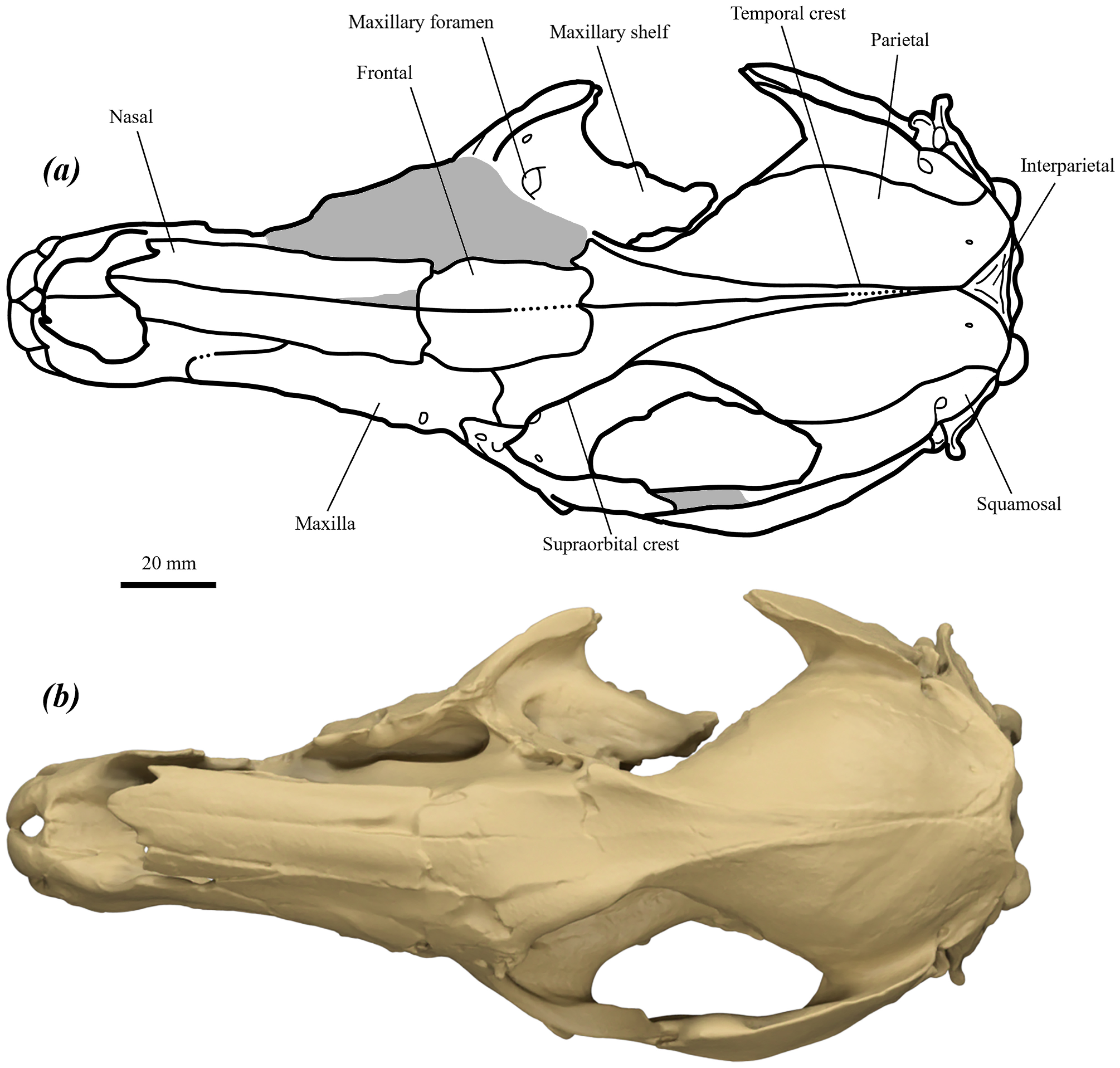
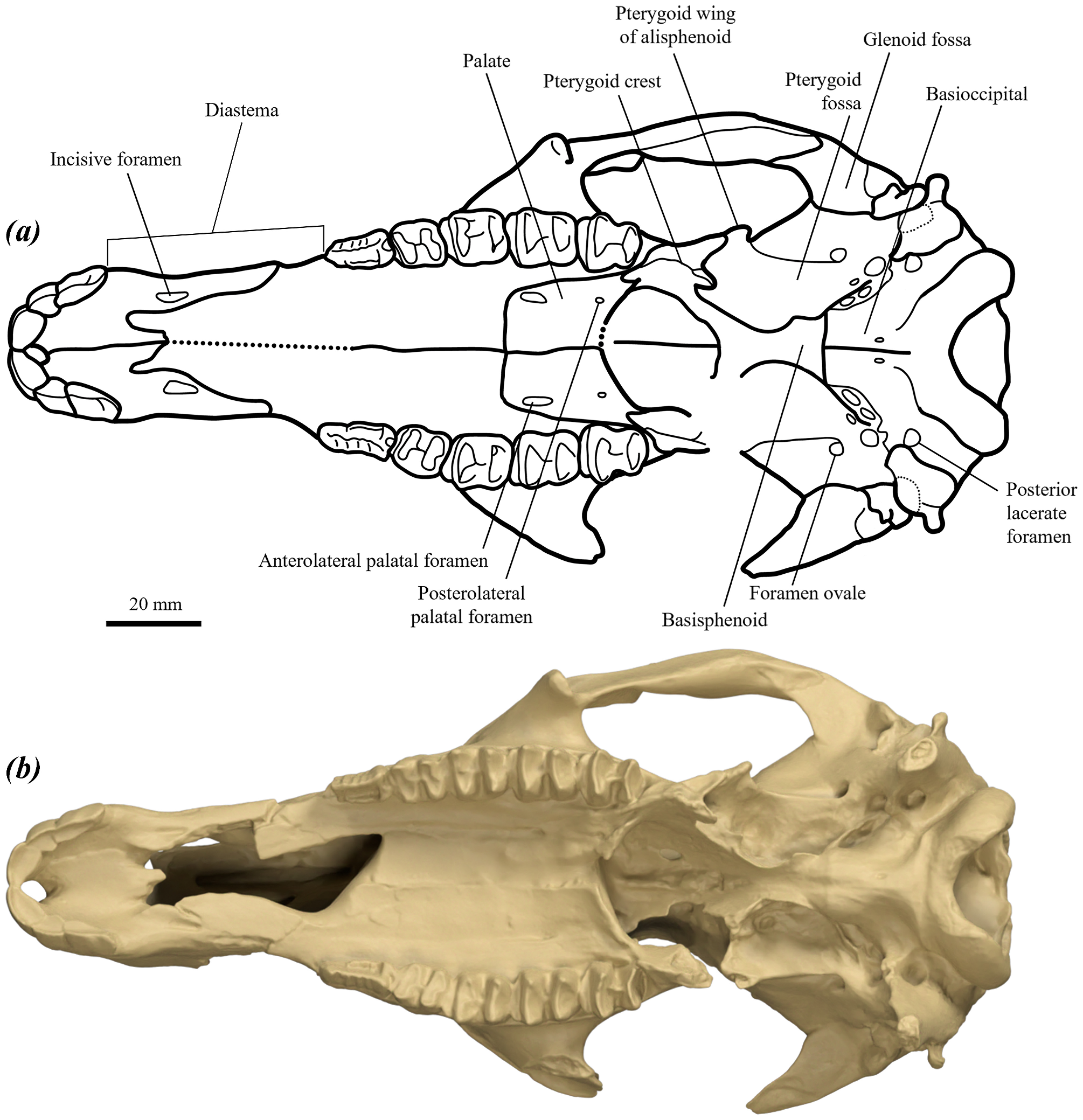
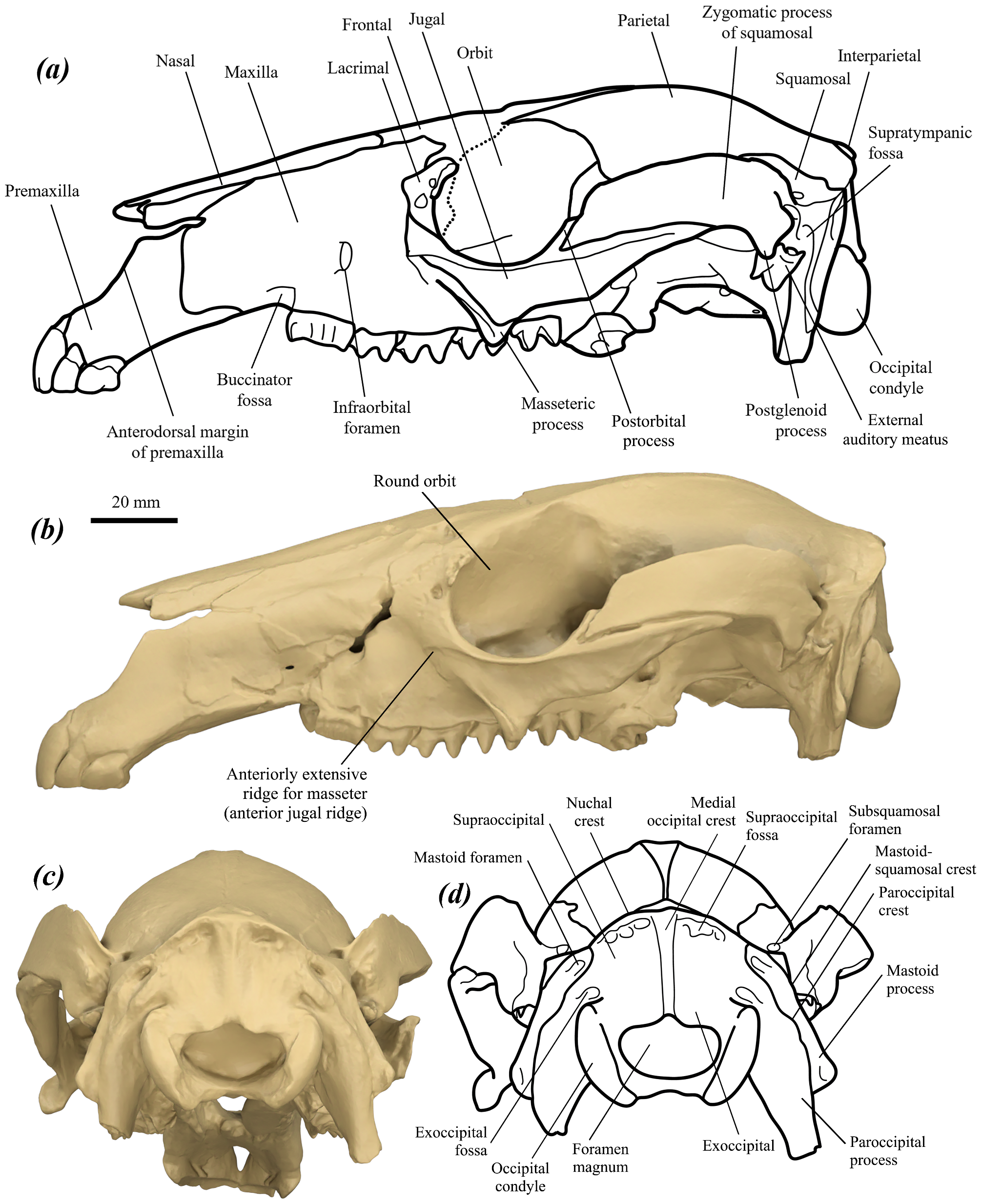
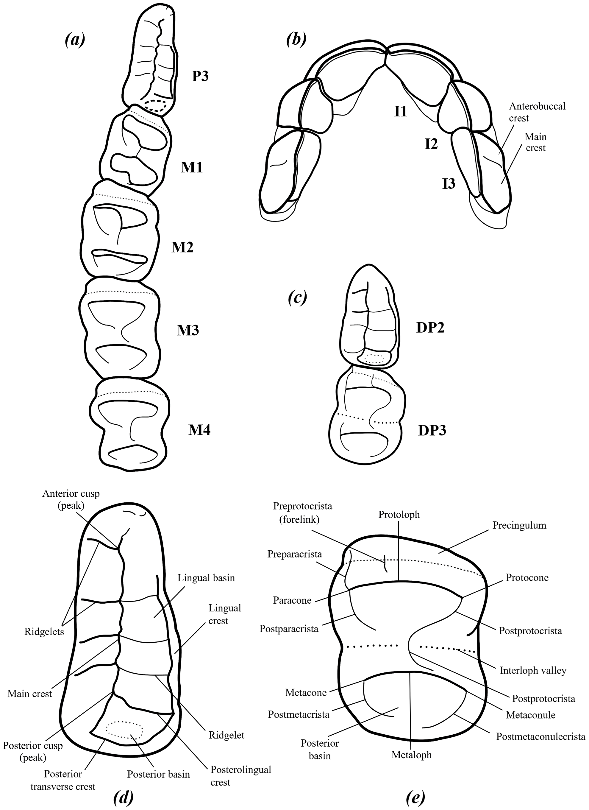

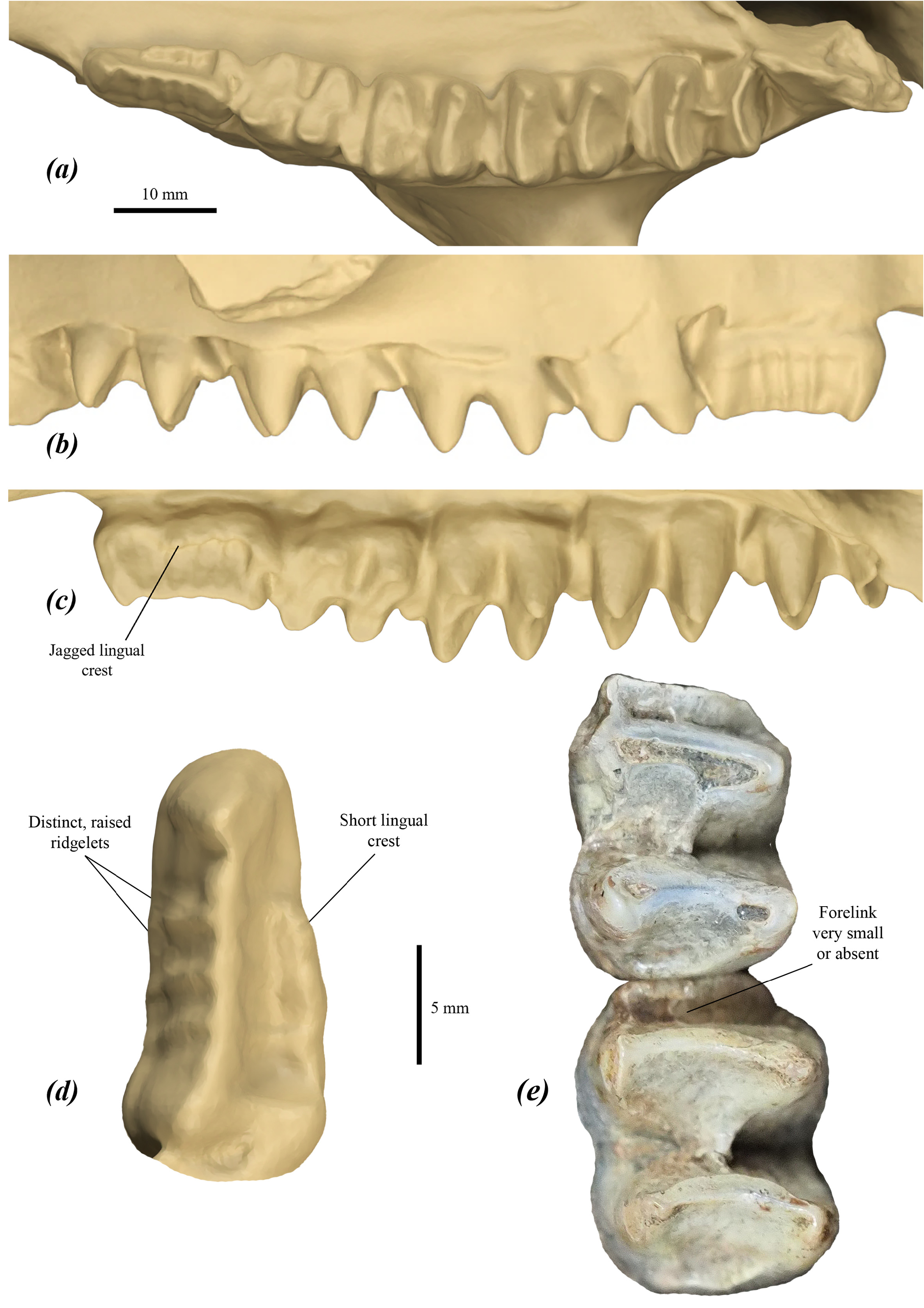
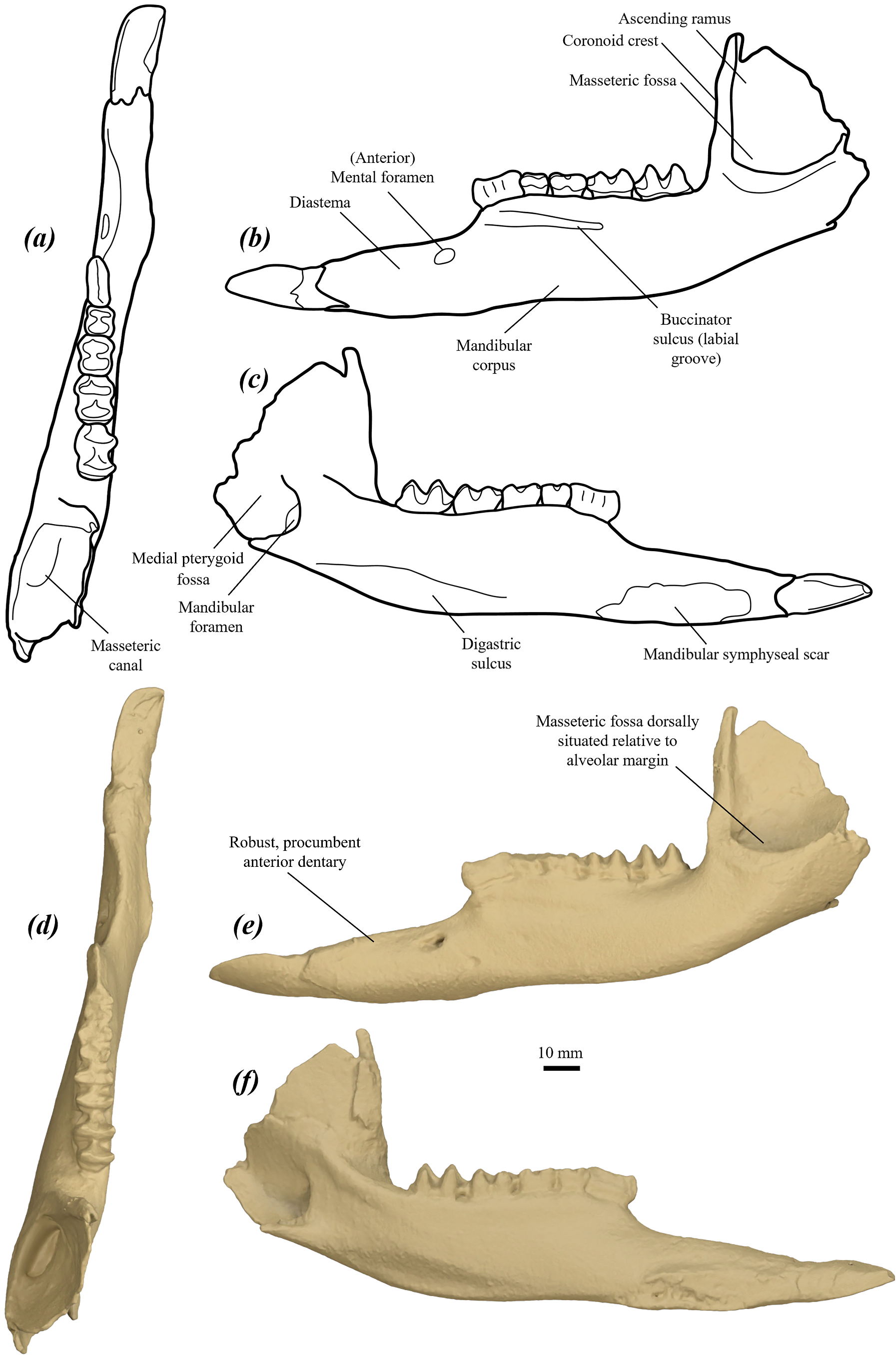
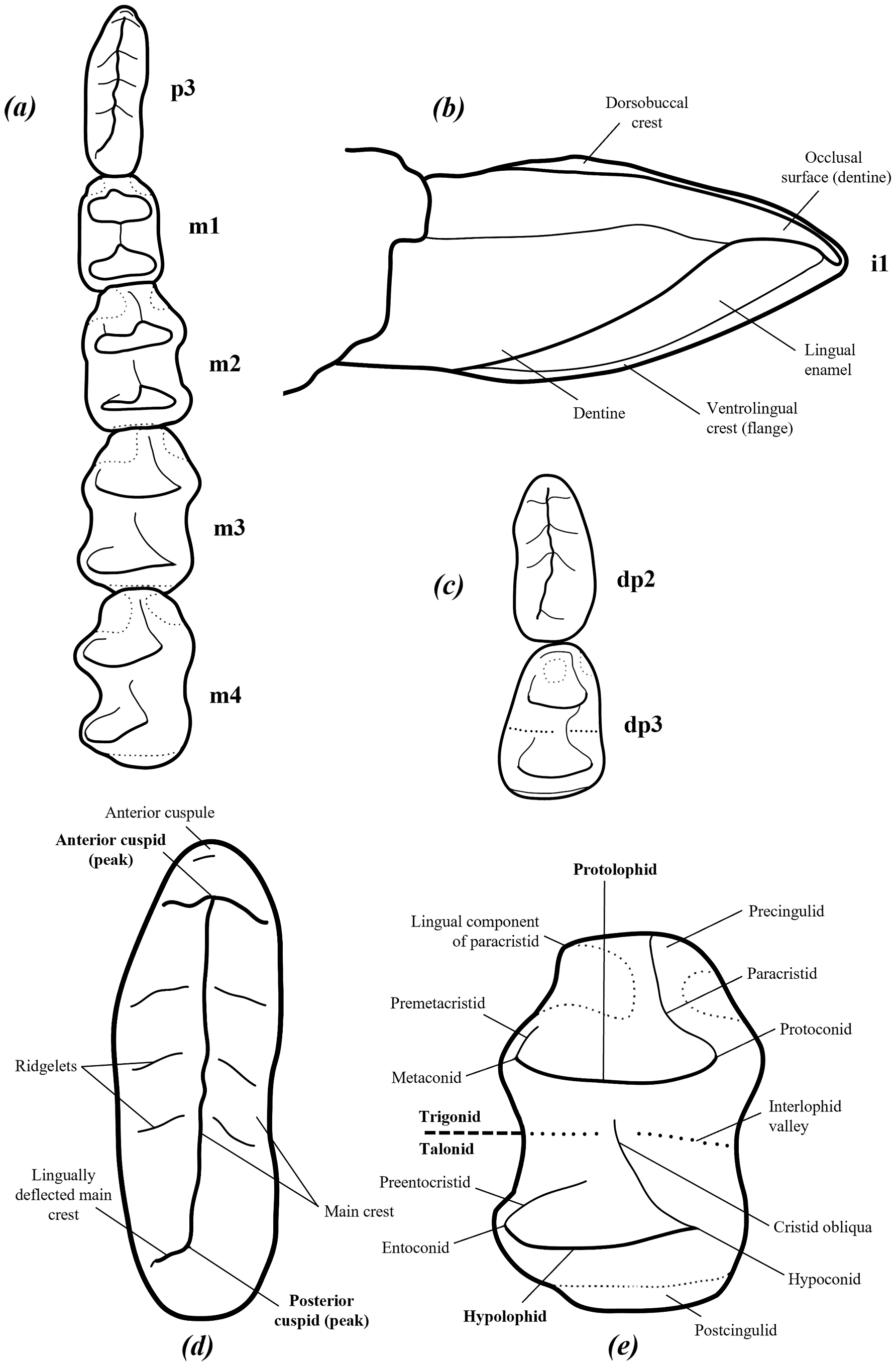
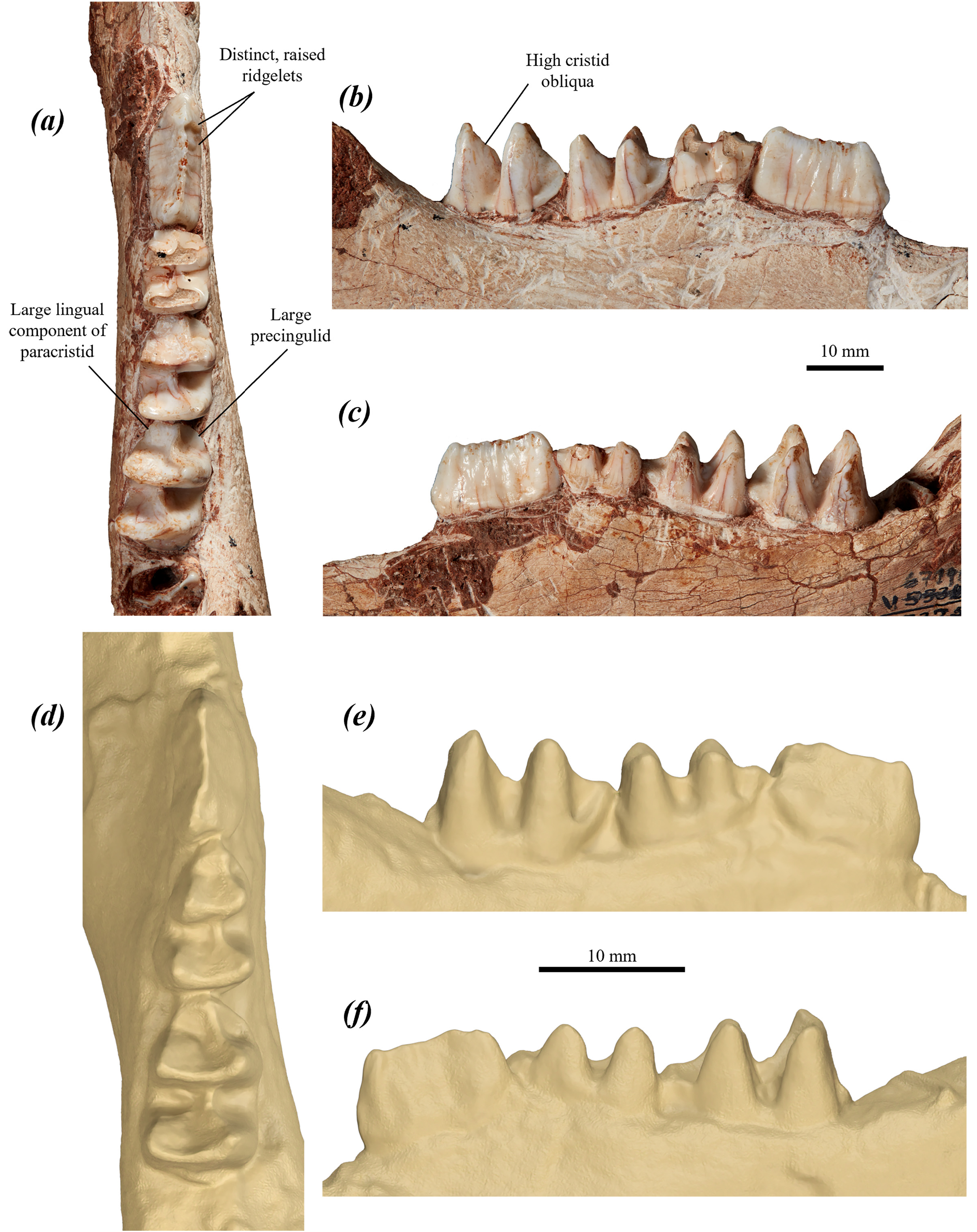
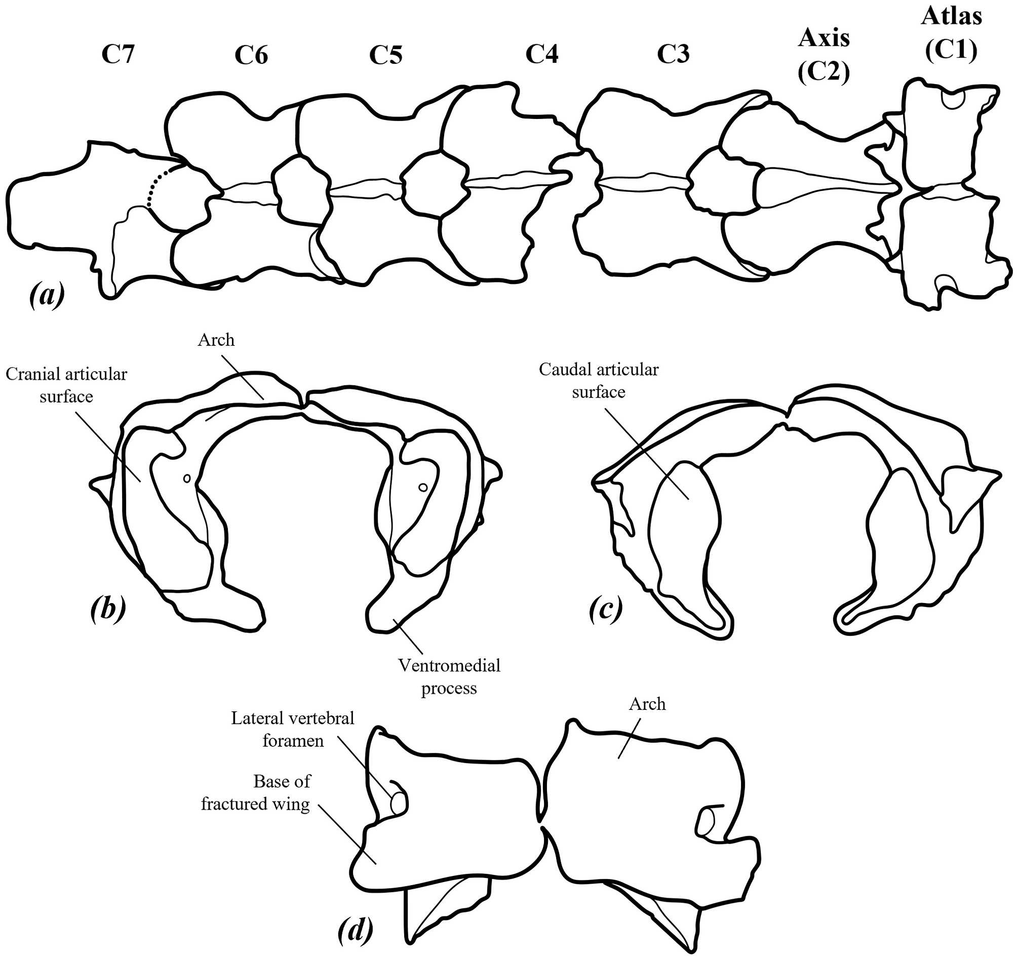
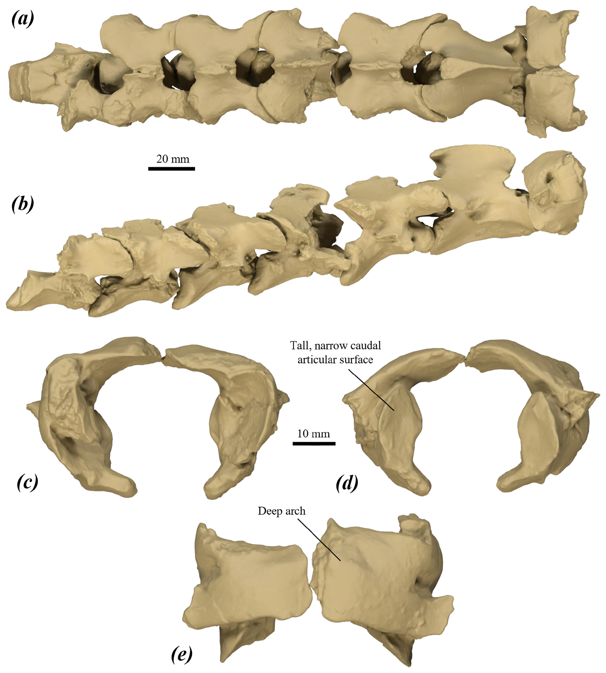
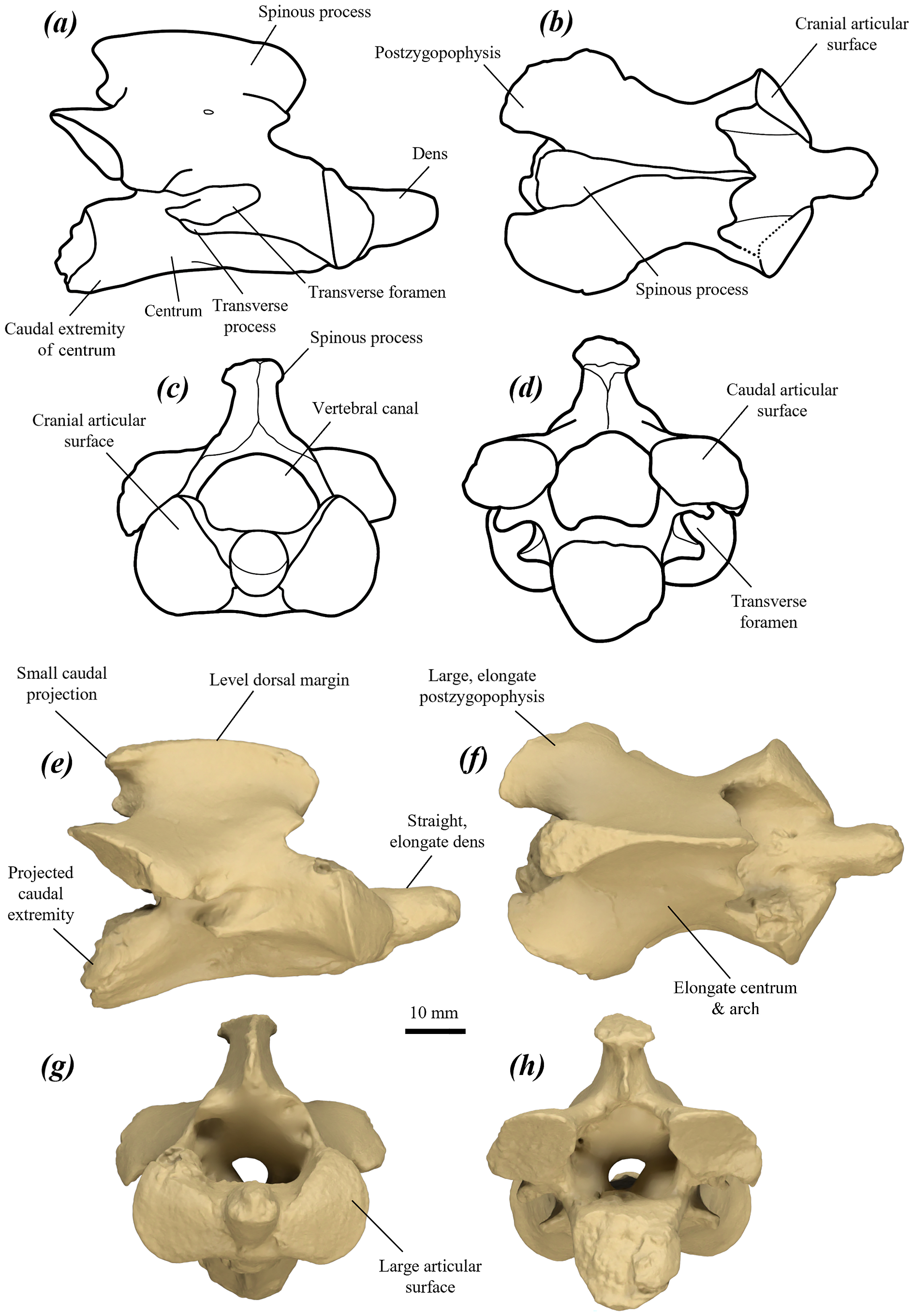
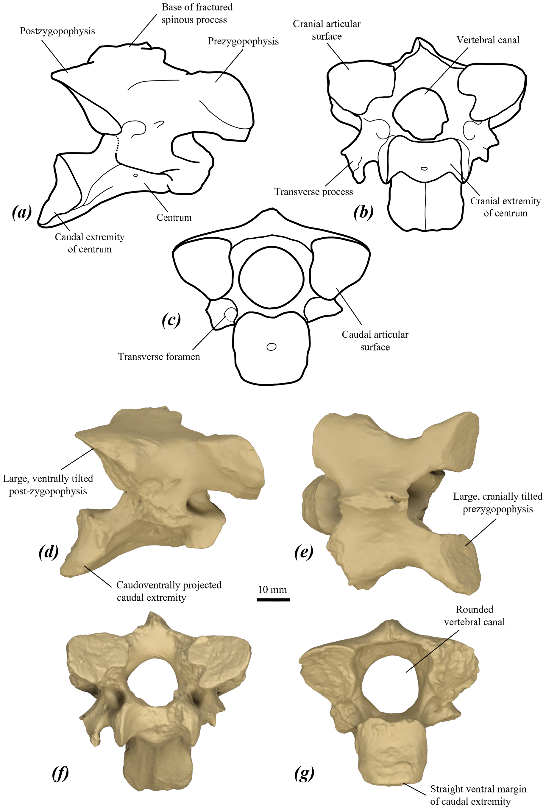
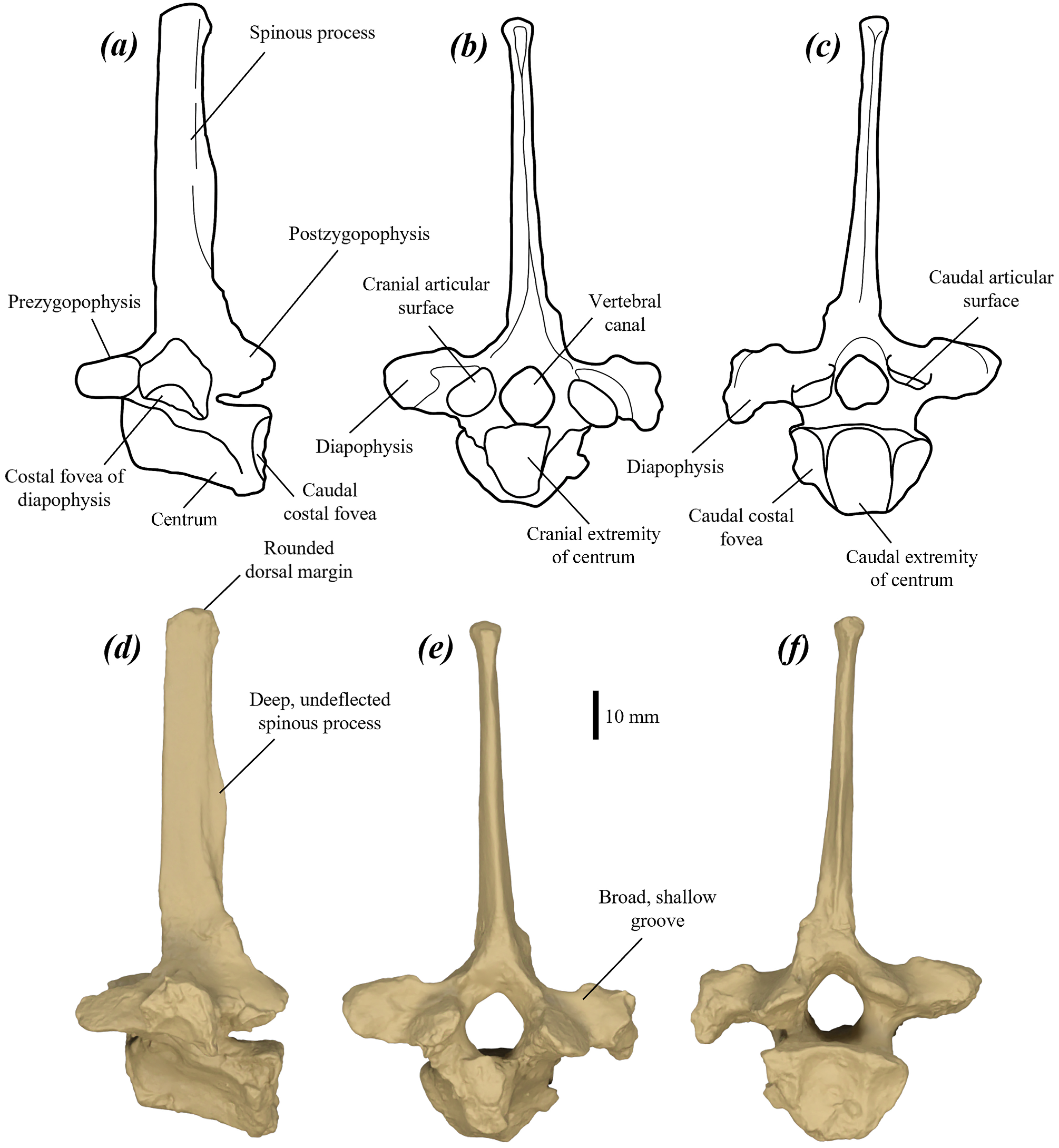
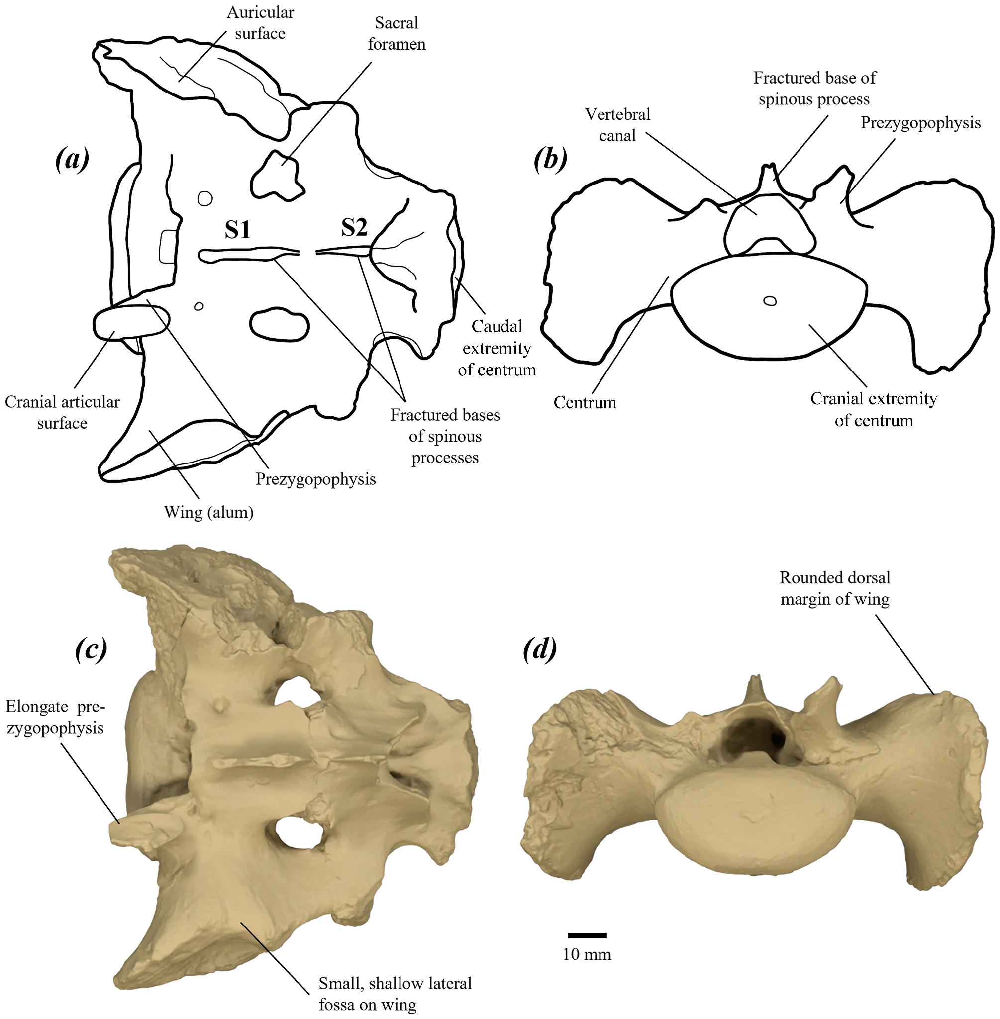
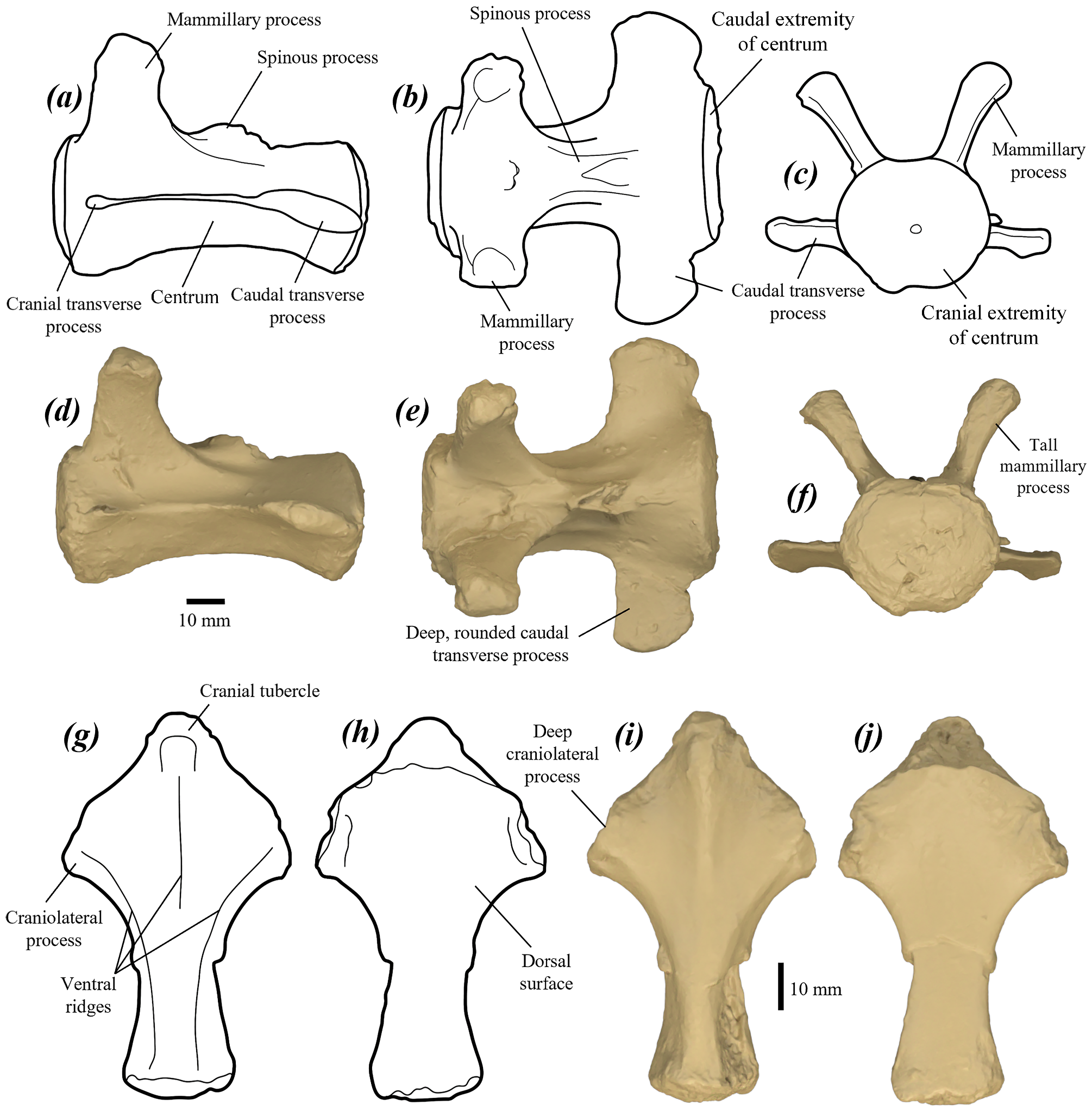
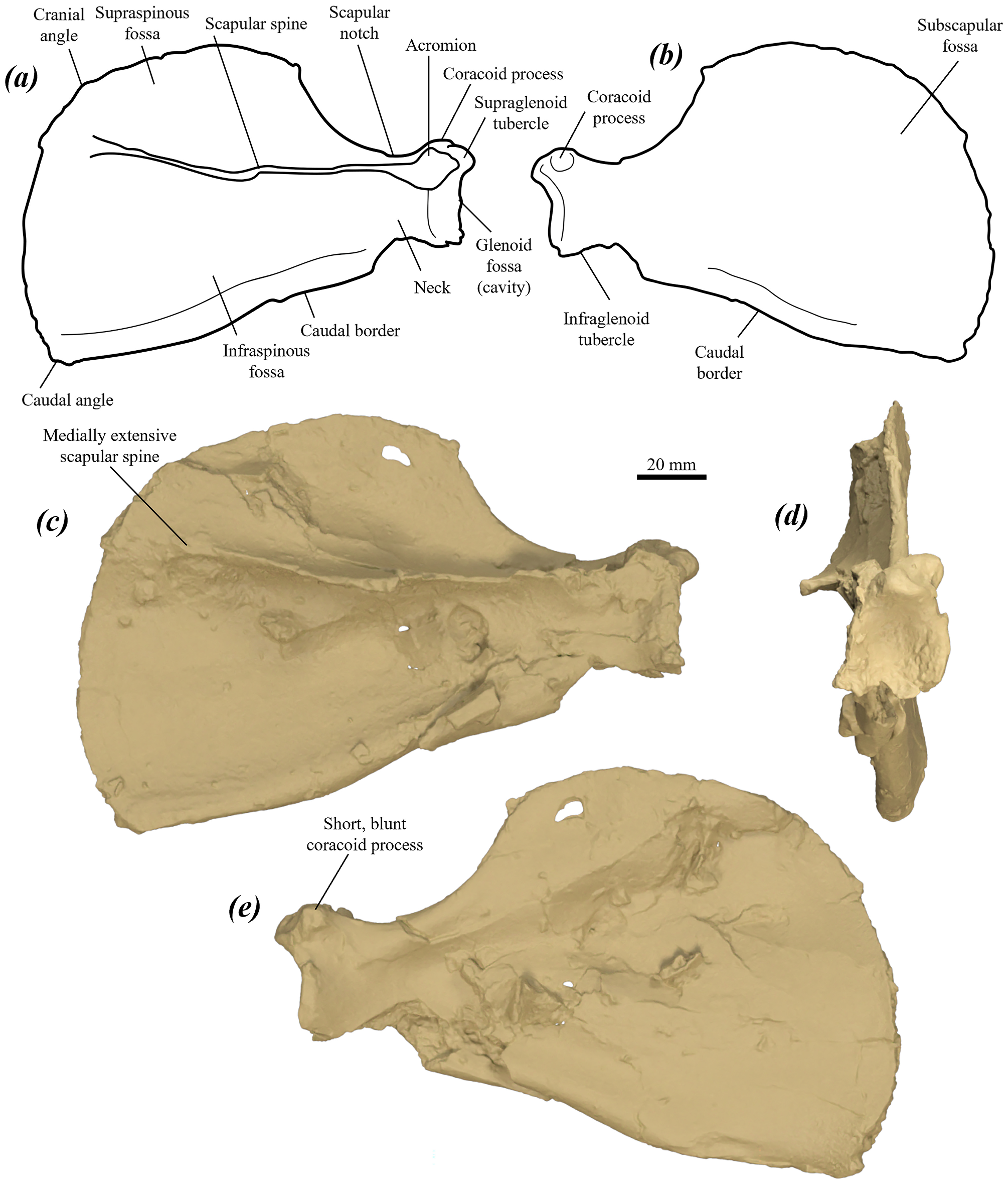
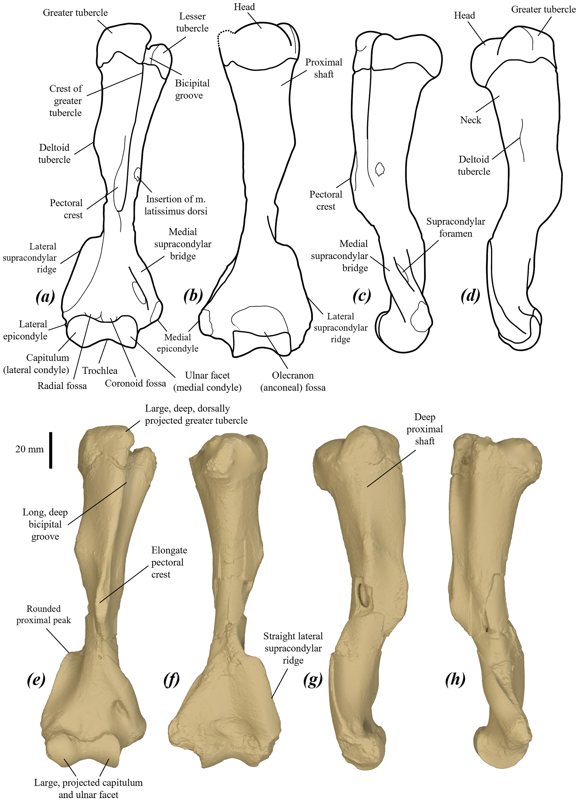
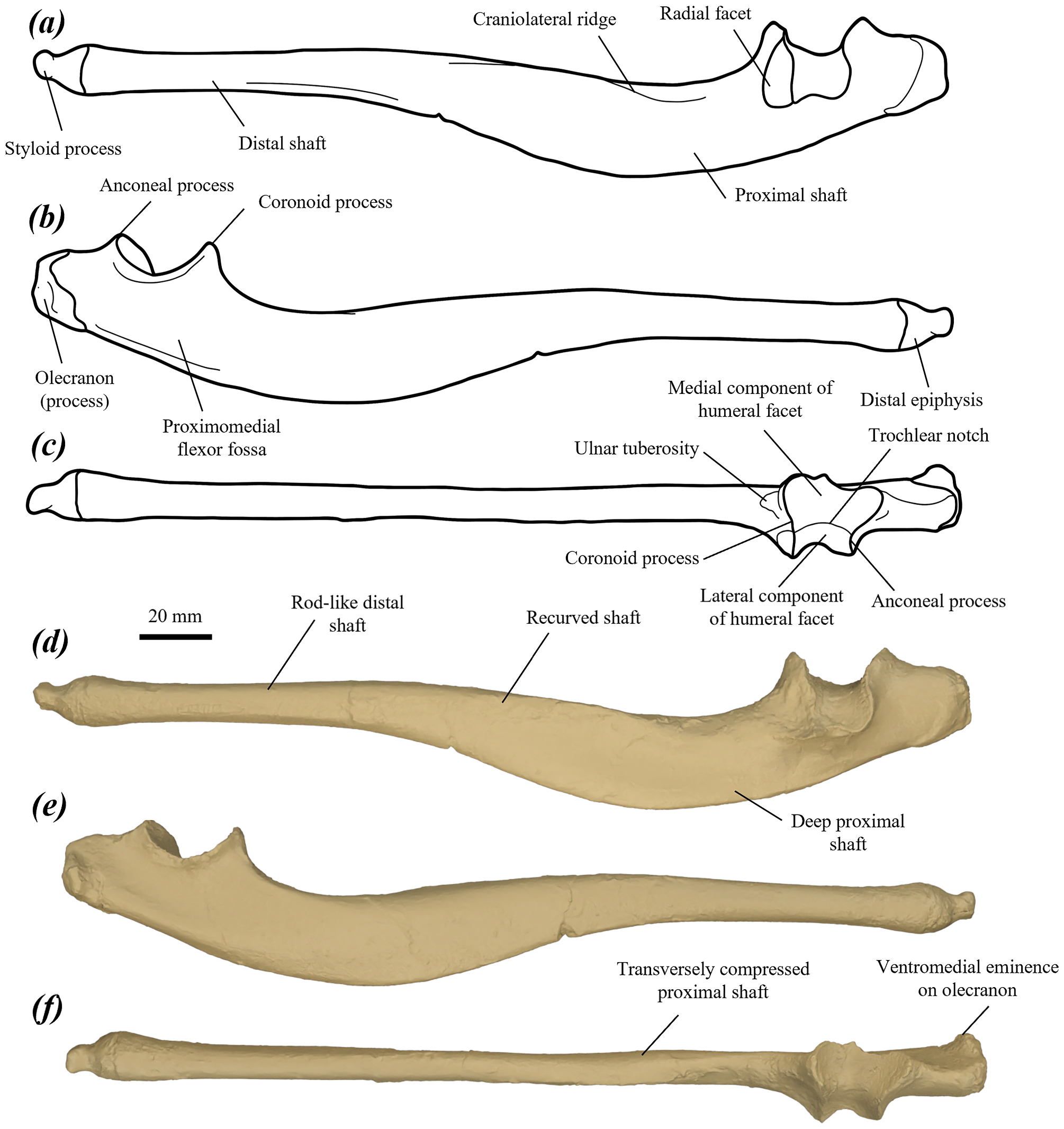
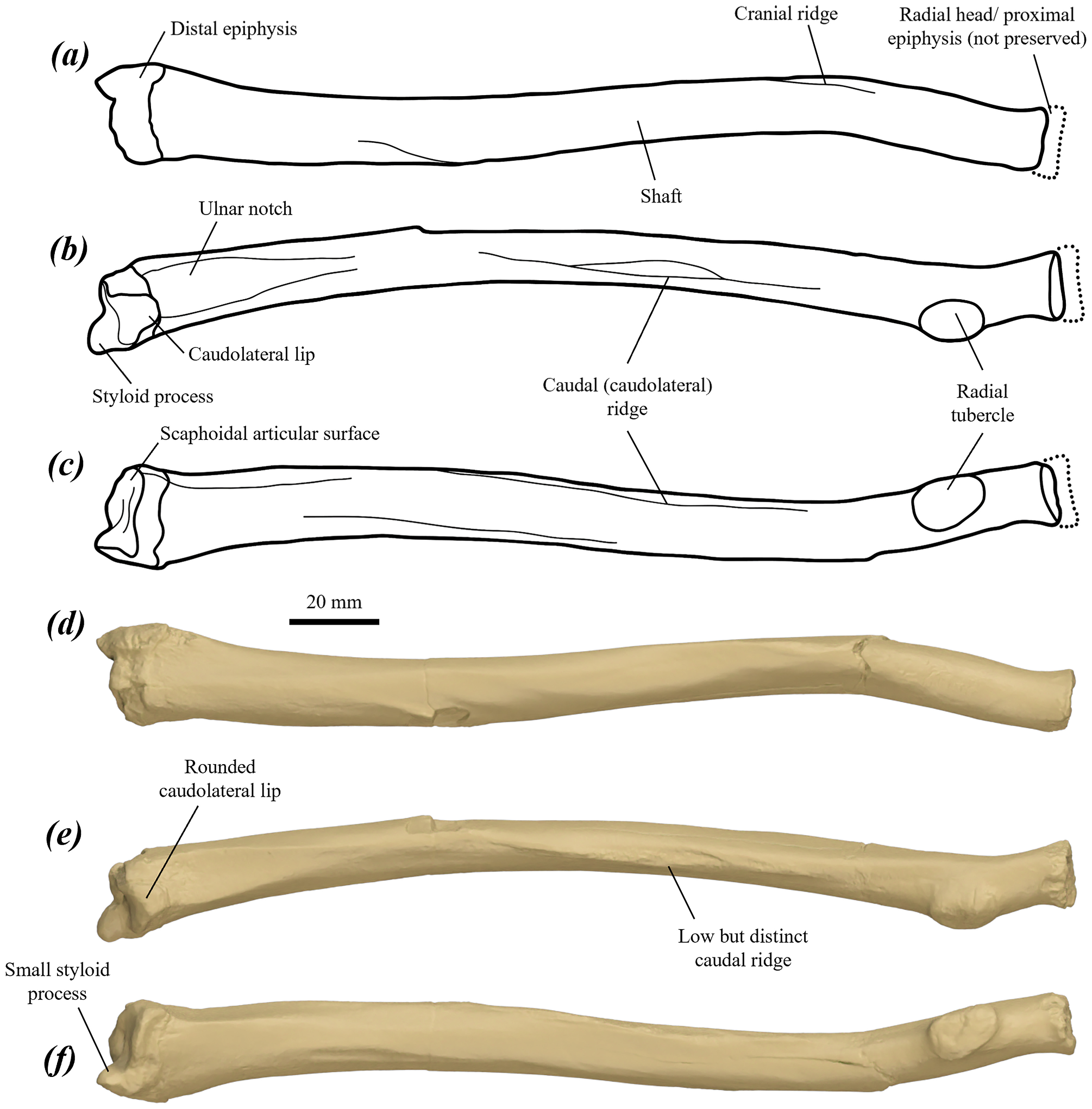
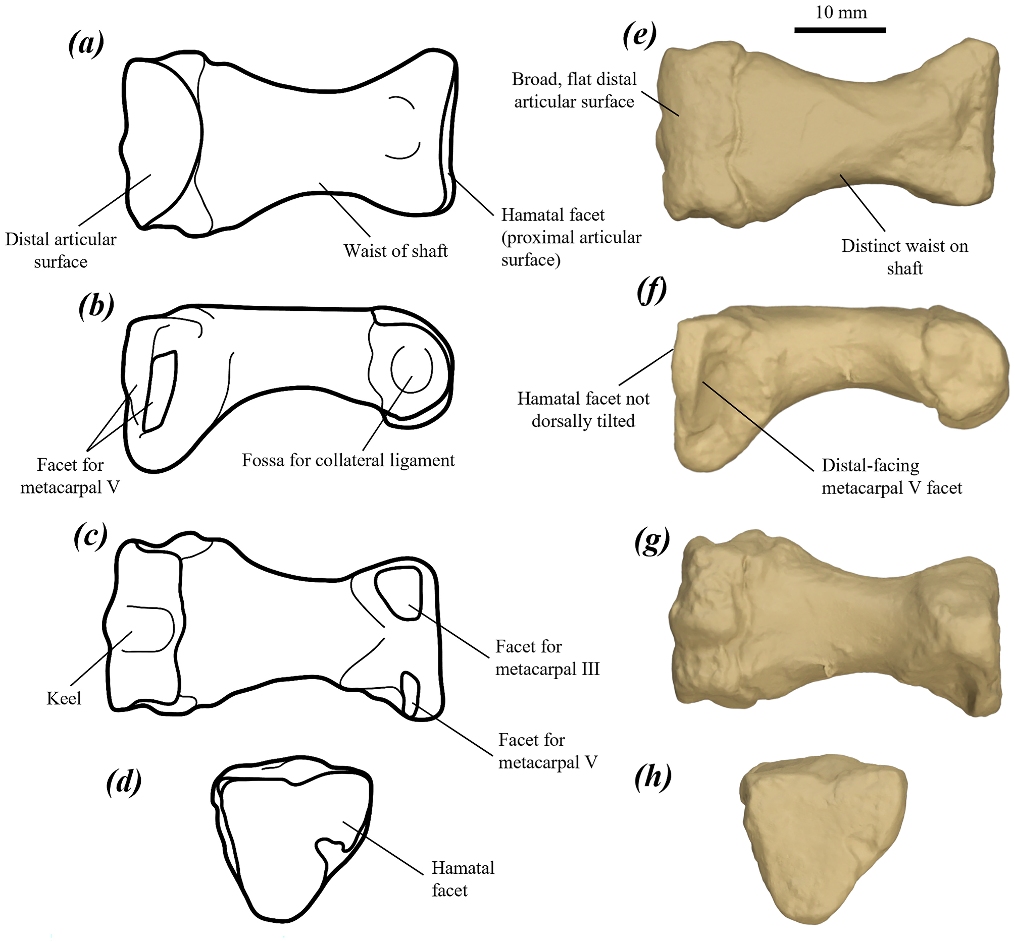
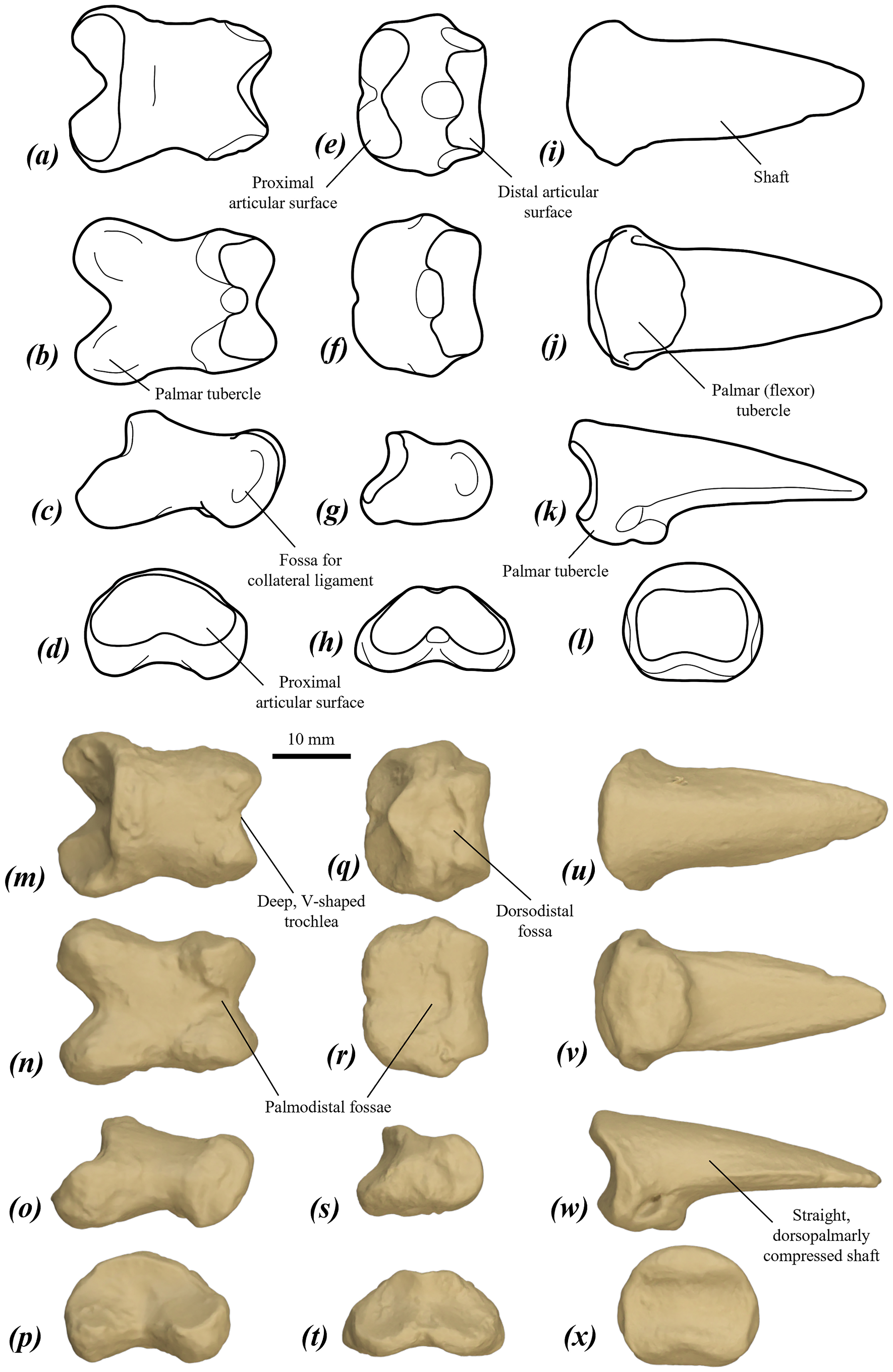
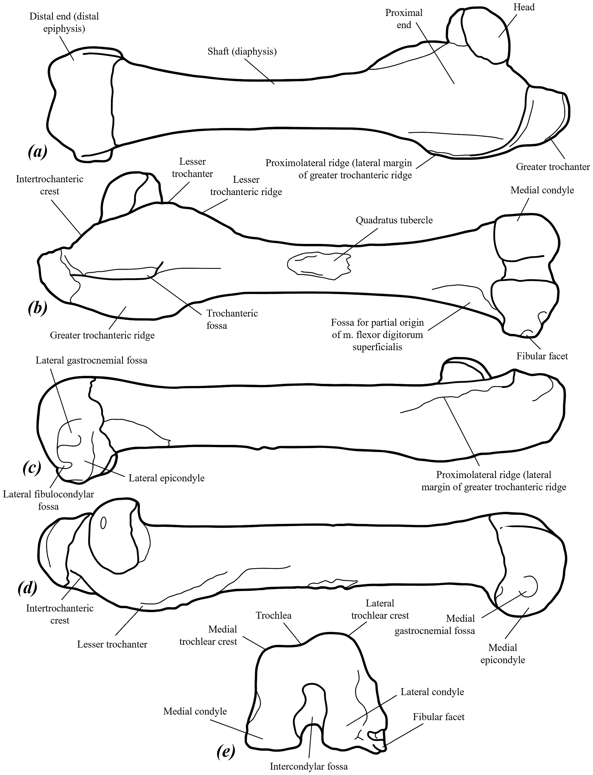
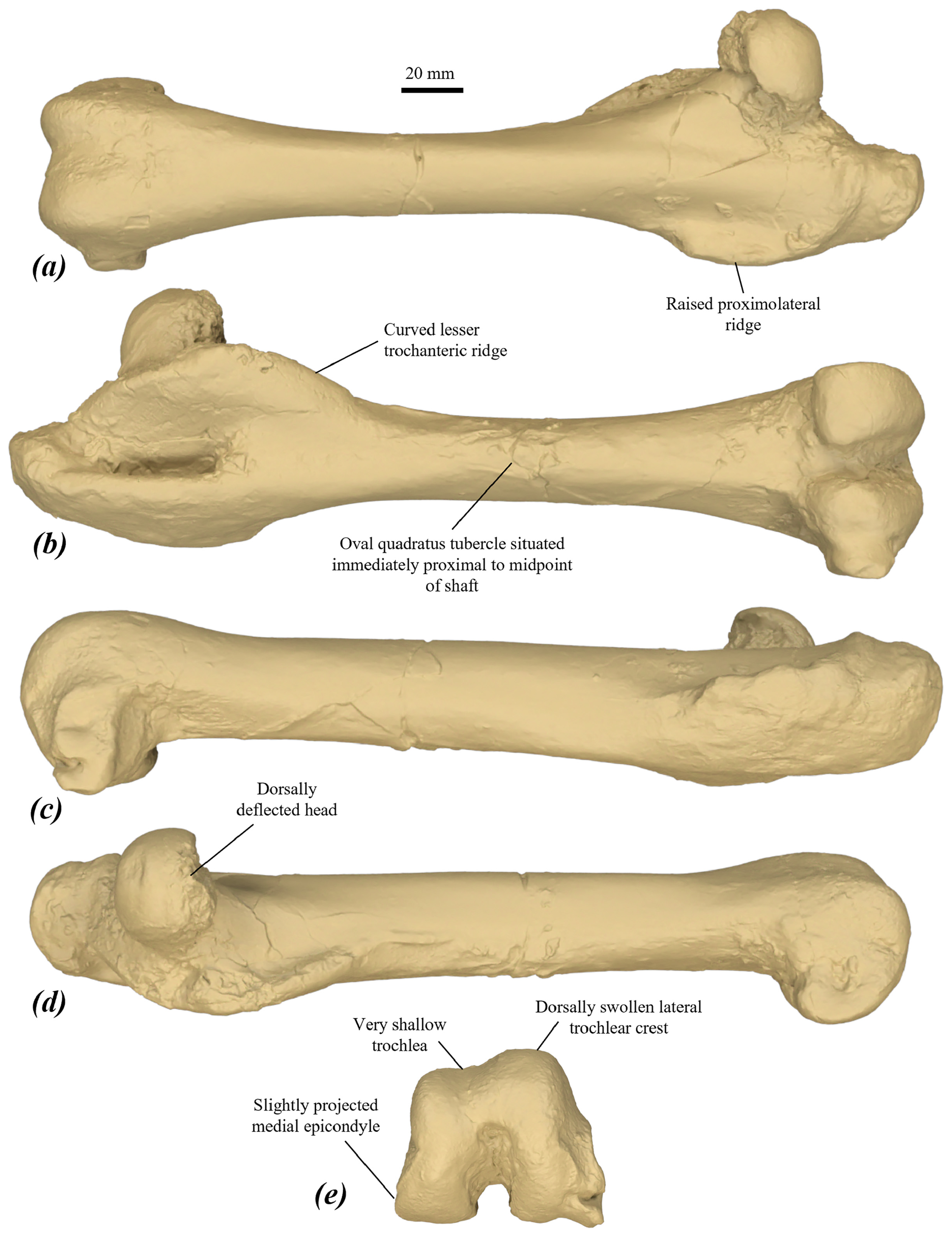
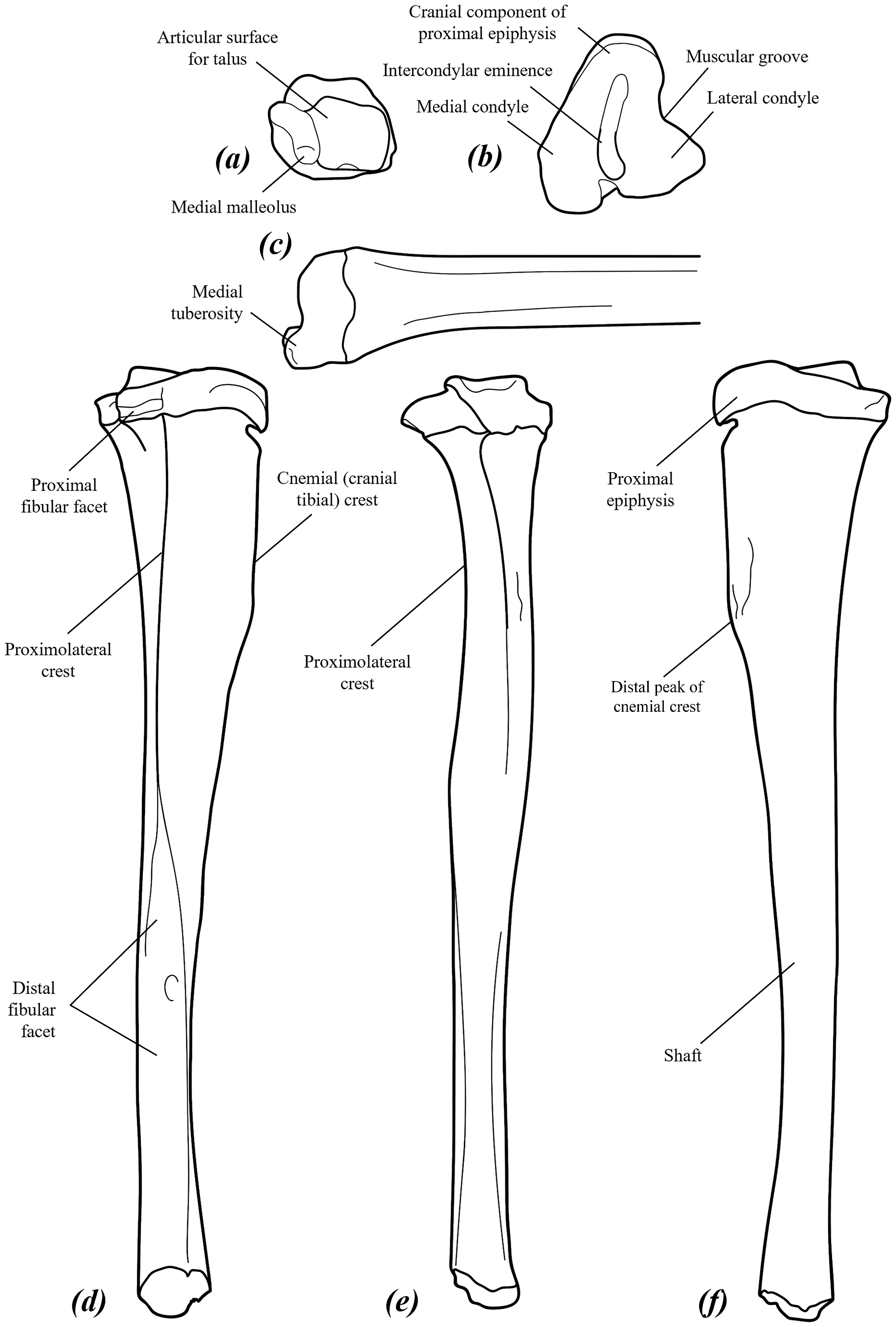
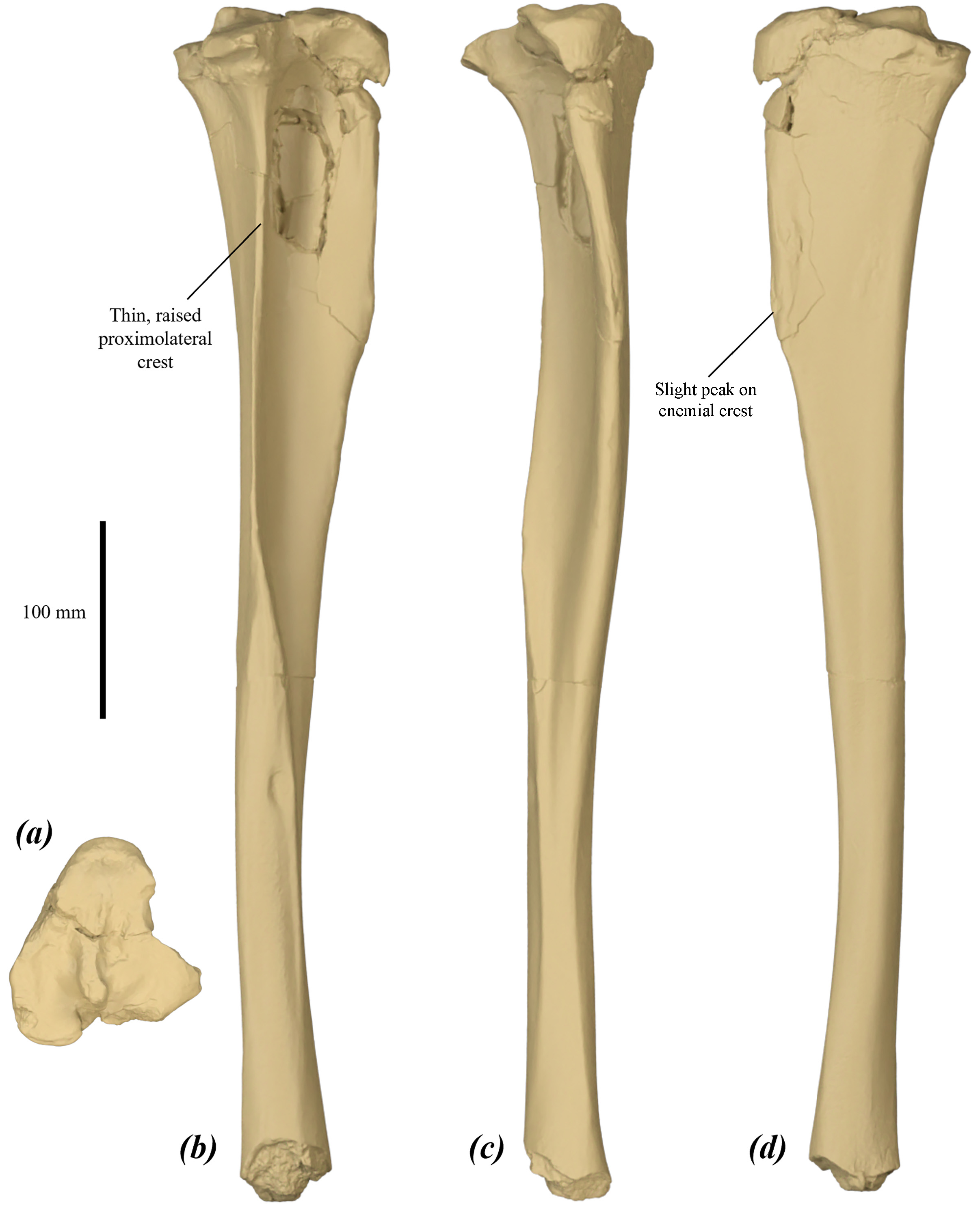
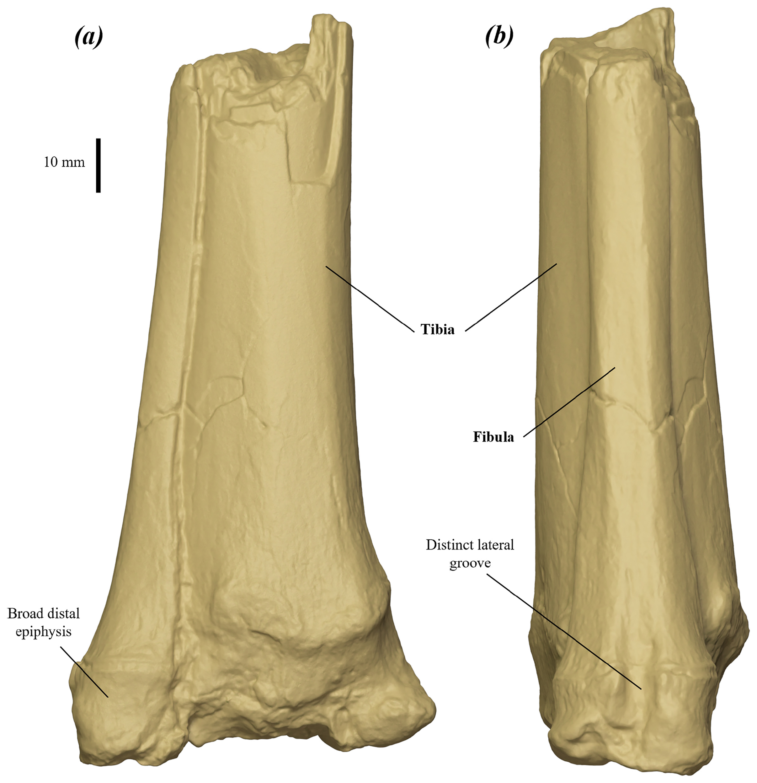
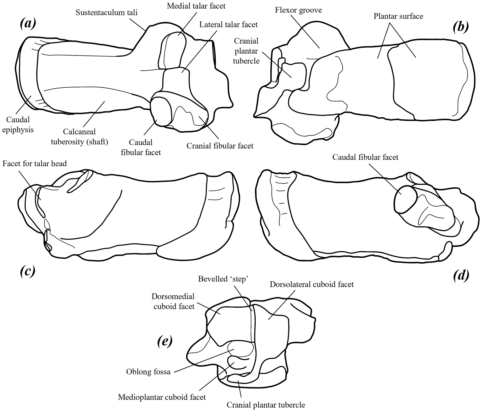
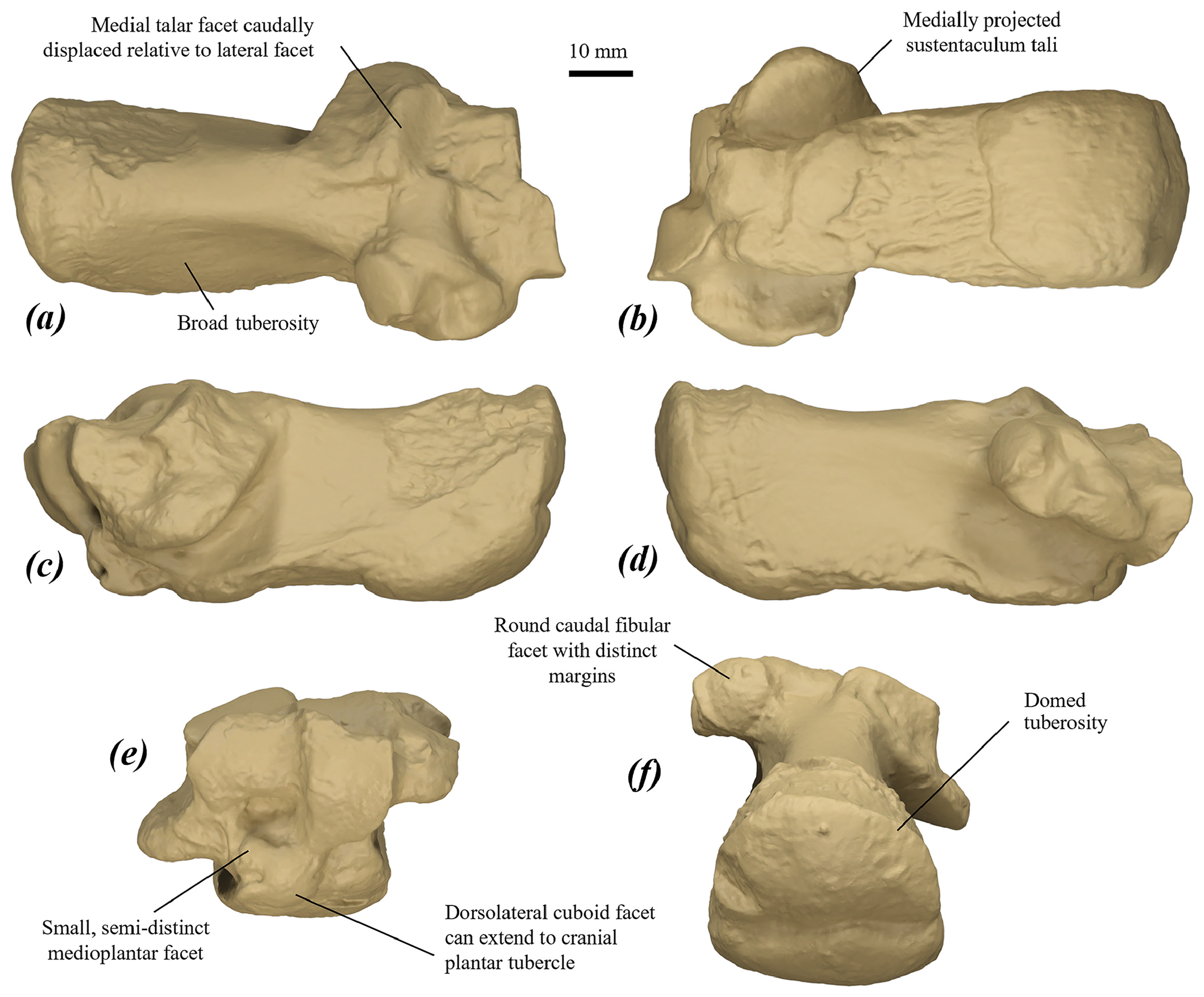
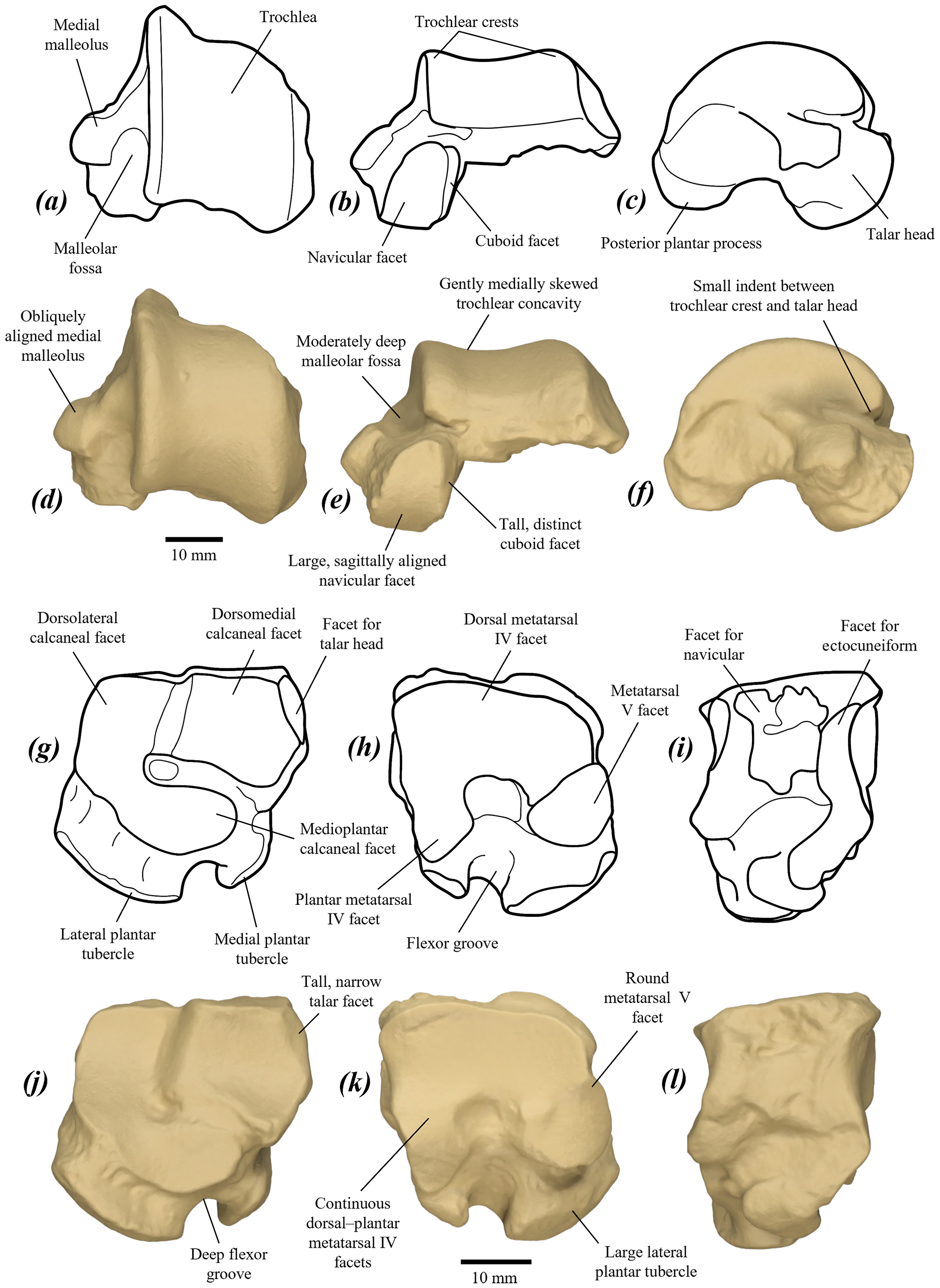
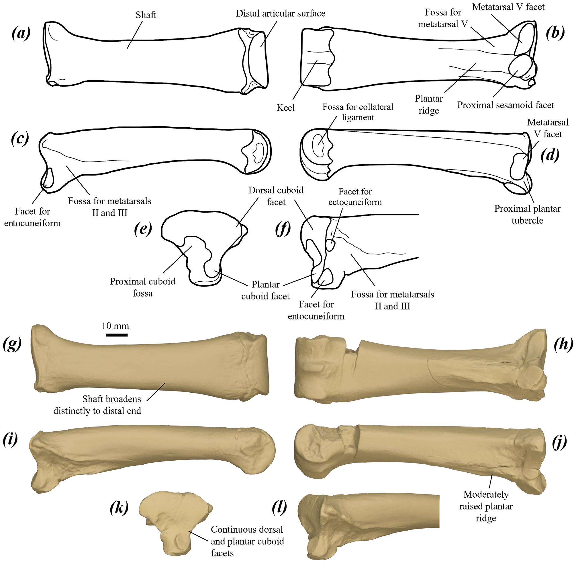
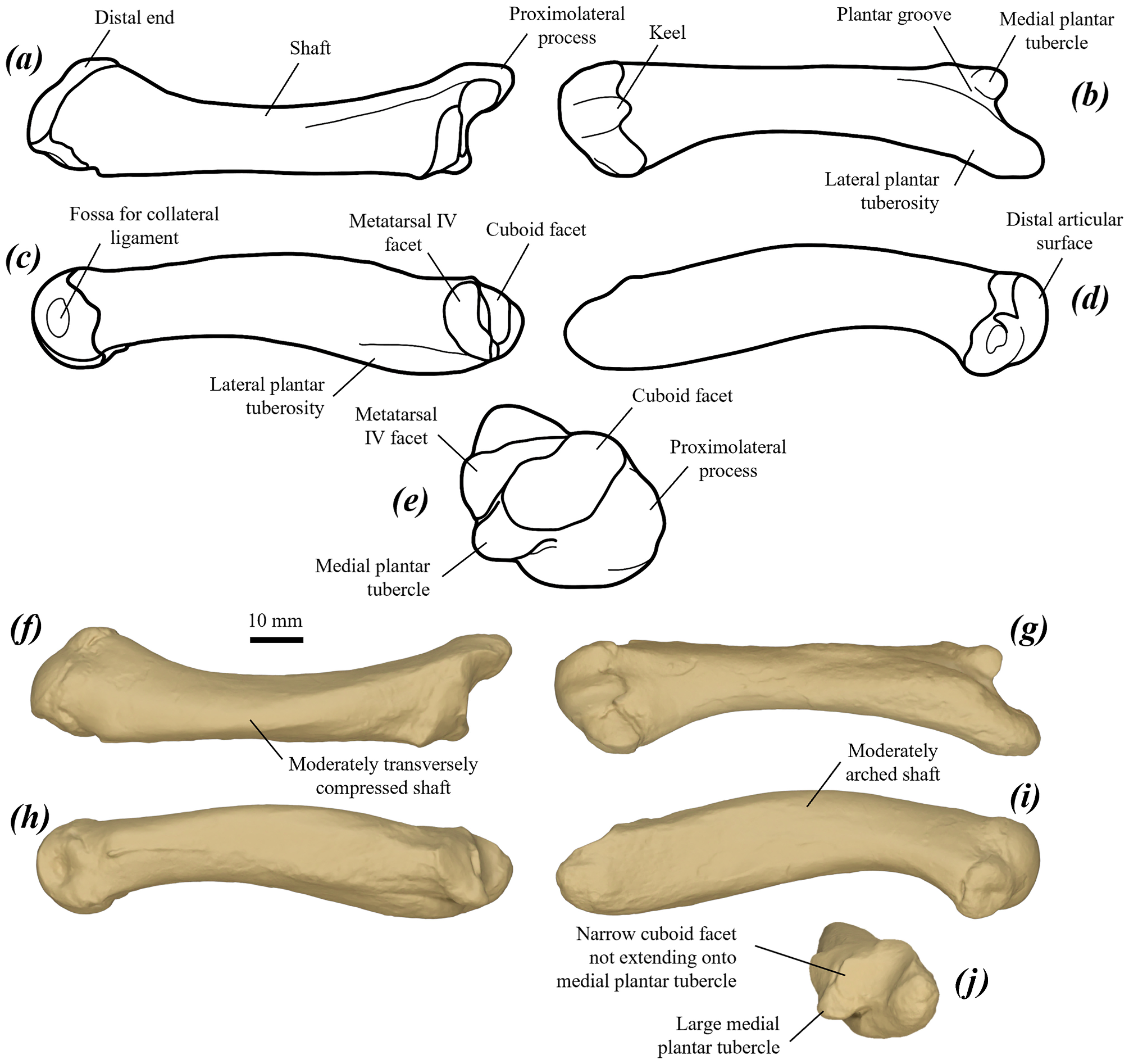
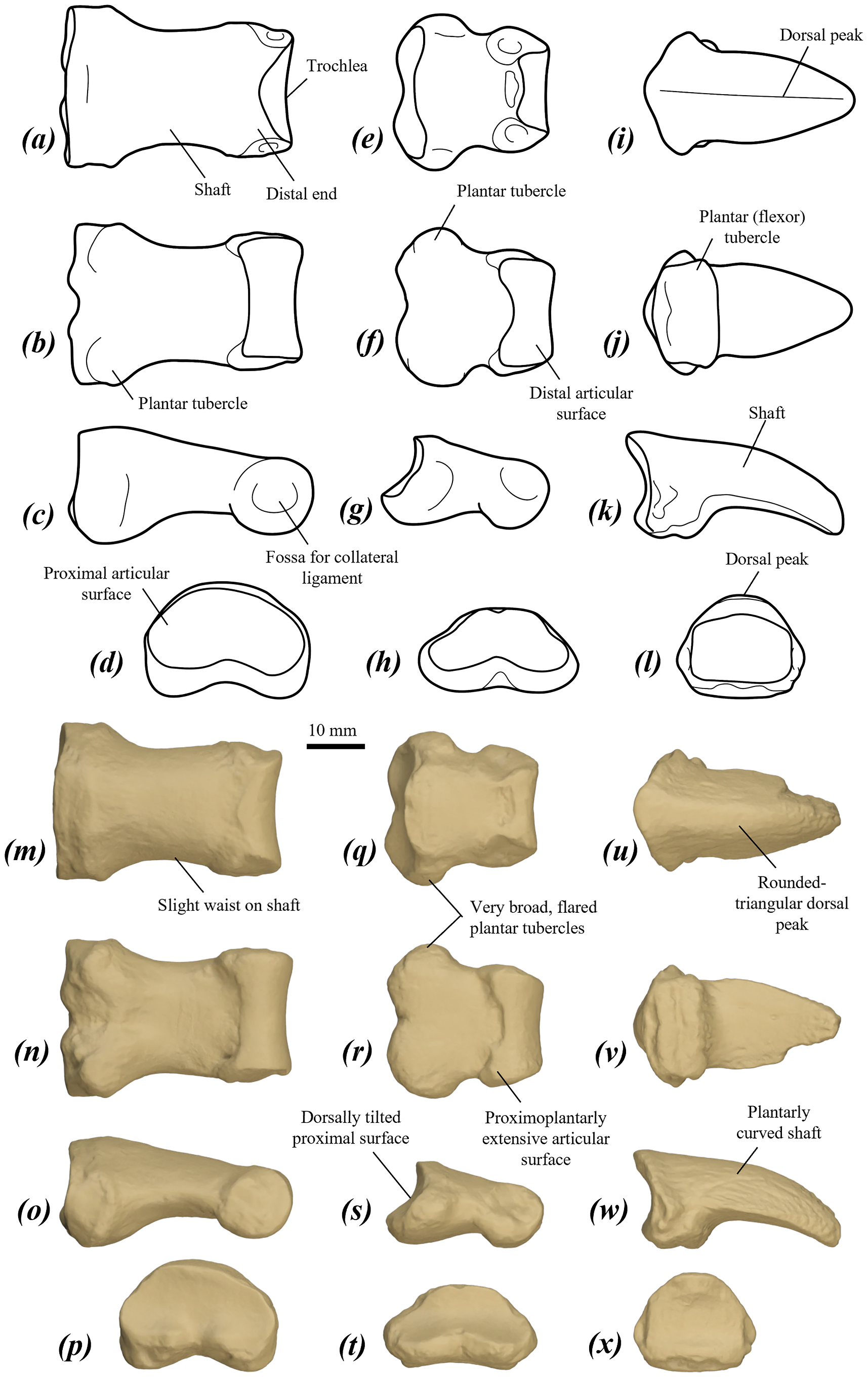
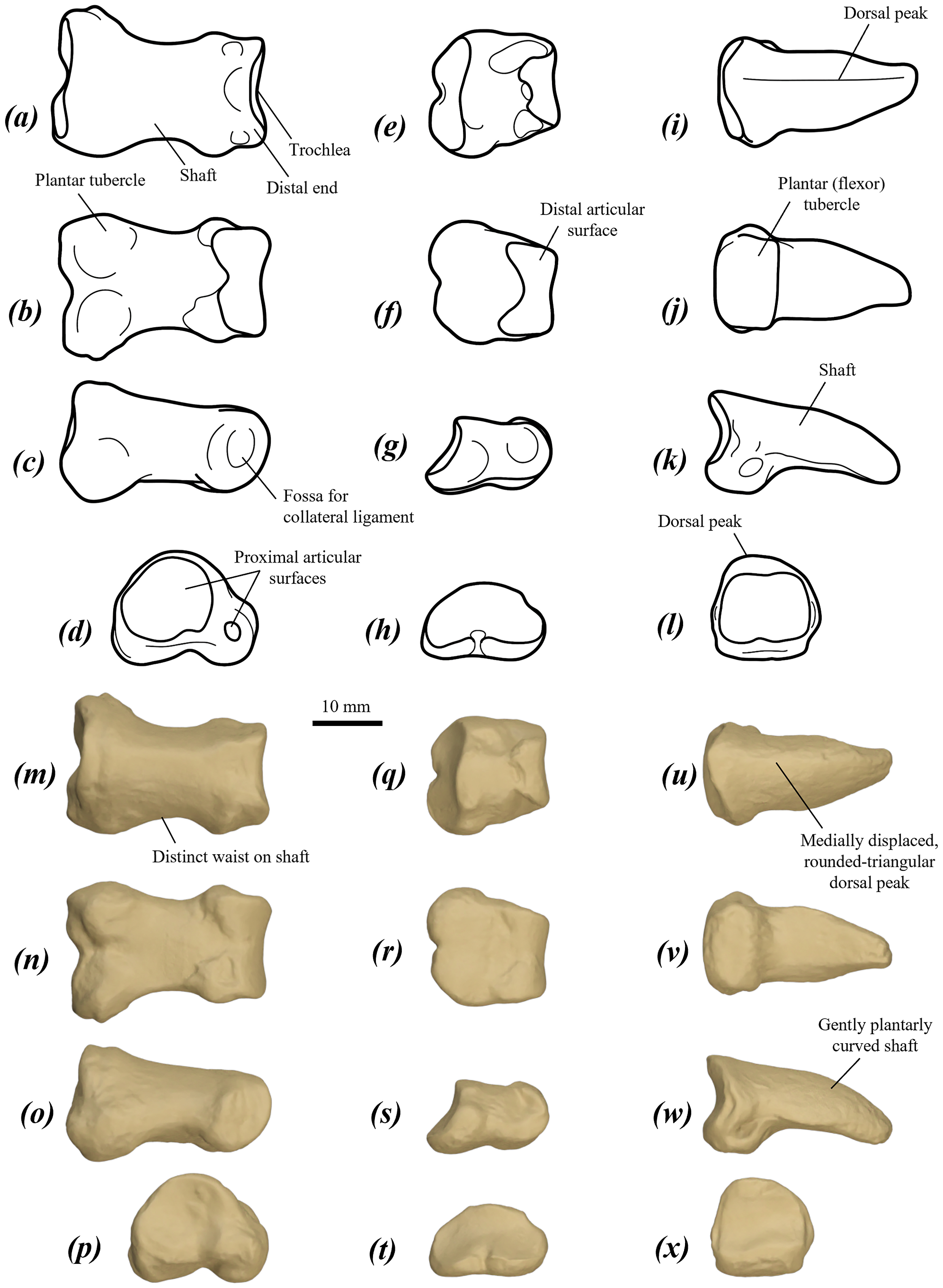
| NHMUK |
NHMUK |
| NHMUK |
Natural History Museum, London |
| AM |
Australian Museum |
| AMNH |
American Museum of Natural History |
| FM |
Department of Nature, Fujian Province Museum |
| QVM |
Queen Victoria Museum |
| QM |
Queensland Museum |
| UCMP |
University of California Museum of Paleontology |
| SAMA |
South Australia Museum |
| NMV |
Museum Victoria |
No known copyright restrictions apply. See Agosti, D., Egloff, W., 2009. Taxonomic information exchange and copyright: the Plazi approach. BMC Research Notes 2009, 2:53 for further explanation.
|
Kingdom |
|
|
Phylum |
|
|
Class |
|
|
Order |
|
|
SubOrder |
Macropodiformes |
|
SuperFamily |
Macropodoidea |
|
Family |
|
|
SubFamily |
Macropodinae |
|
Tribe |
Macropodini |
|
Genus |
Protemnodon anak Owen, 1874
| Kerr, Isaac A. R., Camens, Aaron B., Van Zoelen, Jacob D., Worthy, Trevor H. & Prideaux, Gavin J. 2024 |
Protemnodon anak Owen, 1874
| Jones, B. & Martin-Serra, A. & Rayfield, E. J. & Janis, C. M. 2021: 36 |
| Helgen, K. M. & Wells, R. T. & Kear, B. P. & Gerdtz, W. R. & Flannery, T. F. 2006: 303 |
| Dawson, L. 1985: 66 |
| Stirton, R. A. 1963: 137 |
| Troughton, E. L. G. 1957: 187 |
| Tate, G. H. H. 1948: 297 |
| Tate, G. H. H. & Archbold, R. 1937: 410 |
| Simpson, G. G. 1930: 76 |
| Raven, H. C. 1929: 255 |
| Etheridge, R. & Jack, R. L. 1892: 677 |
Macropus anak (Owen)
| Palmer, T. S. 1904: 883 |
| Lydekker, R. 1896: 257 |
| Lydekker, R. 1894: 257 |
| Flower, W. H. 1884: 715 |
Protemnodon
| Stirton, R. A. 1963: 139 |
| Simpson, G. G. 1930: 76 |
| Palmer, T. S. 1904: 883 |
| Etheridge, R. & Jack, R. L. 1892: 678 |
| Owen, R. 1877: 430 |
Halmaturus anak (Owen)
| Krefft, G. 1875: 208 |