
Protemnodon tumbuna Flannery et al., 1983
|
publication ID |
https://doi.org/ 10.11646/megataxa.11.1.1 |
|
DOI |
https://doi.org/10.5281/zenodo.10993696 |
|
persistent identifier |
https://treatment.plazi.org/id/03E587FD-FF60-D5AE-FF00-710CFCD5FCE9 |
|
treatment provided by |
Felipe |
|
scientific name |
Protemnodon tumbuna Flannery et al., 1983 |
| status |
|
Protemnodon tumbuna Flannery et al., 1983
Protemnodon tumbuna Flannery et al., 1983 : Proc. Lin. Soc. N.S.W., 107, pp. 84–91, figs 4 & 5. Menzies & Ballard (1994), pp. 123–131, figs 6–9. Prideaux et al. (2022), pp. 11–13, 15, figs 7 & 8, table 3.
Protemnodon hopei Flannery, 1992a : Alcheringa, 16, pp. 326–329, fig. 2. See also Helgen et al. (2006), p. 303, Appendix 2.
Holotype: PNG 83-40-8 , a partial R maxilla preserving DP2–M1, with P3 removed from crypt; infraorbital foramen preserved; specimen fractured dorsal to infraorbital foramen, immediately lingual to molar row and posterior to M1. Figured in Flannery (1992a), fig. 4a–d.
Type locality:
Stratum C, Nombe Rockshelter , late Pleistocene Chimbu Province, Papua New Guinea ( Flannery et al. 1983). This rockshelter is situated in mid-Eocene to early Oligocene Chimbu Limestone, on the northeastern face of Mt Elimbari ridge. Altitude is ~ 1720 m above sea level ( Flannery et al. 1983).
Paratype:
PNG 82-40-20, a semi-complete L dentary preserving i1, partial p3 and m1–m4. Anterior of diastema abraded; fractured ascending ramus, coronoid process and ventral masseteric fossa missing fragments; mandibular condyle and angular process absent. Collected during archaeological excavations led by M-J. Mountain from 1978–1980.
Referred specimens:
Papua New Guinea
Nombe Rockshelter , Chimbu Province: NCA X2-13- 241 partial R ulna; NCA A1-18 partial ilium and L metatarsal V; NCA M71-9 partial R ulna; NCA H71- 9 partial R calcaneus; NCA 071 R metatarsal V.
LOG site, Haeapugua Basin, Tari Province: PM 25622 semi-complete skeleton (specimen missing).
LOK site, Haeapugua Basin, Tari Province: PM 25623 partial L dentary.
Central Papua
Kelangurr Cave, West Baliem Valley: AM F83613 maxillae; AM F83413 partial L dentary; AM F83612 R dentary; AM F83614 partial juvenile L dentary; AM F113144 cervical vertebra C3; AM F88917 caudal vertebra (Ca8?); AM F113142 juvenile R humerus; AM F88914 partial L ulna; AM F88925 partial L radius; AM F113140 partial ilium; AM F113143 juvenile ilium; AM F113141 partial L tibia; AM F88922 partial fibula; AM F88921 partial L calcaneus; AM F88929 partial L calcaneus; AM F88927 partial L metatarsal IV; AM F88930 partial R metatarsal IV; AM F113146 L metatarsal V.
West Baliem River, near Ambowak village, West Baliem Valley: AM F83611 L I1; AM F88923, AM F88924 L femur and tibia.
Revised specific diagnosis:
Protemnodon tumbuna is separated from the other species of Protemnodon by several skeletal autapomorphies and by a unique combination of craniodental and postcranial characteristics. The hindlimb of P. tumbuna differs from all species of Protemnodon in having: a more gracile femur with a more elongate, more distally situated quadratus tubercle; a more robust tibia with a particularly low proximolateral crest, especially distally, and a more elongate cnemial crest; a calcaneus with a distinctly convex, caudomedially flared plantar surface that extends dorsolaterally onto the lateral surface of the calcaneal tuberosity; and a shorter, more robust metatarsal V with a relatively shorter lateral plantar tuberosity.
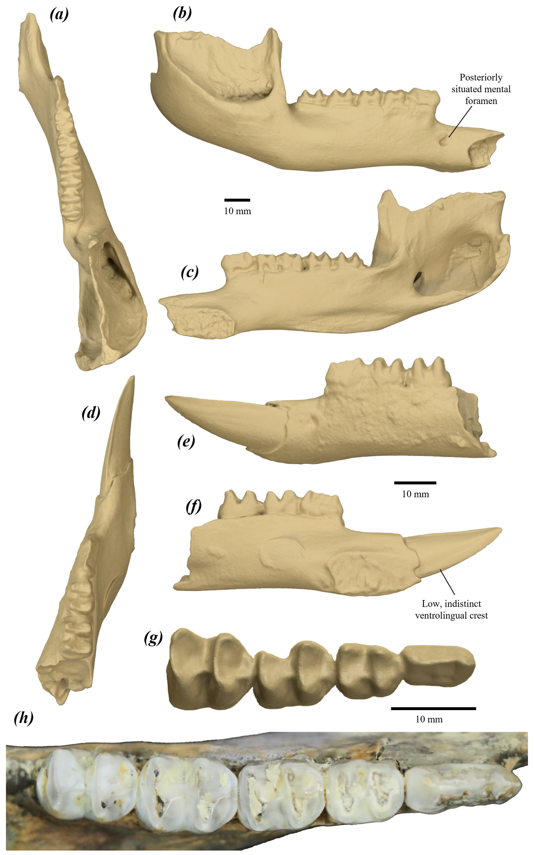
The dentary of P. tumbuna most closely resembles that of P. otibandus , but differs in its more posteroventrally situated mental foramen.The dentition of P. tumbuna is most similar to that of P. otibandus , but differs in having: P3 with three (rather than four or five) less raised and less distinct buccal ridgelets; more rounded molars in occlusal view; and i1 with a less raised, less distinct ventrolingual crest.
The hindlimb of P. tumbuna is morphologically closest to that of P. otibandus . It further differs from that of P. otibandus by having: a tibia with a thicker proximolateral crest; calcaneus with a larger, deeper fossa cranial to the lateral talar facet; and metatarsal V with a taller proximolateral process, a proximodistally shorter metatarsal IV facet, and a smaller medial plantar tubercle.
Etymology:
The name tumbuna means ‘ancestor’ or ‘of the ancestors’ in Tok Pisin (Neo-Melanesian Pidgin). It is used in doubleallusion, first in reference to the primitive morphology of some aspects of the dentition the species, and second, to its association with the Pleistocene human inhabitants of highland Papua New Guinea ( Flannery et al. 1983, p. 84).
Description and comparisons:
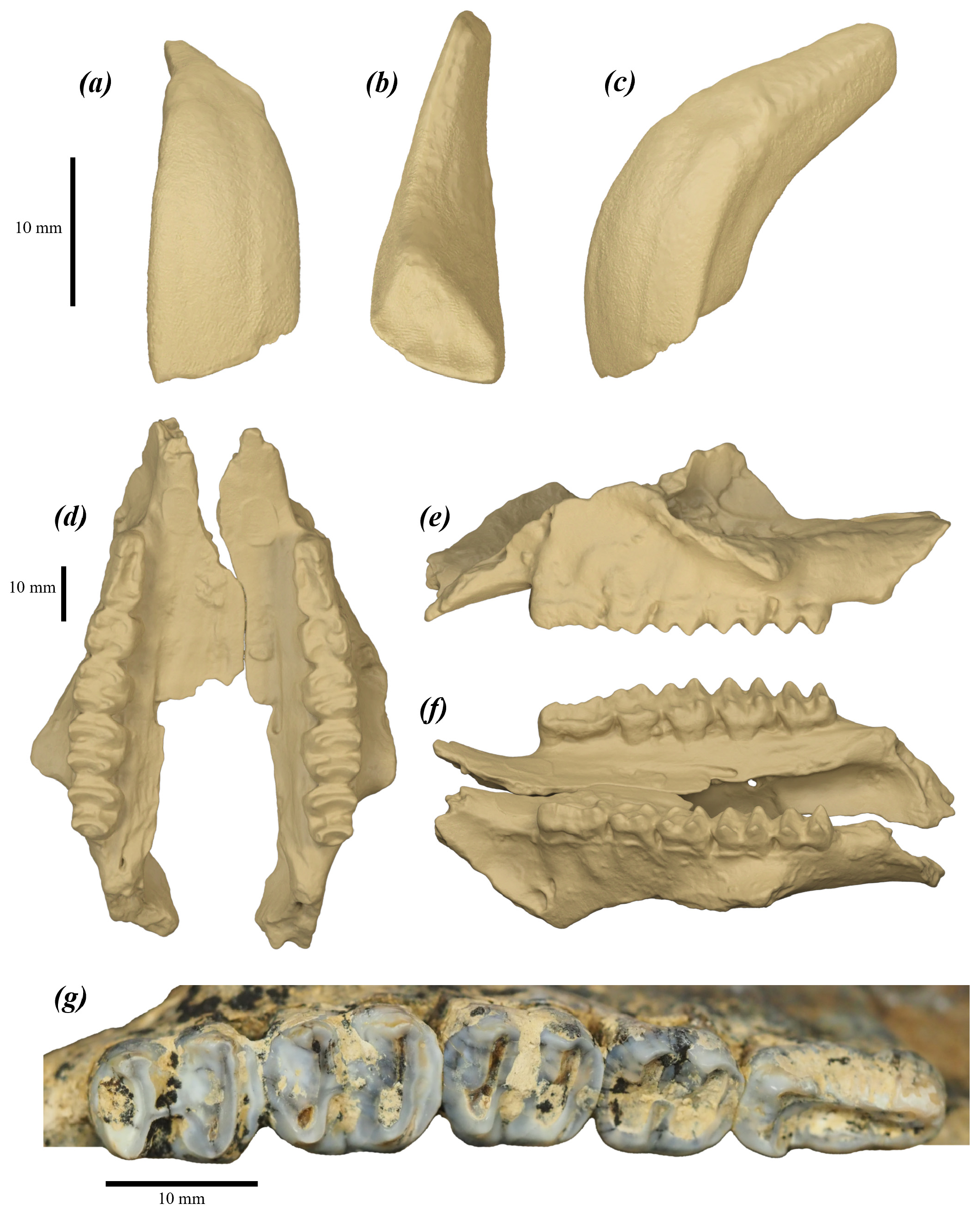
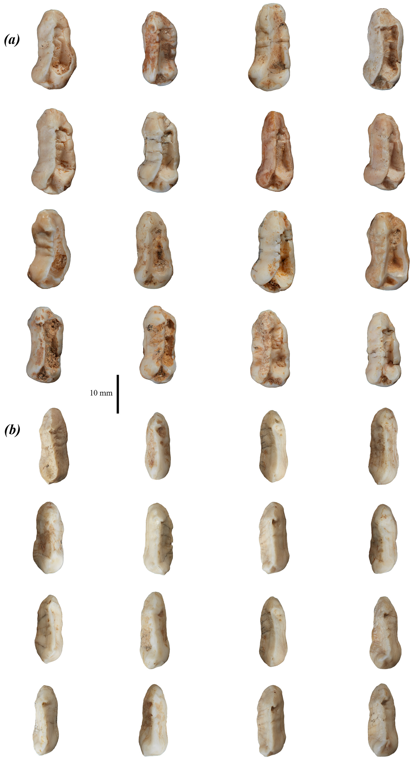
For craniodental description, see original description by Flannery et al. (1983, pp. 87–90). For purposes of comparison, cranium and dentition are figured below ( Figs 81 View FIGURE 81 & 82 View FIGURE 82 ). The semi-complete skeleton of P. tumbuna (PM25622) figured and described by Menzies & Ballard (1994) is currently missing and could not be included in this description.
Axial skeleton
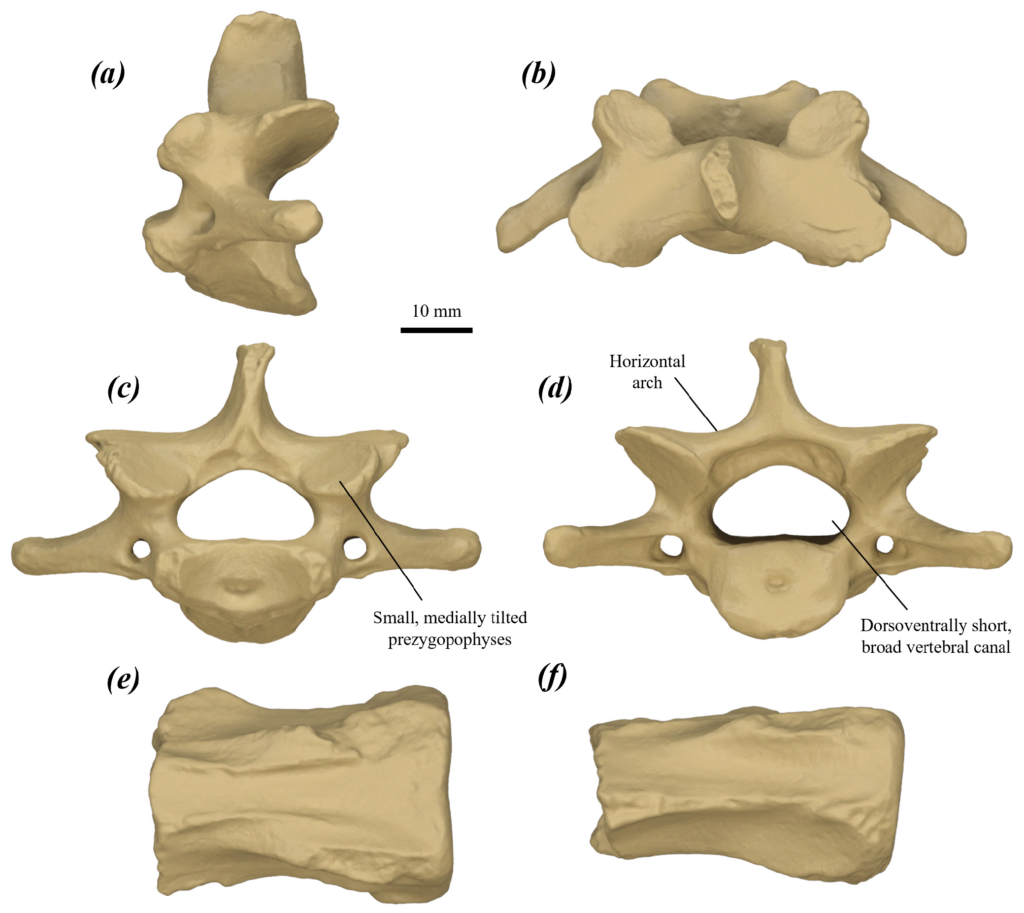
Cervical vertebra (C3?) ( Fig. 83a–d View FIGURE 83 ): craniocaudally quite short, dorsoventrally compressed and broad. Cranial extremity of the centrum gently and smoothly concave with a ventral tilt; broad, dorsoventrally compressed oval in cranial view. Prezygopophyses small and projected cranially subequal to the cranial extremity of the centrum; cranial articular surfaces round, flat and facing dorsally with medial and cranial tilt. Vertebral canal small, quite broad, dorsoventrally low and oval to reniform. Arch thick and low, approaches horizontal in cranial view. Spinous process subequal in depth to the arch, and transversely very compressed; dorsal margin abraded in available specimen. Transverse processes broad and quite gracile, with no ventral deflection, and caudal deflection of ~40° from the transverse plane. Transverse foramina small and round, extends along the caudal surface of the transverse processes as shallow channels facing very slightly ventrally. Postzygopophyses quite small, projected caudally subequal to caudal extremity of the centrum; caudal articular surfaces round, flat and facing ventrally with a lateral and caudal tilt. Caudal extremity of the centrum oval, smoothly convex and slightly caudally projected with a gentle dorsal tilt.
The C3 of P. tumbuna differs from that of P. anak in being much shallower, with smaller, more medially tilted prezygopophyses, smaller, more laterally tilted postzygopophyses, a lower, broader vertebral canal, lower cranial extremity of the centrum and much less caudoventrally projected caudal extremity of the centrum; from P. mamkurra sp. nov. in being less tall relative to width, with relatively narrower cranial and caudal articular surfaces, and the arch much less dorsally deflected and more horizontal in cranial view; from P. viator sp. nov. in being smaller and craniocaudally slightly shorter, with more robust transverse processes; from C. kitcheneri in being shallower, relatively shorter and broader, with longer transverse processes, less elongate, less cranially projected prezygopophyses, lower, broader vertebral canal, broader cranial extremity of the centrum, and a broader, relatively larger, and less caudoventrally projected caudal extremity of the centrum; from O. rufus in being craniocaudally shorter and relatively broader, with smaller, less elongate pre- and postzygopophyses, a broader vertebral canal, and more dorsally situated, less caudoventrally deflected transverse processes; from M. fuliginosus in being craniocaudally shorter and relatively broader, with a more horizontal arch in cranial view, broader, less gracile and less caudally deflected transverse processes, and a broader vertebral canal; and from W. bicolor in being larger, craniocaudally shorter and more robust, with less elongate prezygopophyses, less caudally deflected transverse processes, a broader, oval (rather than reniform) vertebral canal, and the ventral margin of the caudal extremity of the centrum not bilobed.
Caudal vertebra (Ca8?) ( Fig. 83e–f View FIGURE 83 ): single available vertebra is fragmentary; numerical position is not certain. Centrum craniocaudally short and quite broad; cranial extremity oval with small, rounded dorsolateral corners; caudal extremity abraded. Cranial transverse processes extremely small and slightly ventrally situated. Prezygopophyses abraded, appear large, quite elongate at the base and dorsolaterally situated. Caudal transverse processes mostly abraded in the available specimen; they arise from thin craniocaudal ridges along the transverse surfaces, broaden and thicken adjacent to the caudal extremity.
Forelimb
Humerus ( Fig. 84 View FIGURE 84 ): elongate, narrow and quite deep with well-developed ridges for muscle attachments. Head roughly hemispherical, round in dorsal view and moderately caudomedially projected. Greater tubercle mostly abraded; appears broad, quite low and rounded. Lesser tubercule quite low and blunt; oval and deep in dorsal view. Proximal shaft quite broad and deep. Pectoral crest elongate, raised and situated distinctly proximally, such that the distal peak sits just past the midpoint of the shaft. Deltoid tuberosity in joey ( Fig. 84h–k View FIGURE 84 ) elongate, very low and indistinct, situated on lateral surface of the shaft, level with the proximal part of the pectoral crest. The area of insertion of the m. latissimus dorsi is a quite proximally situated, distinct and elongate fossa on the joey, positioned midway between the lesser tubercle and the distal peak of the pectoral crest on the medial surface of the shaft. Shaft narrow and very deep. Distal end narrow, elongate and rotated slightly medially. Lateral supracondylar ridge quite narrow and elongate, extends ~two-fifths of humeral length in joey, with the proximal peak coming to a small point. Capitulum and ulnar facet moderately distally projected, and slightly laterally displaced; combined width is roughly four-fifths of the epicondylar width; capitulum smoothly gently convex; trochlea wide and deep. Olecranon fossa large, broad and deep; radial fossa rounded and shallow; coronoid fossa smaller, shallower and quite broad. Medial supracondylar bridge broad, quite thick craniocaudally and elongate, aligned distinctly proximodistally such that the section over the supracondylar foramen projects medially; continues proximally and slightly laterally as a very low, broad ridge that merges with the distal end of the pectoral crest ( Fig. 84d View FIGURE 84 ). Supracondylar foramen large, oval and moderately flattened craniocaudally; very elongate in the joey.
The humerus of P. tumbuna is not confidently differentiated from that of P. otibandus . It differs from that of P. anak in being smaller, with relatively more proximally situated pectoral crest, more proximally situated insertion of m. latissimus dorsi, more transversely compressed shaft, and relatively narrower and more elongate distal end; from P. mamkurra sp. nov. and P. viator sp. nov. in being more gracile, and in having a more proximally situated insertion of the m. latissimus dorsi, a relatively narrower, more elongate distal end, and a low, broadly rounded ridge linking the pectoral crest and the medial supracondylar bridge; from C. kitcheneri in having a more dorsally raised lesser tubercle, deeper proximal shaft, straighter, less raised pectoral crest, more transversely compressed shaft, relatively more elongate distal end, broader lateral supracondylar ridge, and a broader medial supracondylar bridge; from O. rufus in being more robust, with a deeper proximal shaft, more transversely compressed shaft, a more raised crest of the greater tubercle, and a broader capitulum and ulnar facet relative to the distal width; from M. fuliginosus in being more robust, with a relatively larger head, relatively more elongate distal end, and a broader capitulum and ulnar facet relative to distal width; and from W. bicolor in being larger and less laterally bowed in cranial view, lacking a narrow dorsoventral groove on the medial face of the lesser tubercle, and in having a less medially projected lesser tubercle, more proximally situated insertion of the m. latissimus dorsi, straighter pectoral crest, and a broader capitulum and ulnar facet relative to the distal width.
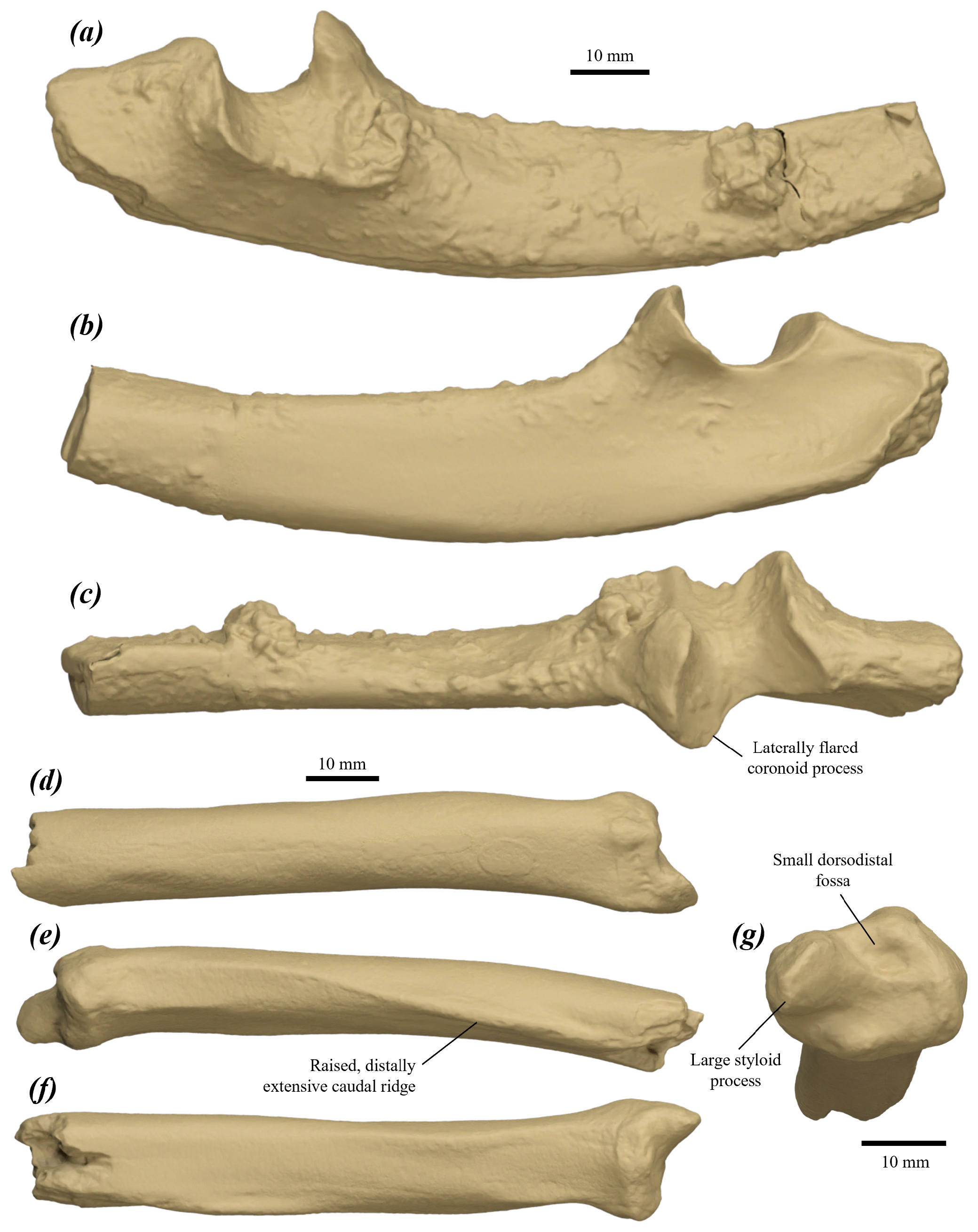
Ulna ( Fig. 85a–c View FIGURE 85 ): olecranon quite large and broad, tapers in height distally; a small eminence is present on the caudomedial margin, extends slightly anteriorly; lateral surface very gently convex. The proximomedial flexor fossa is strongly concave. Humeral facet large, with a smoothly rounded trochlear notch; medial section deeply concave; lateral section broad, very shallow and laterally tilted, with a sinusoidal lateral margin projected slightly laterally; anconeal process low and quite narrow relative to the coronoid process and the radial notch; coronoid process tall, rounded and distinctly medially deflected, with variable breadth. Radial facet broad and gently concave. Ulnar tuberosity indistinct and rugose. Proximal shaft deep and quite transversely compressed, with a slight medial deflection to the cranial section in cross-section; caudal margin very gently convex, approaches straight beneath the olecranon and humeral facet; curvature of the cranial margin increases smoothly distal to the radial facet.
The ulna of P.tumbuna is not confidently differentiated from that of P. otibandus . It differs from that of P. anak in being smaller, with a shallower, less transversely compressed shaft; from P. mamkurra sp. nov. in being smaller; from P. viator sp. nov. in being smaller, with a lower, more laterally flared coronoid process; from C. kitcheneri in being larger and deeper, with a slightly taller, more posteriorly tilted coronoid process and a less laterally tilted lateral section of the humeral facet; from O. rufus in being deeper and more transversely compressed, with a less cranially deflected olecranon, less medially flared anconeal process, taller coronoid process, and a less laterally tilted lateral section of the humeral facet; from M. fuliginosus in having a less tall olecranon, taller anconeal process, a posteriorly deflected coronoid process, and an anteroposteriorly shorter, less laterally tilted lateral section of the humeral facet; and from W. bicolor in being larger, with a relatively longer, cranially undeflected olecranon and a lower, narrower anconeal process.
Radius ( Fig. 85d–g View FIGURE 85 ): quite robust and gently curving caudomedially toward the distal end. Distal shaft moderately compressed obliquely (craniolaterally to caudomedially). Caudal ridge distinct, quite tall, thin and elongate, extends along the caudal then caudolateral surface of the shaft to merge into the caudal margin of the ulnar notch. Distal end broad, rounded and craniocaudally compressed, with a small tubercle on the cranial surface; scaphoidal facet distal facing, broad and gently concave with a craniocaudally short, small, rounded fossa centred in the slightly cranially swollen cranial part of the facet; styloid process quite large and slightly transversely compressed, forms a flattened, rounded tip; ulnar notch extends and tapers proximally along the caudolateral surface as a very shallow, elongate fossa.
The radius of P. tumbuna is not confidently differentiated from that of P. otibandus . It differs from that of P. anak in having a less craniocaudally compressed distal shaft, larger styloid process relative to the scaphoidal facet, more oval distal end in distal view, and the cranial component of distal surface with a small fossa ( Fig. 85g View FIGURE 85 ); from P. mamkurra sp. nov. in having a less craniocaudally compressed distal shaft and a less caudally situated styloid process; from P. viator sp. nov. in being smaller, with a less craniocaudally compressed distal shaft, more raised and more distally extensive caudal ridge, and a larger styloid process relative to the scaphoidal facet; from C. kitcheneri in a having more cranially situated styloid process and a less cranially situated scaphoidal facet; from O. rufus in having a broader distal shaft and more raised caudal ridge; from M. fuliginosus in having a broader, more craniocaudally compressed distal shaft and a more raised caudal ridge; and from W. bicolor in being larger, with a lower caudomedial ridge on the distal shaft and a relatively longer, more cranially situated styloid process.
Hindlimb
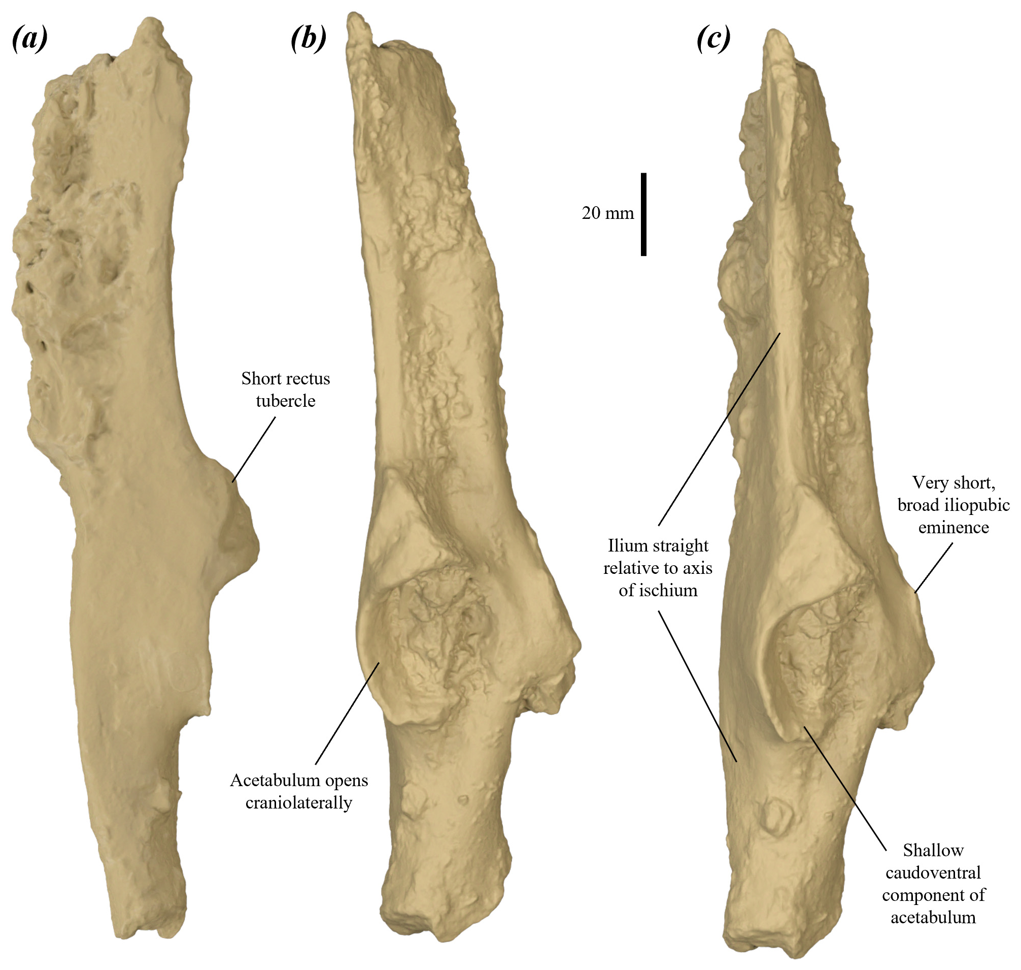
Pelvis ( Fig. 86 View FIGURE 86 ): ilium: elongate, quite straight in ventrolateral view, roughly L-shaped in cross-section and curves gently laterally toward the dorsal end.The epiphysis of the iliac crest is not known; iliac crest deep in dorsal view; ilium increases in depth to maximum at the iliac crest. Iliac fossa narrow and shallow. Gluteal fossa broad, moderately concave, becomes shallower dorsally, extends to the iliac crest. Caudal iliac spine arises abruptly on the caudomedial surface of the base of the ilium opposite the rectus tubercule; thick and tall ventrally for the origin of a large mm. gluteus, becomes thinner and lower dorsally, extends to the iliac crest; in some specimens, a small, pointed eminence is present on the caudoventral shoulder (see NCA-A1-18). Sacral surface very rugose, concave and roughly rounded. Cranial iliac spine present as a very low, short crest, briefly arising opposite the base of the caudal iliac spine and merging into the body of the ilium. Lateral iliac spine tall and slightly thinner than the caudal spine; lateral margin gently concave, extends straight in lateral view to the iliac crest. Rectus tubercule short, protuberant, rugose, roughly triangular, narrows onto the base of the lateral spine.
Acetabulum large and deeply concave, opens laterally and slightly cranioventrally, becomes shallower ventrally, with the height greater than the craniocaudal depth; acetabular fossa not known. Ischium: dorsal part robust, deep, quite transversely compressed, and gently concave medially, with the lateral surface strongly convex; undeflected, aligned with the axis of the ilium in lateral view. Ventral ischium incompletely preserved in available specimens. Iliopubic eminence very low, rounded in outline, quite elongate and rugose. The pubis is not known.
The pelvis of P.tumbuna differs from that of compared species of Protemnodon in having a straighter ilium in lateral view (rather than curving caudally), the acetabulum opening less laterally and more cranioventrally ( Fig. 86b View FIGURE 86 ), and a broader (less transversely compressed) ischium. It further differs from that of P. mamkurra sp. nov. in being generally smaller, with a shorter rectus tubercle, acetabulum with a more deeply concave ventral section, and a smaller iliopubic eminence; from P. viator sp. nov. in having a smaller iliopubic eminence; from P. dawsonae sp. nov. in having a more laterally projected rectus tubercle; from C. kitcheneri , O. rufus , M. fuliginosus and W. bicolor in being more robust, with a broader lateral iliac spine, deeper gluteal fossa, much shallower, less distally extensive iliac fossa, and a less tall, more laterally projected rectus tubercle; and additionally from O. rufus , M. fuliginosus and W. bicolor in having a more ventrally opening acetabulum.
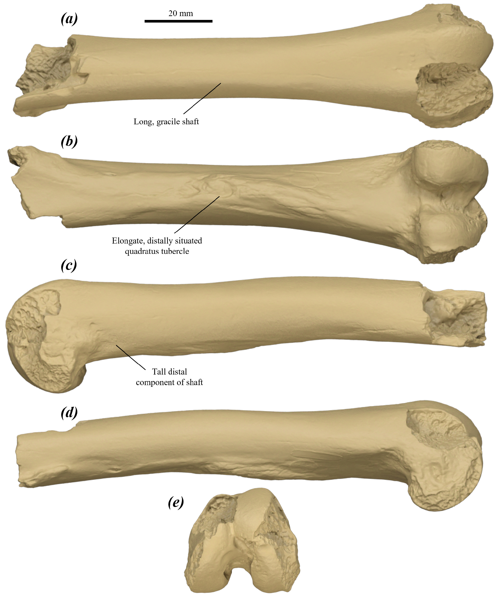
Femur ( Fig. 87 View FIGURE 87 ): long, gracile, although most of the proximal end is not preserved in the available specimen. Lesser trochanteric crest distal margin intact; projects medially from the ventromedial surface of the base of the proximal end; thickened, raised, and interpreted as being quite elongate distally. Shaft straight, broadens slightly and deepens substantially toward the distal end ( Fig. 87c View FIGURE 87 ); ventral surface flattens distally. Quadratus tubercule very elongate, rugose and very raised, extends distally from slightly proximal of the midpoint of the ventral surface of the shaft to just over three-quarters of the femoral length ( Fig. 87b View FIGURE 87 ). The ventrolateral fossa for the partial origin of the m. flexor digitorum superficialis is small, rounded, and situated on the proximal surface of the lateral condyle; extends briefly along the shaft.
Distal end tall. Lateral trochlear crest tall and fairly broad; medial crest not preserved. Trochlea deep, narrow and V-shaped. Intercondylar fossa deep and quite short, such that fossa opening is more distal than ventral. Condyles with the ventral and distal surfaces and the distolateral part of the lateral condyle not preserved; fibular facet abraded. Medial gastrocnemial fossa large, shallow and rounded; lateral gastrocnemial fossa small, shallow and rounded.
The femur of P. tumbuna cannot be differentiated from that of P. otibandus . It differs from all other compared species of Protemnodon in being more gracile, and from all compared taxa in having a taller distal shaft and a more elongate and distally situated quadratus tubercle. Further differs from that of P. anak , P. mamkurra sp. nov. and P. viator sp. nov. in being smaller; from C. kitcheneri in being more elongate, with a less medially displaced quadratus tubercle and a rounder, more proximally situated lateral gastrocnemial fossa; from O. rufus and from M. fuliginosus in having a relatively broader shaft, broader distal epiphysis, lower trochlear crests, and a more proximally situated lateral gastrocnemial fossa; and from W. bicolor in being larger and more robust, with less raised trochlear crests and relatively broader distal condyles.
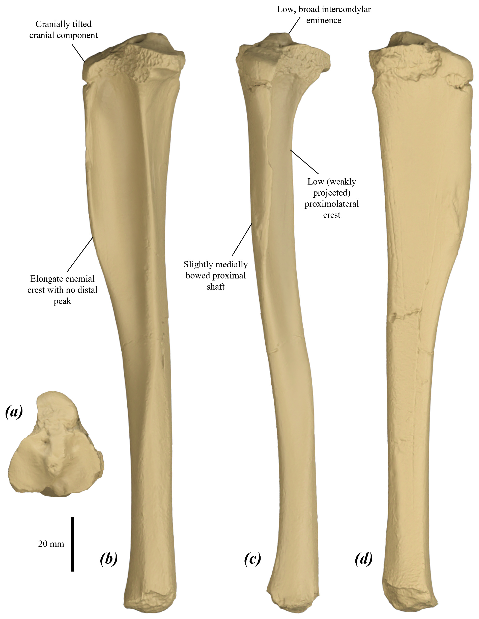
Tibia ( Fig. 88 View FIGURE 88 ): short and robust. Proximal epiphysis width subequal to depth; cranial section slightly convex and distinctly cranially tilted; medial condyle elongate, slightly greater in surface area than lateral condyle; lateral condyle broad and rounded; intercondylar eminence low and broad. Proximal fibular facet mostly abraded. transversely quite broad.
Proximal shaft bowed very slightly medially and distal shaft bowed slightly laterally in cranial view. Cnemial crest deep, thickened and relatively elongate, extending just over one-third of the tibial length ( Fig. 88b View FIGURE 88 ); lateral surface moderately concave and medial surface moderately convex; variably large muscle-scarred area on the craniodistal margin of medial surface for the partial origin of the m. gastrocnemius medialis or for the attachment of the medial superficial fascia. Proximolateral crest thick and quite low ( Fig. 88c View FIGURE 88 ), particularly distally; merges smoothly with the shaft around the midpoint of the tibia. Distal shaft narrows very slightly before expanding slightly to the distal end, rounded to squarish in cross-section, becomes more rounded distally. Distal fibular facet deep, flattened, extends down the distal one-third of the lateral surface of the shaft. The distal epiphysis is not known.
The tibia of P. tumbuna differs from all compared taxa in being more robust, with a particularly low proximolateral crest, especially distally, and a relatively more elongate cnemial crest, and differs from all species except some specimens of P. mamkurra sp. nov. in having no distinct distal peak on the cnemial crest. It further differs from that of P. anak in being shorter, with lower, broader intercondylar eminence, cranially tilted cranial section of the proximal epiphysis, and a thicker proximolateral crest; from P. mamkurra sp. nov. in being shorter, with a thicker proximolateral crest and deeper, more flattened distal fibular facet; from P. viator in being smaller, shorter, and more bowed in cranial view, with a broader intercondylar eminence and cranially tilted cranial section of the proximal epiphysis; from P. otibandus in having thicker proximolateral crest; from P. snewini in being shorter; from C. kitcheneri in being shorter, with a less straight shaft in cranial view, a lower, broader intercondylar eminence, thicker cnemial crest that is slightly more laterally curved in cross-section, thicker proximolateral crest, deeper, more distinct distal fibular facet, and the distal shaft expanding more to the distal end; from O. rufus and M. fuliginosus in being shorter and much more robust, with a less straight shaft in cranial view, lower, broader intercondylar eminence, cranially tilted cranial section of the proximal epiphysis, thicker cnemial crest that is slightly more laterally curved in cross-section, thicker proximolateral crest, and deeper and more distinct distal fibular facet; and from W. bicolor in being larger and more robust, with a larger, broader intercondylar eminence, a more cranially tilted cranial section of the proximal epiphysis, relatively thicker cnemial crest, thicker proximolateral crest, particularly distally, and a deeper, more distinct distal fibular facet.
Pes
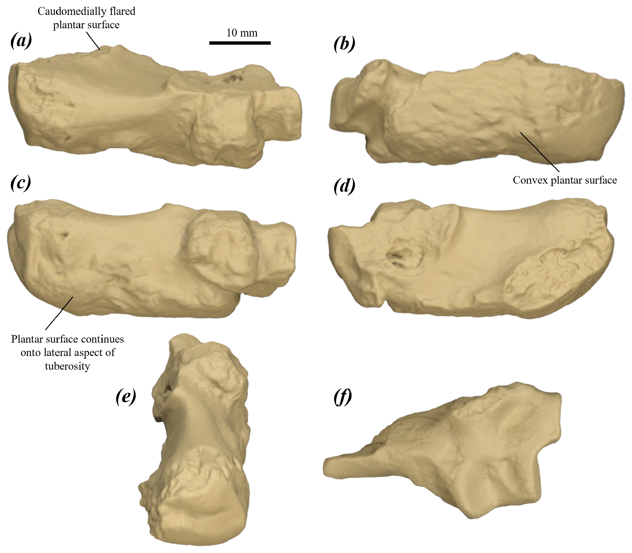
Calcaneus ( Fig. 89 View FIGURE 89 ): available specimens are fragmentary and badly abraded; small, low and robust. Calcaneal tuberosity quite broad, low and rounded-triangular in cross-section; broadens caudally, with the caudoplantar part flaring medially ( Fig. 89a View FIGURE 89 ). Caudal epiphysis rounded and robust, with a narrow transverse valley across the centre of the caudal surface. Plantar surface broad, rugose, oblong in plantar view and distinctly convex, with the medial margin increasingly medially flared toward the caudal end, and the lateral component wrapping around the plantolateral margin of tuberosity onto a convex lateral surface. Calcaneal head broad; the midline of the head in the sagittal plane is medially offset relative to that of the calcaneal tuberosity, particularly visible in the relatively medial placement of the medial margin of the lateral talar facet. Sustentaculum tali broad, thick and medially projected; flexor groove very broad and deep. Fibular facet mostly abraded, appears small, slightly laterally projected, rounded and bulbous dorsally. Lateral talar facet broad, with slight medial tapering; smoothly convex in the sagittal plane; a large, rounded, shallow fossa is present immediately cranial to lateral talar facet; medial talar facet small, oval, orientated caudomedially to craniolaterally and strongly cranially tilted; caudally displaced relative to the lateral facet.
Facet for the talar head; very small, rounded, abuts the dorsomedial margin of the dorsomedial facet on the medial surface of the calcaneal head. Dorsomedial cuboid facet gently convex with a slightly rounded dorsomedial margin, separated from the dorsolateral facet by a deep, bevelled step, plantar section not preserved in available specimens; dorsolateral cuboid facet subequal in width to the dorsomedial facet, very cranially projected and gently plantarly tilted, with a slightly rounded dorsal margin; tapers slightly and curves plantomedially into the broad, dorsoplantarly short, medially situated and slightly medially tilted plantomedial cuboid facet.
The calcaneus of P. tumbuna differs from that of all compared taxa in having a caudomedially flared, distinctly convex plantar surface, extending dorsolaterally onto the lateral surface of the tuberosity, and a more medially displaced calcaneal head. It further differs from that of P. anak in being smaller and lower; from P. mamkurra sp. nov. in being smaller and lower, with a smaller, more caudally displaced medial talar facet and a relatively broader, less convex medial cuboid facet; from P. viator sp. nov. in being smaller and more robust, with a relatively narrower dorsomedial cuboid facet and a more medially displaced calcaneal head; from P. dawsonae sp. nov. in being smaller; from P. otibandus in having a larger, deeper fossa cranial to the lateral talar facet; from C. kitcheneri in having a more medially rotated head; from O. rufus in being much shorter, lower and relatively broader, lacking an articular surface for the posterior plantar process of talus, and in having a larger, more medially projected sustentaculum tali, more medially rotated head, less triangular, more rounded calcaneal tuberosity in cross-section, more transversely aligned, less cranially tilted medial talar facet, the lateral talar facet with a shallower cranial fossa, and a relatively broader dorsomedial cuboid facet; from M. fuliginosus in being slightly larger, much shorter, lower and relatively broader, and in having a larger, more medially projected sustentaculum tali, more medially rotated head, less triangular, more rounded calcaneal tuberosity in cross-section, more transversely aligned, less cranially tilted medial talar facet, and the lateral talar facet with a shallower cranial fossa; and from W. bicolor in being larger, broader and more robust, with broader medial talar facet.
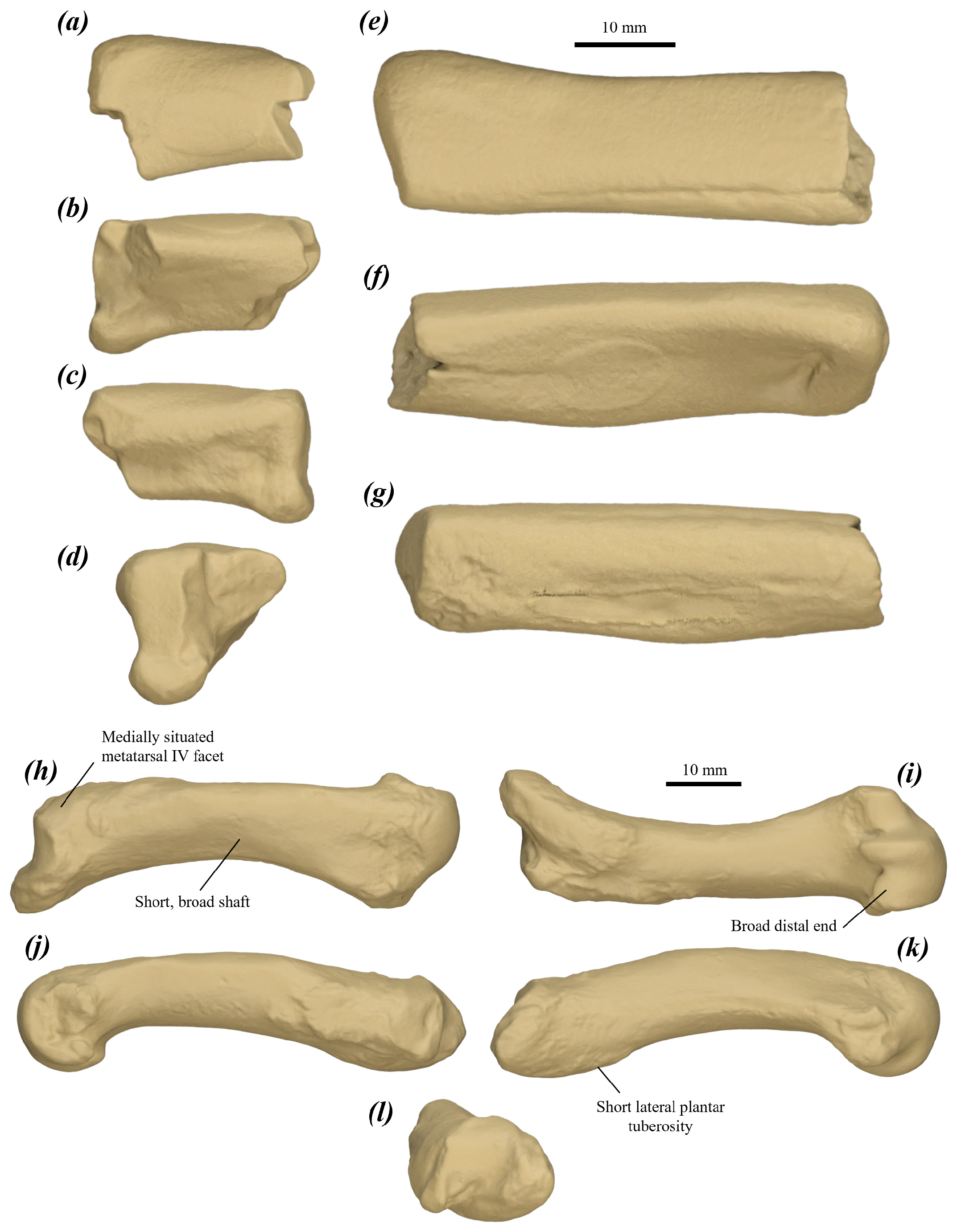
Metatarsal IV ( Fig. 90a–g View FIGURE 90 ): two available specimens fragmentary and extremely abraded. Proximal surface medially tilted; dorsal cuboid facet mostly abraded, interpreted as broad; plantar cuboid facet mostly abraded, situated on the proximal surface of the plantar tubercle. Plantar tubercle quite large, plantarly projected and slightly proximally deflected. Metatarsal III fossa tall, elongate, shallow, and slightly rugose.Facet for metatarsalVpartially abraded; tall, quite deep, gently concave and oblong. Plantar ridge large, rugose, rounded in cross-section, extends distally from the base of the plantar tubercle to slowly merge with the plantar shaft; bordered medially by the fossa for metatarsal III and laterally by the fossa for metatarsal V. Shaft very slightly rounded dorsally; height tapers to the midpoint.
The metatarsal IV of P. tumbuna cannot be differentiated from that of P. otibandus . It differs from that of P. anak , P. mamkurra sp. nov., P. viator sp. nov., P. dawsonae sp. nov. and P. snewini in being smaller; from C. kitcheneri in having a larger proximal plantar tubercle and a more raised plantar ridge; from O. rufus and M. fuliginosus in being relatively broader and more robust, and a having less plantarly raised plantar ridge relative to the proximal plantar tubercle; and from W. bicolor in being larger and more robust.
Metatarsal V ( Fig. 90h–l View FIGURE 90 ): very short and robust; curves slightly laterally distally; variably arched in lateral view. Proximolateral process tall, blunt, rugose, curves slightly medially, proximodistally quite short, and transversely compressed. Cuboid facet large, quite tall, rounded and smoothly concave, with the dorsal margin slightly raised; extends from the medial margin of the posterior surface across the majority of the medial surface of the proximolateral process. Metatarsal IV facet quite small, broad, gently convex, slightly raised and oblong; situated on the medial and dorsomedial surfaces of the shaft, abuts the posterior margin of the shaft. Lateral plantar tuberosity short, low, broad and rugose ( Fig. 90k View FIGURE 90 ), separated from the medial plantar tubercle by a narrow,very shallow channel. Medial plantar tubercle small, smoothly rounded and plantarly and proximomedially projected. Shaft rounded in cross-section; broadens significantly from the midpoint to the distal end, particularly on the lateral margin. Distal end very broad; fossae for collateral ligament large, rugose and shallow.
The metatarsal V of P. tumbuna differs from all species of Protemnodon in being more robust, with a shorter lateral plantar tuberosity. It further differs from that of P. anak in being shorter and not transversely compressed, with a taller cuboid facet that is more extensive along the proximolateral process, a more medially situated metatarsal IV facet, and a smaller medial plantar tubercle; from P. mamkurra sp. nov. in being shorter and more robust, with a more medially situated metatarsal IV facet and a broader distal end; from P. viator sp. nov. in being shorter and not transversely compressed, with a lower lateral plantar tuberosity, more medially situated metatarsal IV facet, and a lower, broader distal end with a more rounded keel; from P. dawsonae sp. nov. in being smaller, with a shorter, broader proximolateral process and a smaller medial plantar tubercle; from P. otibandus in not being transversely compressed, and in having a taller proximolateral process, proximodistally shorter metatarsal IV facet, and a smaller medial plantar tubercle; from C. kitcheneri in being shorter and relatively much broader, lacking a slight kink in the arch of the shaft immediately proximal to the midpoint in lateral view, with a larger, more rounded cuboid facet, a plantar groove present, and a larger medial plantar tubercle and lateral plantar tuberosity; from O. rufus and M. fuliginosus in being much shorter, much more robust, and not transversely compressed, with larger medial plantar tubercle and deeper, more distinct plantar groove; and from W. bicolor in being larger, much more robust, and not transversely compressed, and in having a larger proximolateral process and a more convex cuboid facet.
Remarks:
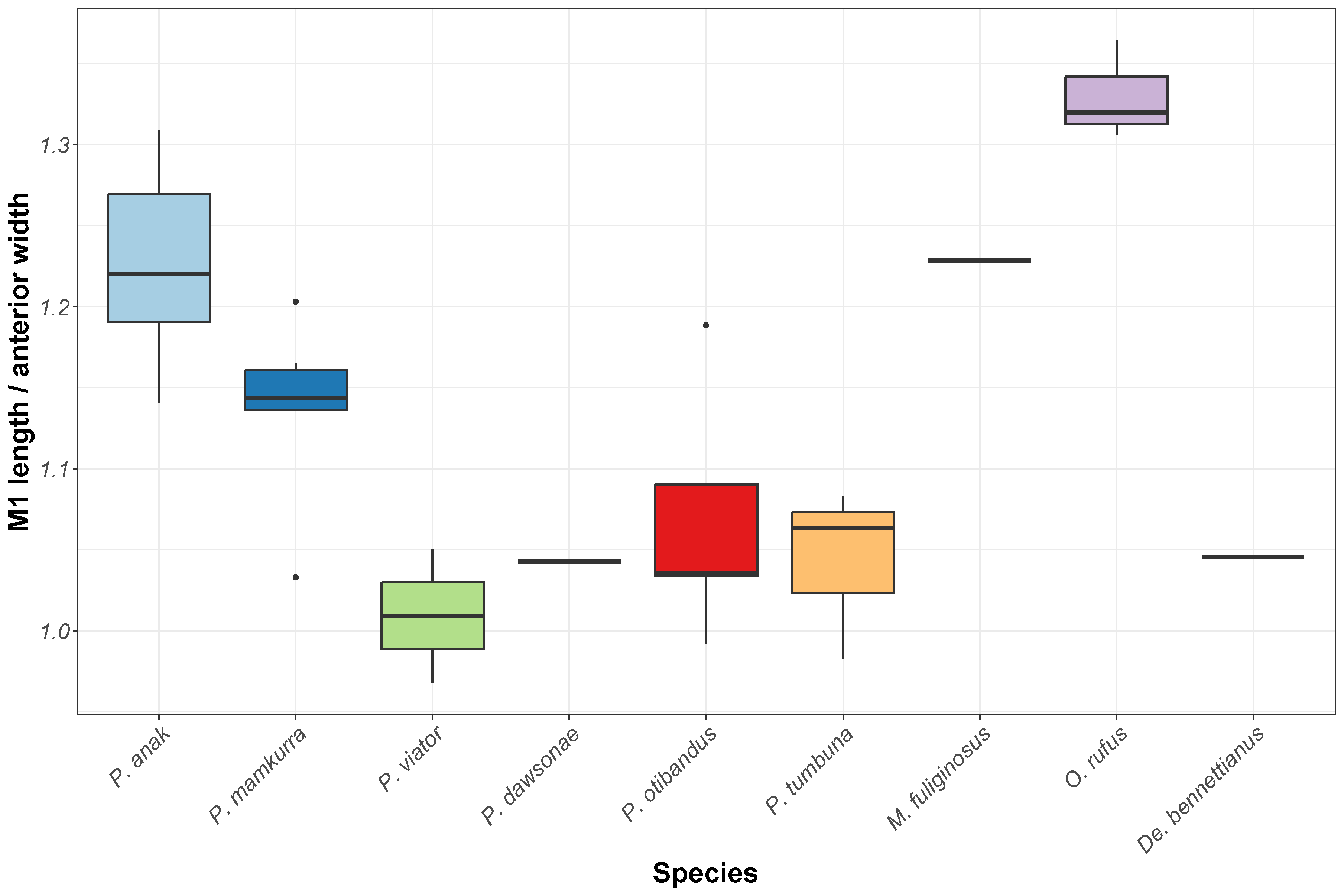
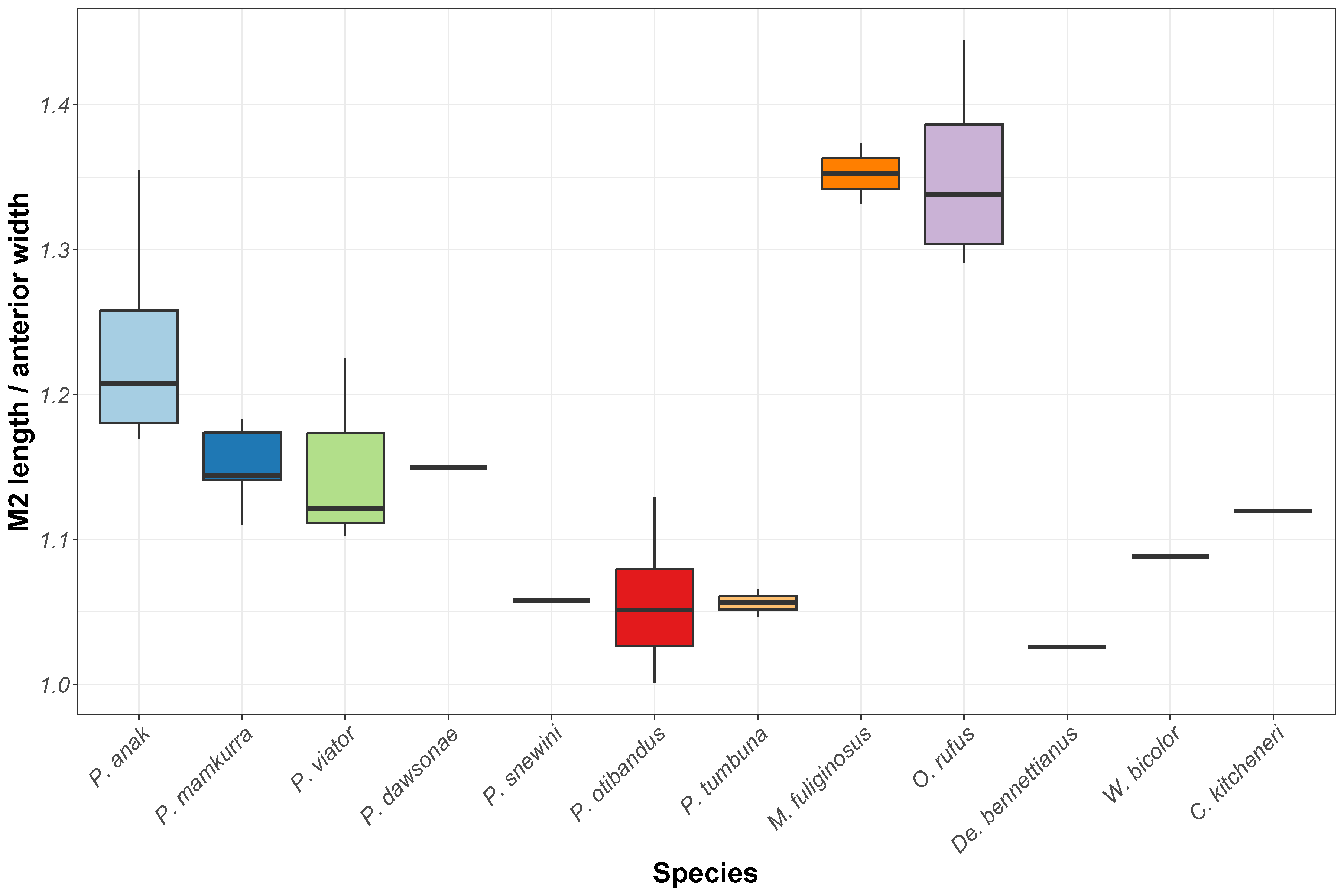
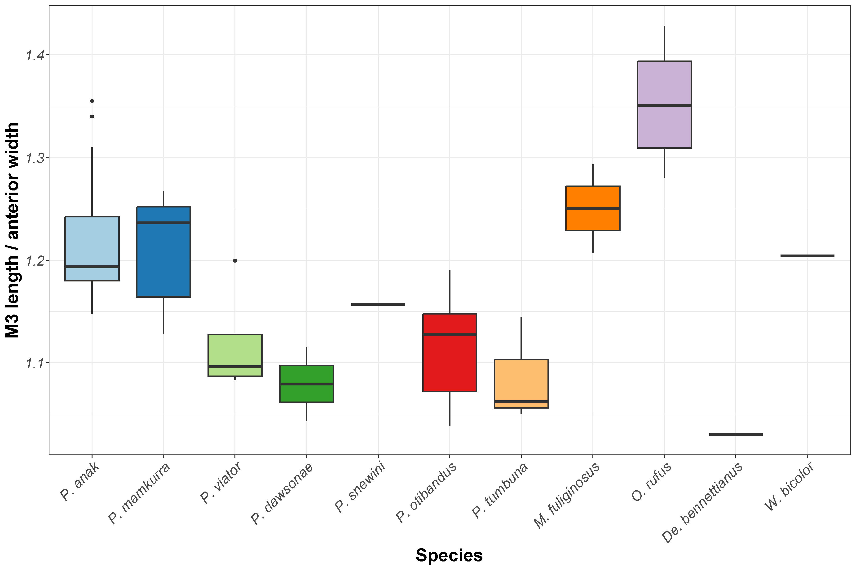
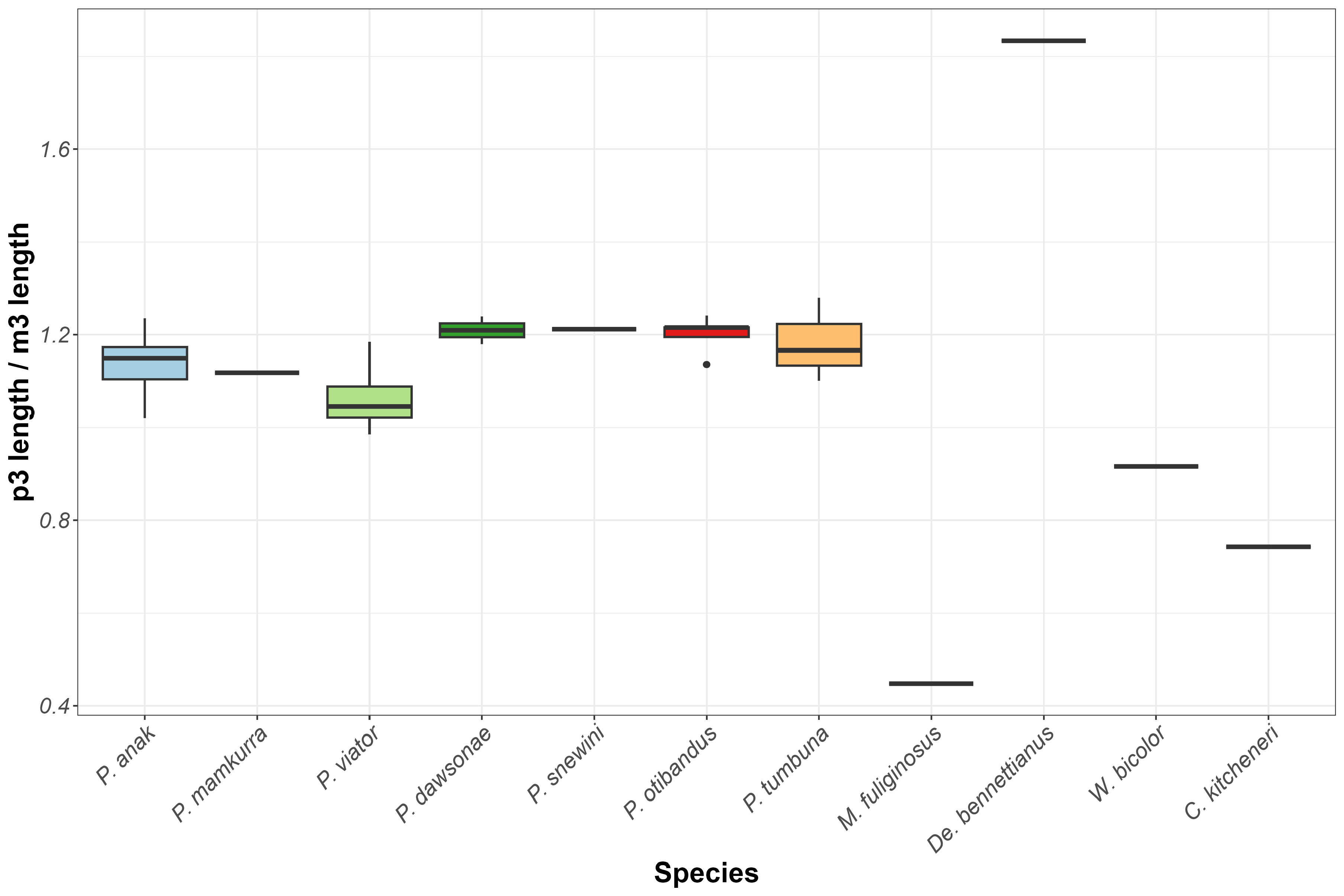
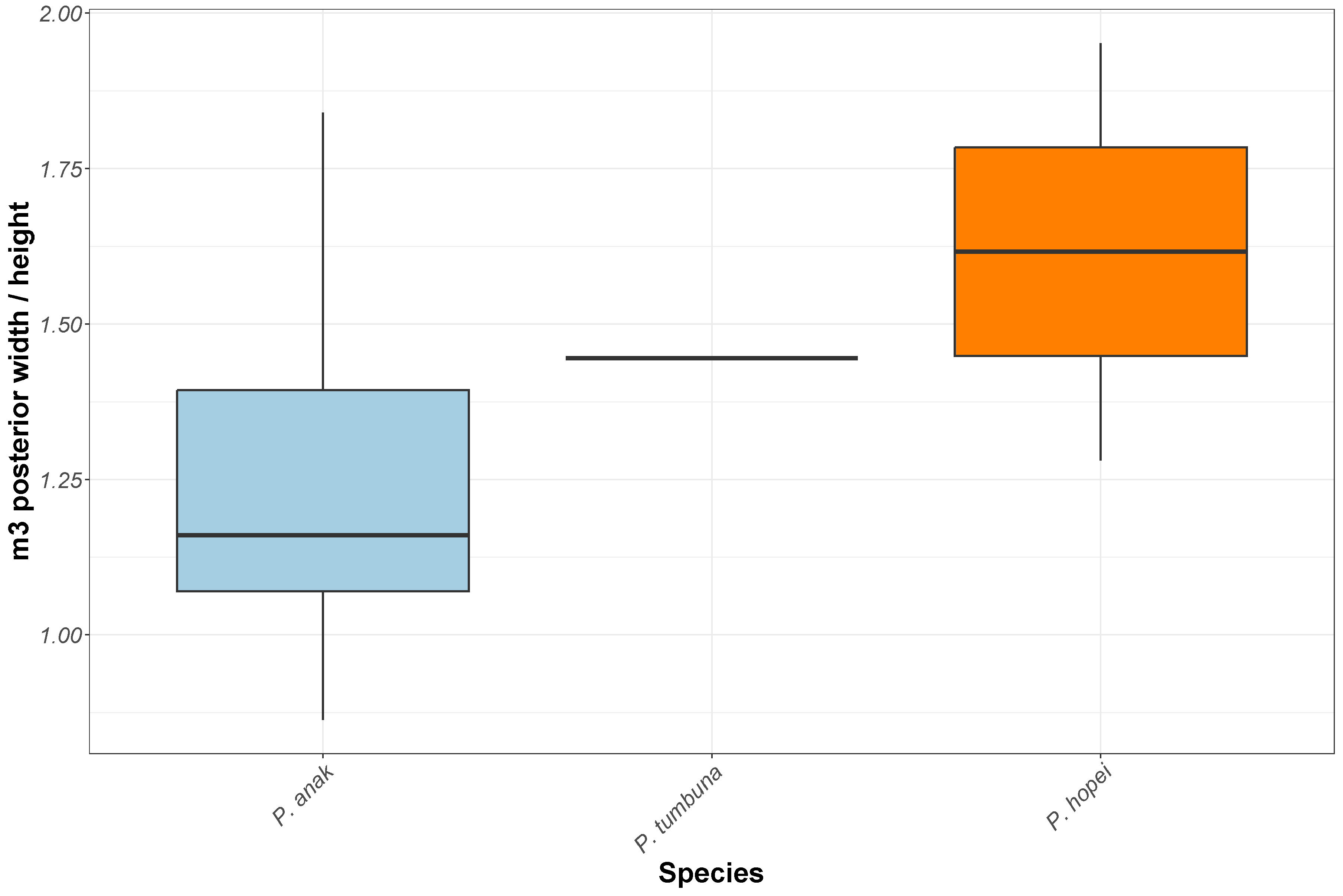
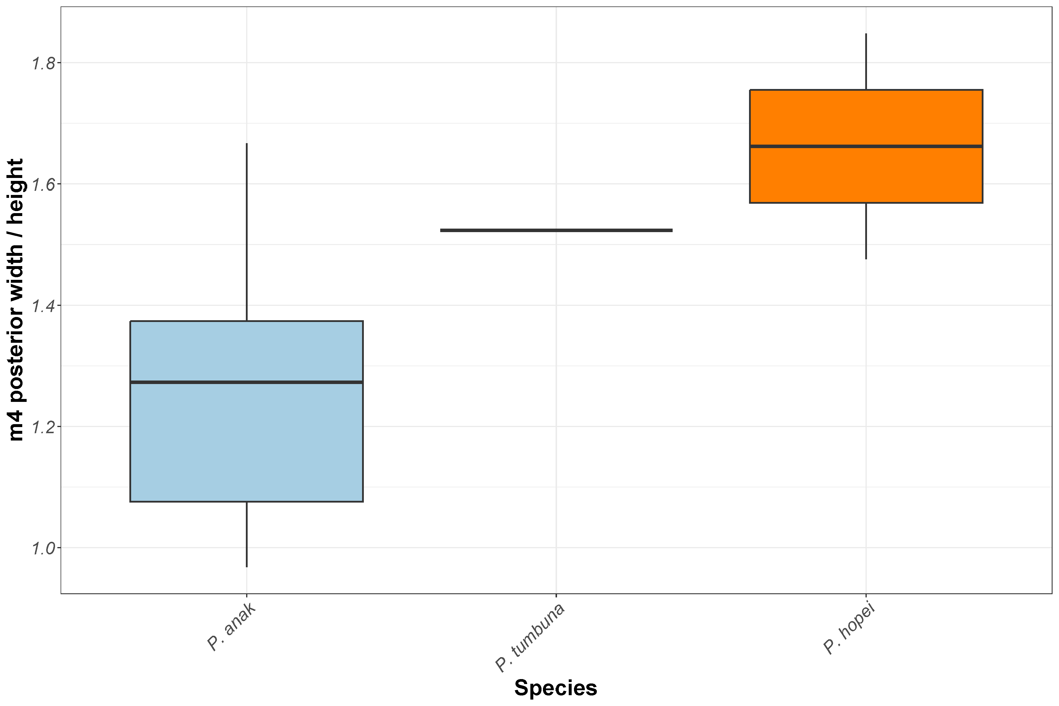
Protemnodon hopei Flannery, 1992a , was described from craniodental material of four or five individuals from Kelangurr Cave and from sediments in the nearby West Baliem River, West Papua. The taxon was described as having most in common morphologically with P.otibandus and P. tumbuna . Some of the purportedly diagnostic features ( Flannery 1992a), however, vary significantly within species (see Figs 113–115 View FIGURE 113 View FIGURE 114 View FIGURE 115 & Figs 121–125 View FIGURE 121 View FIGURE 122 View FIGURE 123 View FIGURE 124 View FIGURE 125 ). The following is an extract from the specific diagnosis, where the morphology of P. hopei is contrasted with that of other species of Protemnodon : ‘differing from P. tumbuna in being larger, with proportionately longer premolars, P3 with a straight rather than concave [buccal face of main] crest, and in having relatively narrower molars’ ( Flannery 1992a, p. 326). Although it was stated ( Flannery 1992a) that P. hopei possesses relatively narrower molars than topotypic specimens of P. tumbuna , they are actually slightly broader relative to length ( Flannery et al. 1983, table 3; Flannery 1992a, table 2; SI Measurement Dataset). Protemnodon hopei has also been described as differing from P. tumbuna by having ‘better developed anteroposterior links on the molars, indicating adaptation to a more abrasive diet’ ( Hope et al. 1993, p. 124). However, we do not find that the taxa can be diagnosed on the basis of these characteristics.
The differences between relative and absolute dental dimensions of specimens referred to P. hopei and P. tumbuna are not sufficient to support these representing two species (see Figs 113–115 View FIGURE 113 View FIGURE 114 View FIGURE 115 & Figs 121–125 View FIGURE 121 View FIGURE 122 View FIGURE 123 View FIGURE 124 View FIGURE 125 ). The larger sample sizes of other species of Protemnodon , for example P. anak , illustrate the degree of variation that may be manifested within species of the genus. Thus, although the holotype palate of P. hopei , the only specimen preserving the upper dentition of this taxon, does possess relatively and absolutely wider molars than are seen in the holotype of P. tumbuna , the only upper dental material known at the time that P. hopei was described, the difference is considerably less than that seen within the sample of P. anak . The same is true of the premolar length relative to the molars. Although this does cluster specimens into loose taxonomic groups (see Fig. 121 View FIGURE 121 ) it varies to such an extent in the better-sampled P. anak and other Australian Pleistocene species as to render the difference seen between the two samples in question here insignificant. The concavity of the buccal crest of the P3 is highly variable within Protemnodon species, for example within P. mamkurra sp. nov. (see Fig. 109 View FIGURE 109 ). The crown height of the lower molars of P. tumbuna is lower than that of P. hopei when allowing for differing wear stage. However, given the greater range of relative molar crown heights within the better-sampled P. anak , this feature is insufficient to separate the two taxonomically (see Figs 121–123 View FIGURE 121 View FIGURE 122 View FIGURE 123 ).
Comparison of the postcranial material of P. hopei with that of P. tumbuna from Nombe Rockshelter and that figured in Menzies & Ballard (1994, pp. 130–131, figs 8 & 9) shows strong similarities. Both samples show an elongate ilium, an acetabulum rotated cranioventrally, a long, gracile femur paired with a short, robust tibia, and a very short, broad, arched metatarsal V. It is noted that the three dentary specimens of P. hopei do possess a deeper digastric sulcus than that seen in P. tumbuna , but again, this character varies greatly within the better-sampled material from the Australian Pleistocene. In the absence of reliable characters to separate the two holotypes, and since no features were found in the postcranial elements that served to differentiate between the two taxa, P. hopei is here considered to be a junior synonym of P. tumbuna .

| PM |
Pratt Museum |
| AM |
Australian Museum |
No known copyright restrictions apply. See Agosti, D., Egloff, W., 2009. Taxonomic information exchange and copyright: the Plazi approach. BMC Research Notes 2009, 2:53 for further explanation.
|
Kingdom |
|
|
Phylum |
|
|
Class |
|
|
Order |
|
|
SubOrder |
Macropodiformes |
|
SuperFamily |
Macropodoidea |
|
Family |
|
|
SubFamily |
Macropodinae |
|
Tribe |
Macropodini |
|
Genus |
Protemnodon tumbuna Flannery et al., 1983
| Kerr, Isaac A. R., Camens, Aaron B., Van Zoelen, Jacob D., Worthy, Trevor H. & Prideaux, Gavin J. 2024 |
Protemnodon hopei
| Helgen, K. M. & Wells, R. T. & Kear, B. P. & Gerdtz, W. R. & Flannery, T. F. 2006: 303 |