
Myrmozercon burwelli
|
publication ID |
https://doi.org/ 10.5281/zenodo.186168 |
|
DOI |
https://doi.org/10.5281/zenodo.6214925 |
|
persistent identifier |
https://treatment.plazi.org/id/03E487BC-E574-FFB4-4680-F9A2FC2B68DD |
|
treatment provided by |
Plazi |
|
scientific name |
Myrmozercon burwelli |
| status |
|
Myrmozercon burwelli n. sp
( Figs 1–3 View FIGURE 1 View FIGURE 2 View FIGURE 3 )
Material examined. Holotype female (QMS 83733) and 3 female paratypes (QMS 83734-6), ex nest of Polyrhachis (Campomyrma) flavibasis Clark, Boombana National Park, 27º 24' 07"S, 152º 47' 23"E, 1 Aug 2004, Chris Burwell coll. subtropical rainforest; 2 paratype females and 1 paratype male (QMS 83737-9) ex nest of P. flavibasis, Lamington National Park, 28° 8' 31" S, 153° 7' 59" E, 17 Oct 2006, Chris Burwell coll., altitude 248 metres, subtropical rainforest, day hand collection. Holotype female, paratype male and 4 female paratypes are deposited in QM. One female paratype to be deposited in the Australian National Insect Collection, Canberra.
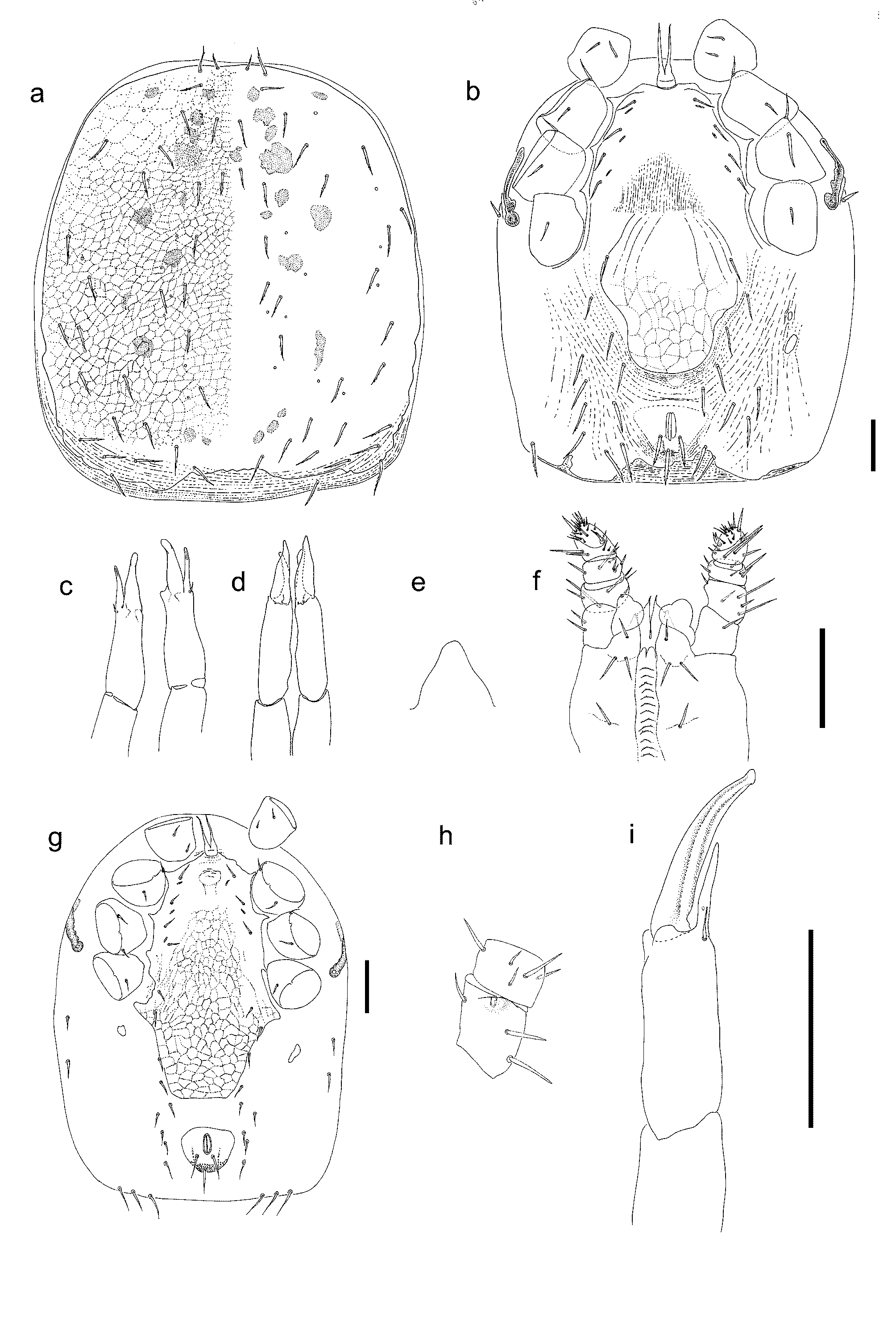
Description of female (n = 6). Dorsum: Broad dorsal shield 780–890 x 670–755 covering all of dorsum, subcircular. Shield finely reticulate. Muscle insertions prominent as desclerotised circular patches. Hypotrichous, with 12–13 podonotal and 12 opisthonotal setal pairs. Many setae lightly barbed in apical third or less. Posterior of dorsal shield squarish. Posterior edge irregular where dorsal shield grades into striate cuticle.
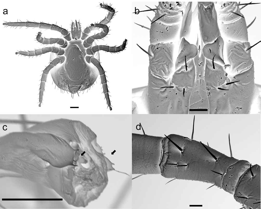
Ve n te r: Tritosternum with short (13–17) broad base. Tritosternal base free from sternal shield. Laciniae forked a short distance above suture (9–11). Laciniae with smooth edges, strap-like and broad at base, tapering towards tips, 98–110 long. Anterior edge of sternal shield extending anteriorly beyond level of st1 as a biconvex margin, presternal striae absent. Posterior margin of sternal shield obscure but shield apparently bearing three pairs of setae (st1 35–40, st 2 31–37, st 3 30–37) and two pairs of lyrifissures. Posterolateral corners fused with endopodal shield. Setae st 4 in soft cuticle, sometimes absent on one side. Genito-ventral shield broad, 200–217 long, 243–297 wide at level of st5, maximum width 245–307. Anterolateral portion of genito-ventral shield formed of elongate cells which flare laterally, forming a lobed outline immediately posterior to st5. Remainder of shield ornamented by squat irregular cell-like markings. Sperm induction pore visible on coxa IV of one specimen; tubuli annulati in one specimen contain dense spheres (= unused sperm?). Opisthogaster with sparse setae in soft cuticle, all barbed at least lightly. Setae in R series absent. Principal metapodal plates elongate, 37–46 long by 8–16 wide. Inner secondary metapodal plate small and slender in one specimen (11 x 6) but absent in others. Peritremes short (137–146), extending to level of mid coxa III.
Post-stigmatal plate and pores absent. Anal shield broad with strongly produced anterolateral corners, 200–201 maximum width, 145–168 at level of mid-anal opening. Anal valves narrow. Field of cribral spicules forming a dense narrow band without discernible rows, which project within shield as lateral arms to level of insertion of para-anal setae. Cribral pores not seen. Para-anal and post-anal setae barbed apically.
Gnathosoma: Tectum rounded, projecting to level of mid palp femur, mostly unornamented, but basally with some transverse striae. Corniculi 23–28 long, well-spaced, bearing broad membranous lobes which extend inward over malae. Internal malae separate, fine and pointed, essentially smooth although edges sometimes with one or two denticles. Lateral arms absent. Subcheliceral shelf squat, with simple broadlyrounded tip. Deutosternal groove a broad channel with 14 denticulate rows of 20–30 very fine denticles, topped anteriorly by a pair of broad lobes. Hypostomal setae h 1 20–28, h 2 29–33, h 3 28–32; palp coxal seta 30–34. Palp with variable hypotrichy; trochanter: femur: genu: tibia with 1–2:4–5:5–6:11–13 setae. On the left palp trochanter of one specimen there are two setae ( Fig. 1 View FIGURE 1 h). Dorsodistal edge of palp femur with a low swelling, immediately internal of condyle. All palp setae setiform, undistinguished. Palp apotele 2-tined. Chelicerae edentate, fixed digit parallel-sided and narrow, pilus dentilis apparently absent. Cheliceral seta 14 long, positioned dorsally. Movable digit long and tapering, 62–68 long, arthrodial corona absent, not visible on slide mounts or scanning electron micrographs. First cheliceral segment 52–61, second cheliceral segment 140–156 to tip of fixed digit.
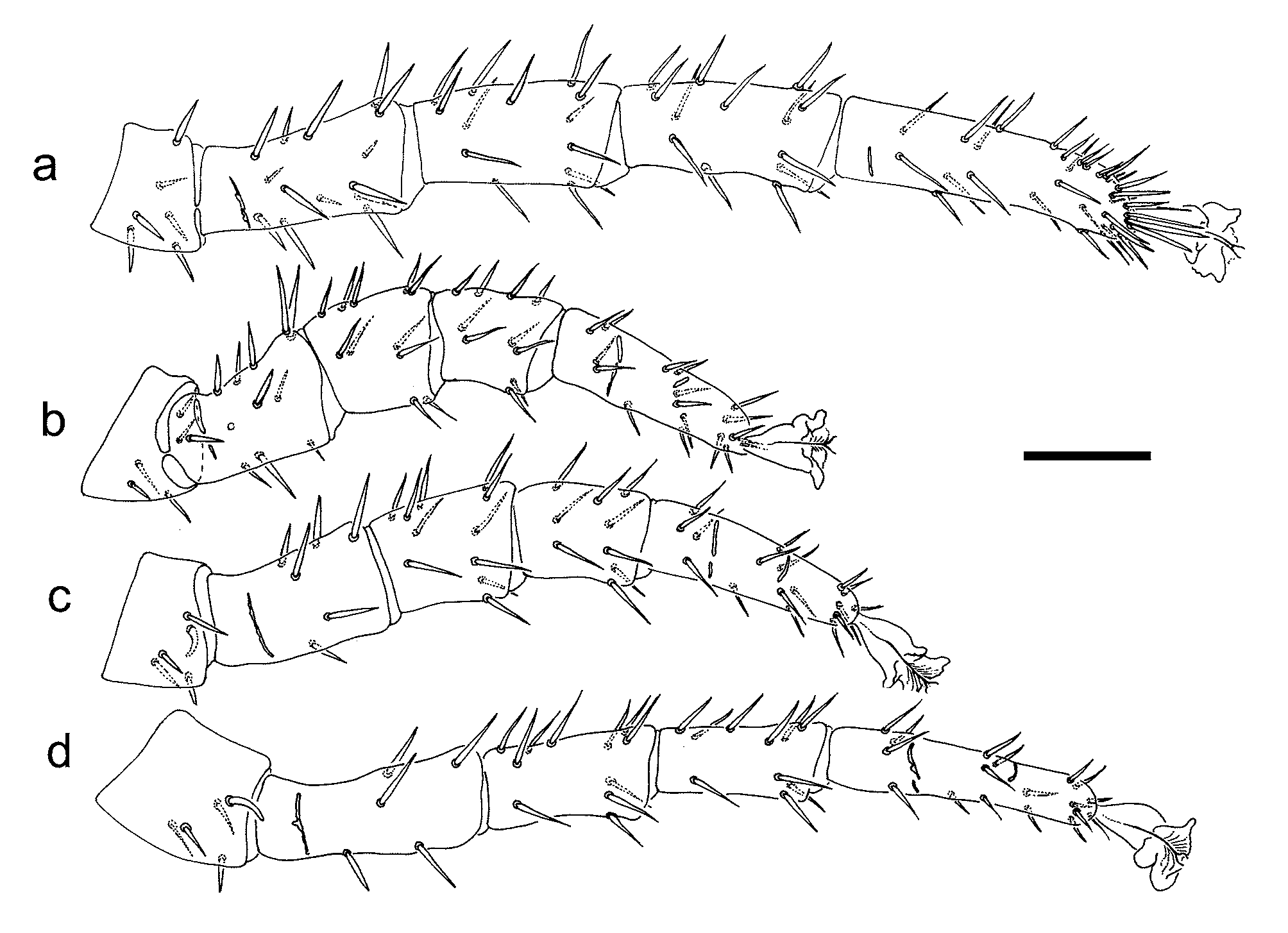
Legs: Cuticle and all setae of legs smooth. Chaetotaxy: Coxae 2-2-2-1. Trochanters 6-5-5-5. Leg I femur 2, 3/2, 2/3, 2, genu 2, 3/2,3/1, 2, tibia 2, 3/2, 3/1, 2. Leg II femur 2, 3/2, 2/1, 1, genu 2, 3/1, 2/1, 2, tibia 2, 2/1, 2/1, 2. Leg III femur 2, 2/0, 2/0, 0, genu 2, 2/1, 3/1, 2, tibia 2, 1/1, 2/1, 2. Leg IV femur 1, 4/1, 0, genu 2, 2/1, 3/1, 2, tibia 2, 1/1, 3/1, 2. Femur II ad1, pd1, and ad1, ad2 on femora III–IV, are relatively stout. Claws apparently absent; ambulacral pads well-developed; pretarsal opercula bear a reduced but variable number of weak tines (n= 2–6; Fig. 2 View FIGURE 2 c); mostly ca. 4 tines. Leg segment lengths as in Table 1.
I II III IV
Femur 159–175 127–135 127–130 165–175 Genu 161–170 96–104 102–118 134–141 Tibia 170–177 90–101 103–115 135–140 Tarsus 230–255 167–178 172–187 210–215 Description of male (n = 1). As for females except as noted below.
Dorsum: Dorsal shield 680 x 550. Narrow band of striate cuticle posteriorly not covered by shield.
Ve n te r: Sternoventral shield bears st1–3, st5 and 1–2 pairs setae; st4 present on one side only. Setae st1 30, st2 26 and st3 28. Genital opening at level of st1, 18 wide x 13 deep. Anal shield broad and separate from sternoventral shield. Para-anal setae 52; post-anal 55.
Gnathosoma: Palp setation 1:4:6:11–12. Corniculi 15 long. Deutosternum with 13 rows of denticles. Hypostomal setae h1 20, h2 25, h3 25, palp coxal seta 26. First cheliceral segment 65, second segment 138. Movable digit completely fused with spermadactyl.
Legs: Leg setation as for holotype female. Leg measurements are ca. 80% of those for females.
Etymology. This new species is named for its collector, Dr Chris Burwell (QM).
Remarks. This new species differs from other Myrmozercon species except M. beardae sp. nov. in having hypotrichy on both the dorsal shield and the venter. It differs from M. beardae in having a single seta on coxa IV and in having barbed dorsal and circum-anal setae. Also the anal opening is close to the anterior margin of the shield, and the opisthogastric setae are of uniform thickness. This new species conforms to the diagnosis given for Myrmozercon by Walter (2003) except that the soft ventral cuticle lacks hypertrichy. The legs are holotrichous as given by Evans & Till (1965) save for hypertrichy on genu IV with an additional pl and a pv seta, on genu and tibia III with an additional pl seta, and on femur I with an additional ventral seta. However one specimen has just the typical four ventral setae on femur I on one side. Other variations include one specimen with femur IV lacking a proximodorsal seta on one side. The addition of a ventral seta on femur I also occurs in Laelaspulus flexuosus (Michael) ( Evans & Till 1965) . The low swelling on the palp femur is in the same position as an identical structure in Myrmozercon iainkayi Walter. The flaring of the spermadactyl tip recalls Pneumolaelaps hyatti Evans & Till, 1966 .
Polyrhachis (Campomyrma) flavibasis Clark is arboreal and lignicolous, nesting within hollow branches and twigs. Of 15 Lamington NP nest collections of P. flavibasis , only one yielded mites. Another positive collection was that from Boombana NP. Reference specimens of P. flavibasis from a Lamington NP M. burwelli -infested colony are deposited in the Queensland Museum (T153537 – T153542). Reliable descriptions of host-parasite relations require the ability to refer back to the original host specimens as the taxonomy of host species can change. Frey et al. (1992) provide a valuable discussion of relevant concepts, although the term they propose for host reference specimens (“symbiotypes”), would be misleading if readers inferred that a reference specimen has some nomenclatural status.
No known copyright restrictions apply. See Agosti, D., Egloff, W., 2009. Taxonomic information exchange and copyright: the Plazi approach. BMC Research Notes 2009, 2:53 for further explanation.
|
Kingdom |
|
|
Phylum |
|
|
Class |
|
|
Order |
|
|
Family |
|
|
Genus |