
Argoravinia (Argoravinia) catiae, Filho, Fernando Da Silva Carvalho & Esposito, Maria Cristina, 2012
|
publication ID |
https://doi.org/10.5281/zenodo.280654 |
|
DOI |
https://doi.org/10.5281/zenodo.6174654 |
|
persistent identifier |
https://treatment.plazi.org/id/03E387E7-E746-231C-FF7A-FD08FE37FD8A |
|
treatment provided by |
Plazi |
|
scientific name |
Argoravinia (Argoravinia) catiae |
| status |
sp. nov. |
Argoravinia (Argoravinia) catiae View in CoL sp. nov.
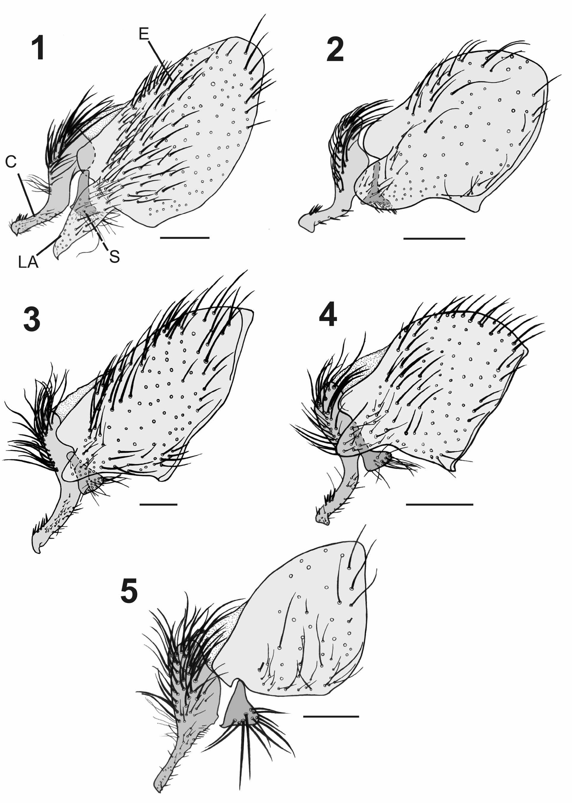
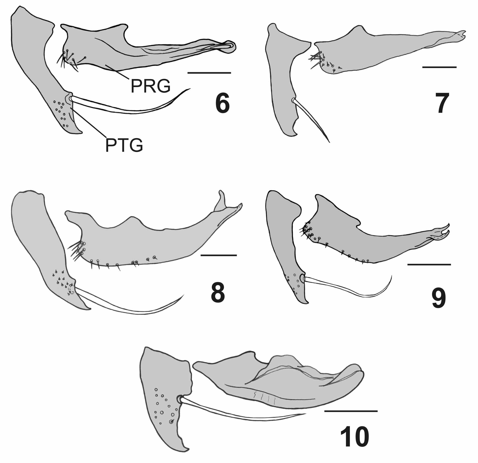
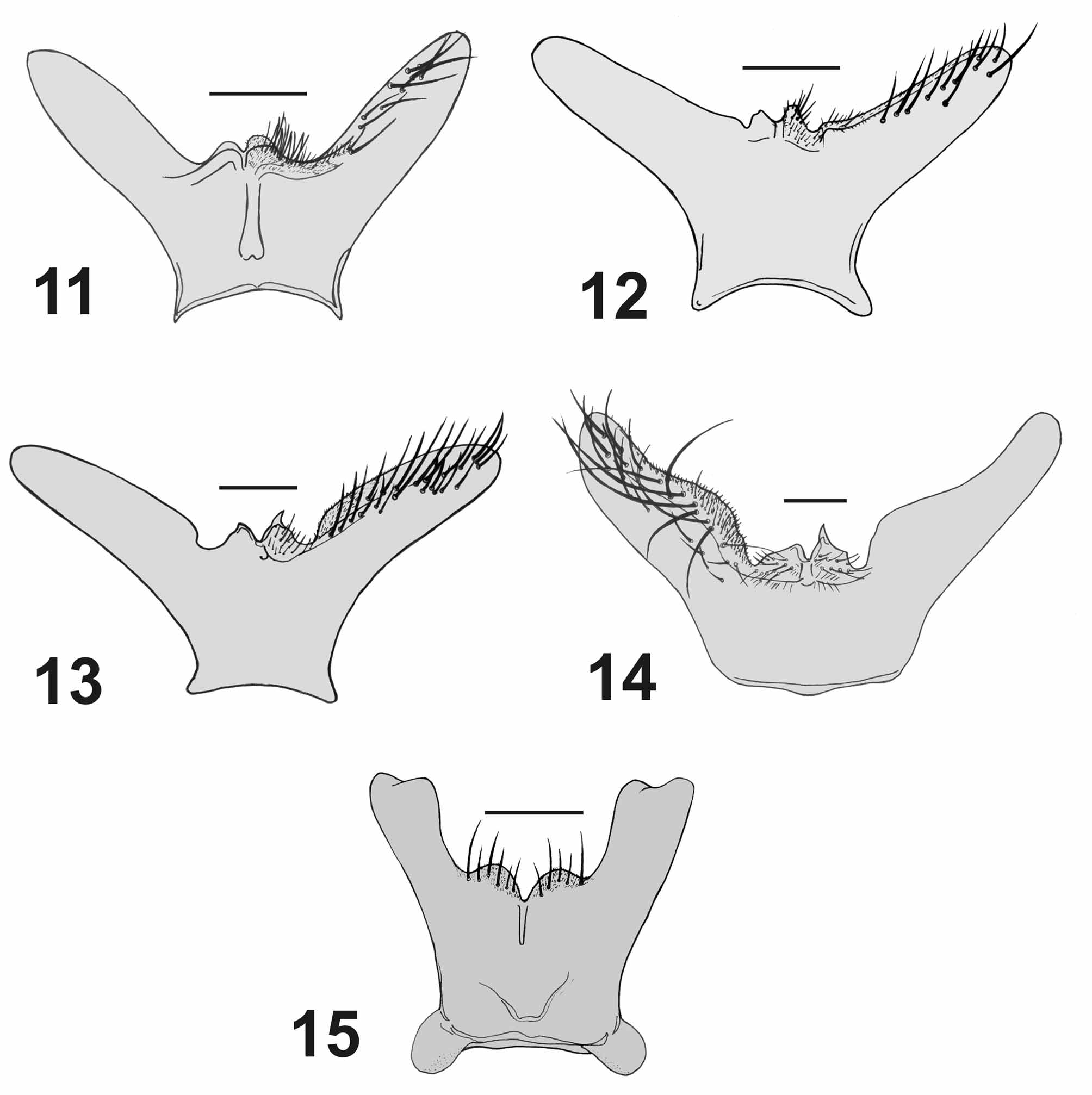
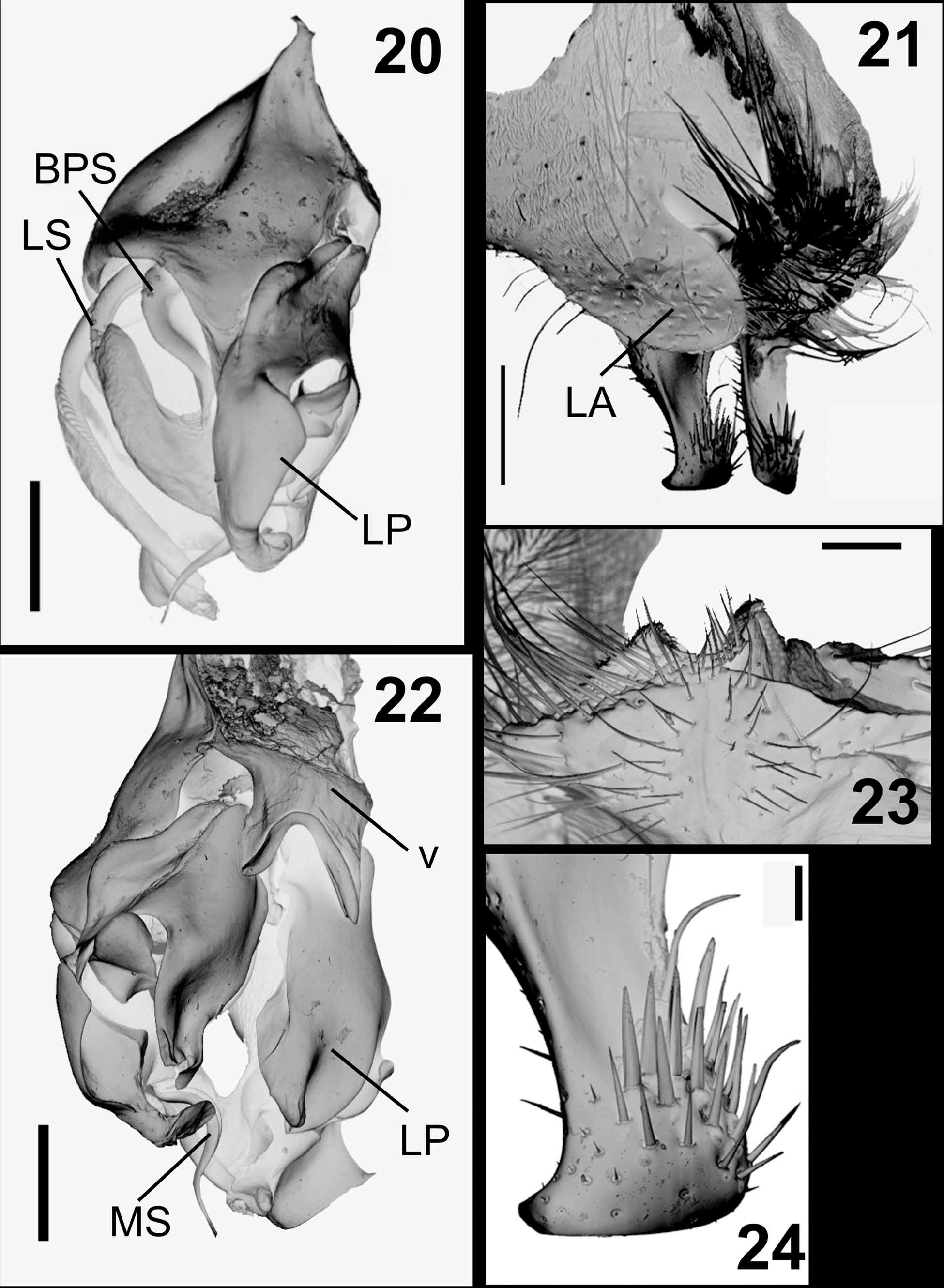
( Figs. 2 View FIGURES 1 – 5 , 7 View FIGURES 6 – 10 , 12 View FIGURES 11 – 15 , 20–24 View FIGURES 20 – 24 , 63 View FIGURES 61 – 66 )
Male —Length = 6.4−7.0 mm (n = 6).
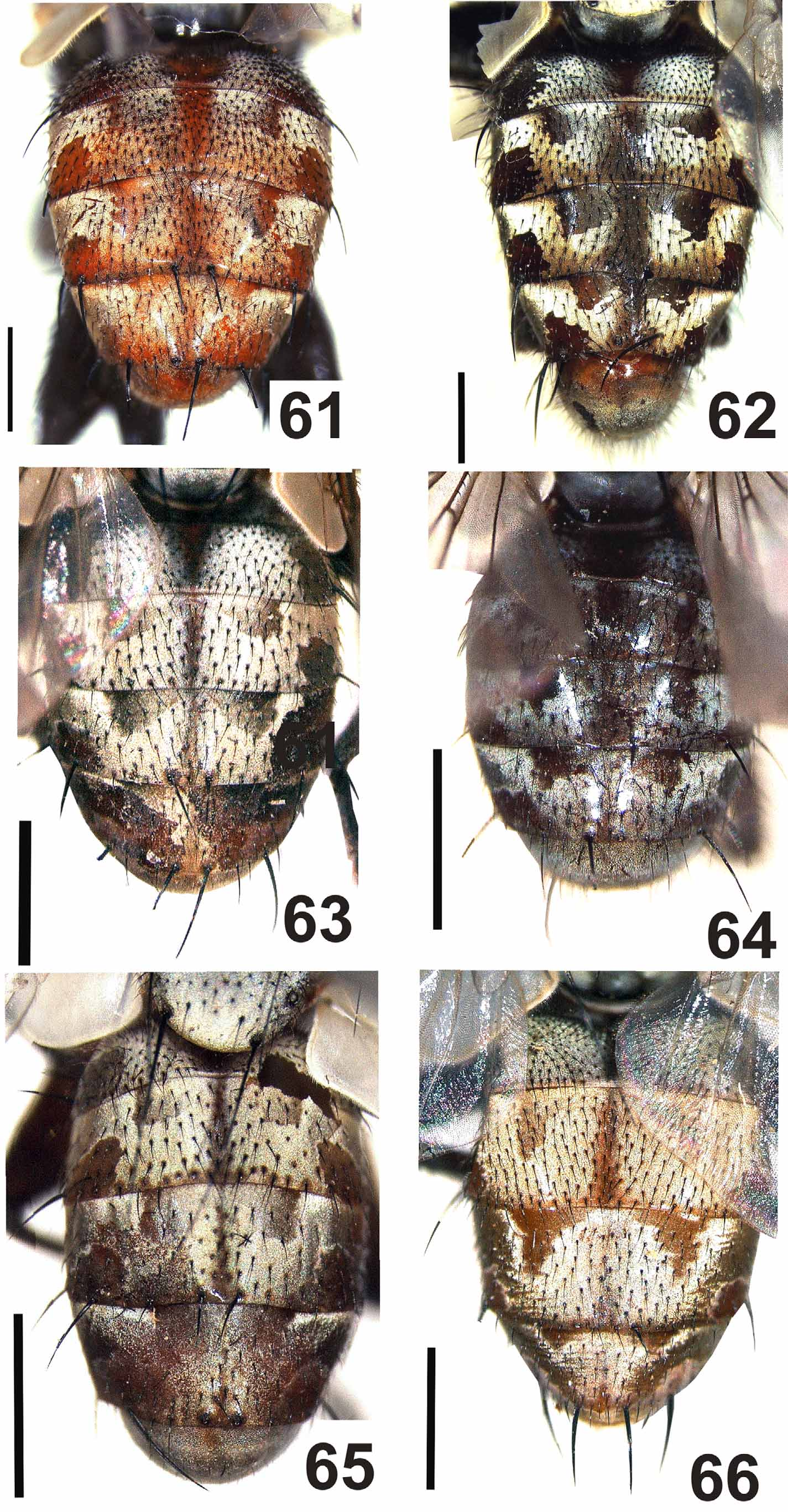
Similar to A. alvarengai male but differing as follows: Head. Frons at vertex 0.26x head width; frontal row of six bristles, first four bristles convergent and upper two reclinate. Abdomen. Reddish brown with usual pattern of silvery gray microtomentum ( Fig. 63 View FIGURES 61 – 66 ); ST5 with posterior arm long and strongly divergent with rounded apex, and with long setae on inner lateral margin, posterior margin of ST5 with two short median lobes, covered with minute setae ( Figs. 12 View FIGURES 11 – 15 , 23 View FIGURES 20 – 24 ).
Terminalia. Syntergosternite 7+8 very large and globulous, reddish with grayish microtomentum, scattered short black setulae, and five marginal bristles; epandrium reddish with short black setulae, cercus short and strongly bent backwards ( Figs. 2 View FIGURES 1 – 5 , 21 View FIGURES 20 – 24 ), with short apical projection on outer lateral margin ( Figs. 21, 24 View FIGURES 20 – 24 ), apex of cercus with a cluster of long spines on dorsal surface, close to tip and reaching inner lateral margin ( Fig. 24 View FIGURES 20 – 24 ), outer lateral and apical margin bearing minute spines ( Fig. 24 View FIGURES 20 – 24 ), ventral surface of cercus with many scattered spines; lateral apophyses rounded and very large, completely covering surstylus, lateral margin and dorsal surface of cercus ( Figs. 1 View FIGURES 1 – 5 , 21 View FIGURES 20 – 24 ). Surstylus with narrow base and enlarged apex, and long and slender setae at apex. Postgonite short and almost straight, with pointed apex, and with a strong and short pre-apical bristle on anterior margin ( Fig. 7 View FIGURES 6 – 10 ). Pregonite long and narrow, apically bifid with inconspicuous, pointed tips ( Fig. 7 View FIGURES 6 – 10 ). Phallus reddish; distiphallus with narrow base and enlarged apex; basal process of lateral stylus slightly sinuous ( Fig. 20 View FIGURES 20 – 24 ); lateral plate large, folded and with apophyses; vesica short and membranous, U-shaped, with short and wide stem, and anterior margin finely thick ( Fig. 22 View FIGURES 20 – 24 ); lateral stylus long and striated at apex ( Fig. 22 View FIGURES 20 – 24 ); median stylus very long and slender.
Female. Unknown
Type material. Holotype 3 ( MPEG): BRAZIL: Pará: São Geraldo do Araguaia, Serra das Andorinhas, Sta. Cruz, 6°12'58.8"S 48°26"1.6"W, 1–10.XII.2001, Cerrado, Malaise [= Malaise trap], I.S. Gorayeb, A. Tavares, J.M.F. Ribeiro and L.A.S. Sousa leg.
Paratypes: 5 3 with same data as holotype (3 in MPEG and 2 in MNRJ).
Distribution. NEOTROPICAL: Brazil (Pará)
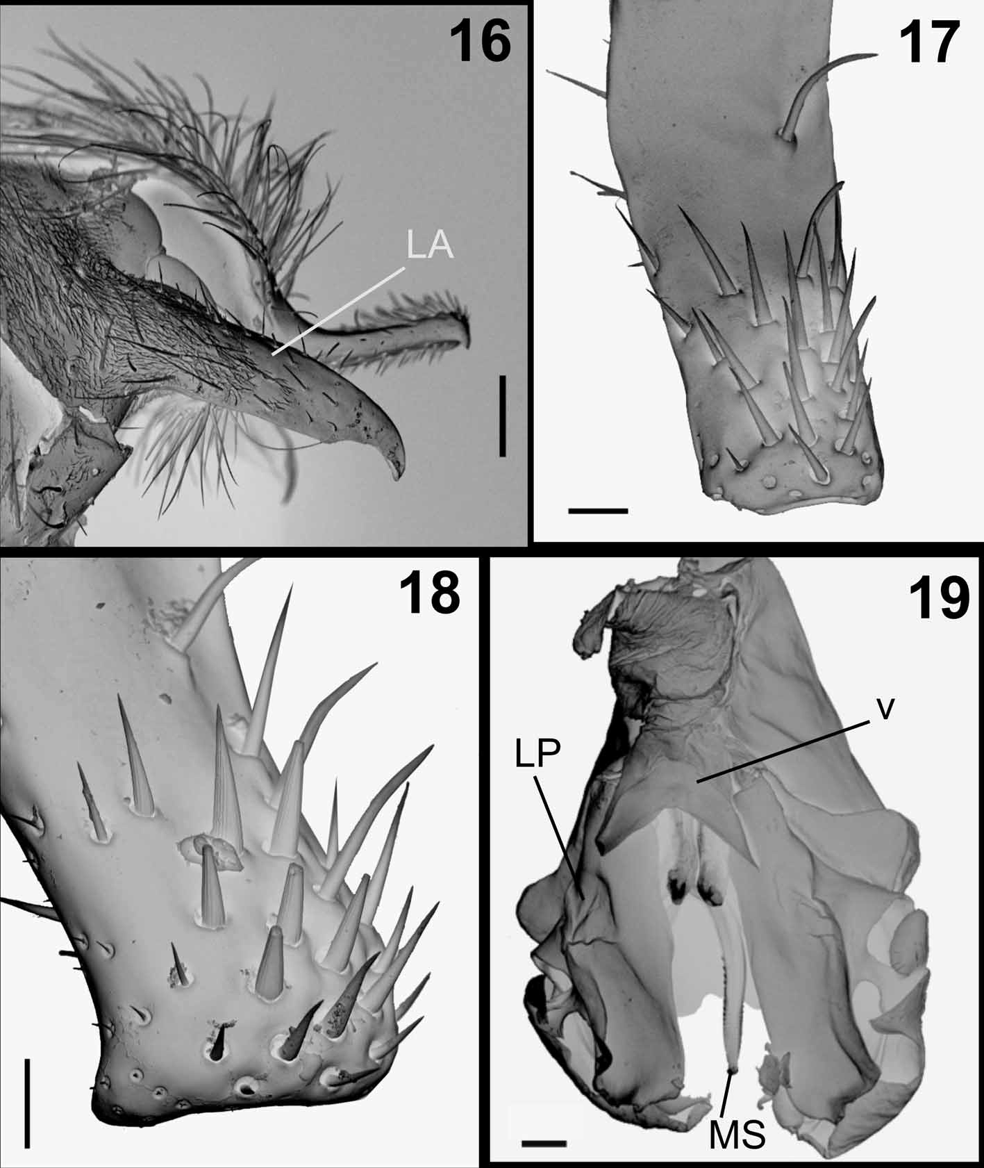
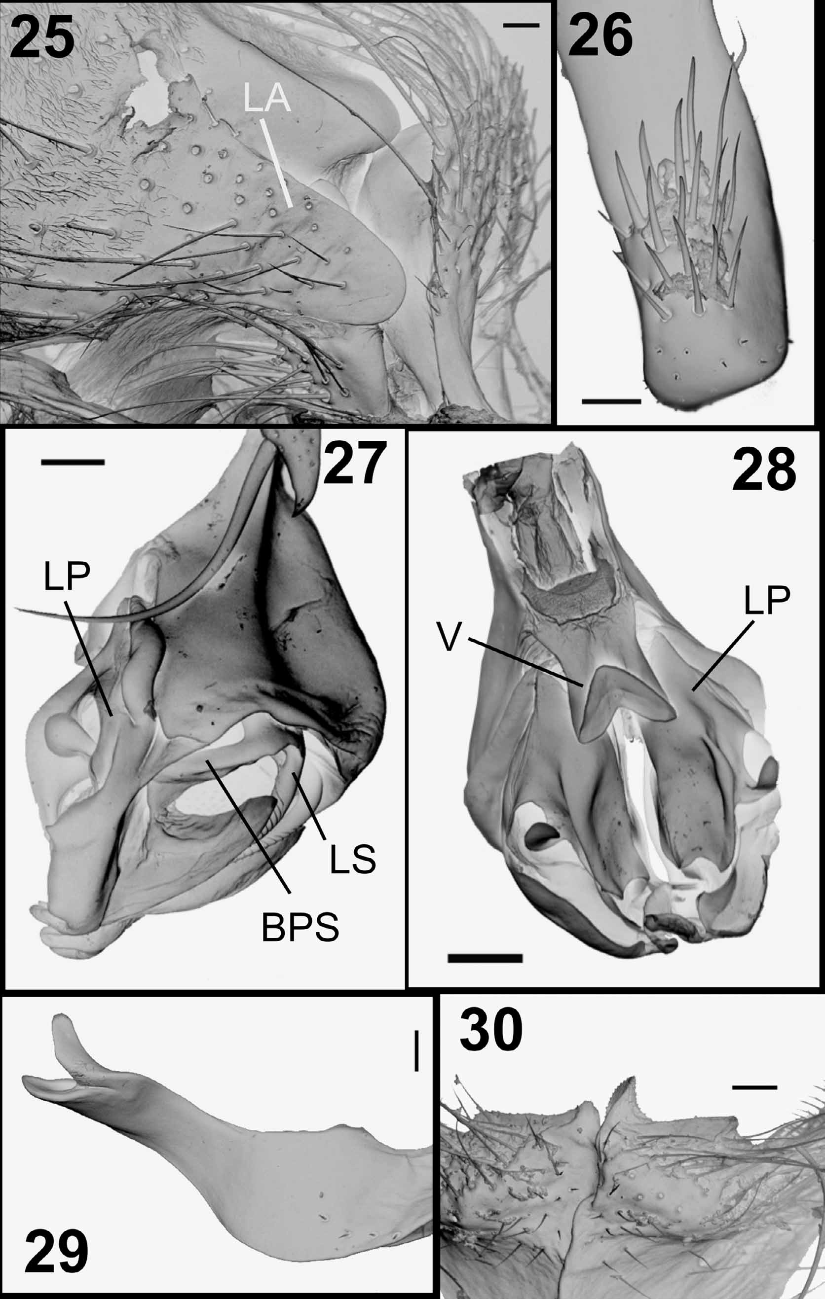
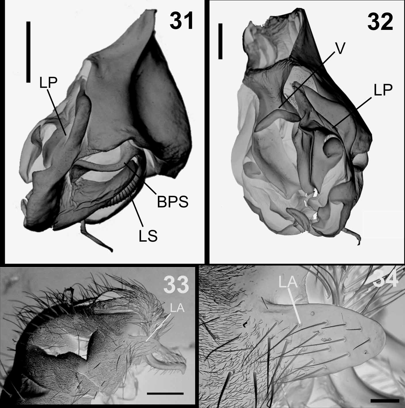
Remarks. This species is similar to A. alvarengai , A. rufiventris and A. paraensis , all of which have the cercus strongly bent backwards, spines on dorsal surface of cercus, and epandrium with a lateral apophysis. Argoravinia catiae differs from A. alvarengai by having the lateral apophysis shorter, very broad and with rounded apex ( Figs. 2 View FIGURES 1 – 5 , 21 View FIGURES 20 – 24 ). The lateral apophysis is very long, narrow and with pointed apex in A. alvarengai ( Fig. 16 View FIGURES 16 – 19 ). The lateral apophysis has a rounded apex also in A. rufiventris and A. paraensis , but in these species the apophysis is short and covers only the basal portion of the surstylus ( Figs. 3, 4 View FIGURES 1 – 5 , 25 View FIGURES 25 – 30 , 33 View FIGURES 31 – 34 , 34), while in A. catiae the lateral apophysis completely covers the surstylus and upper lateral portion of the cercus ( Figs. 2 View FIGURES 1 – 5 , 21 View FIGURES 20 – 24 ).
The redescription of Sarcophaga rufiventris by Aldrich (1930: 5, fig. 27), based on study of Wiedemann’s type, includes a figure of the male epandrium and cercus in lateral and posterior views. The epandrium does not have a conspicuous broad lateral apophysis covering the dorsal surface of the cercus, which is a distinctive feature of A. catiae . This structure is very large in A. catiae and can be seen even in pinned specimens under a stereomicroscope. All examined specimens of A. catiae are from Serra das Andorinhas, an area in Brazil within the Cerrado, a vast region characterized by primary forest bearing patches of savanna, with waterfalls and caves. Some species of animals and plants collected in Serra das Andorinhas are endemic to the Cerrado. Argoravinia catiae seems to be restricted to this kind of environment, since it was not found among the material collected in other regions of Amazonia and Brazil.
Etymology. Dedicated to the Brazilian dipterist Dr. Cátia Antunes de Mello-Patiu (MNRJ) for her invaluable contribution to the study of the flesh fly fauna of Brazil.
Biology. Unknown.
No known copyright restrictions apply. See Agosti, D., Egloff, W., 2009. Taxonomic information exchange and copyright: the Plazi approach. BMC Research Notes 2009, 2:53 for further explanation.
|
Kingdom |
|
|
Phylum |
|
|
Class |
|
|
Order |
|
|
Family |
|
|
Genus |