
Vrijenhoekia balaenophila, Pleijel & Rouse & Ruta & Wiklund & Nygren, 2008
|
publication ID |
https://doi.org/ 10.1111/j.1096-3642.2007.00360.x |
|
persistent identifier |
https://treatment.plazi.org/id/03E287CA-170C-FFCC-FCD1-15882AD6687E |
|
treatment provided by |
Felipe |
|
scientific name |
Vrijenhoekia balaenophila |
| status |
sp. nov. |
VRIJENHOEKIA BALAENOPHILA View in CoL SP. NOV. ( FIGS 1–4 View Figure 1 View Figure 2 )
Holotype: Off California, Monterey Canyon, 36°36.8 ′ N, 122°26.0 ′ W, 2891 m, RV Western Flyer, ROV Tiburon dive T 742, on grey whale carcass (see Goffredi et al., 2004; Rouse et al., 2004, for further descriptions of this whale fall), coll. F. Pleijel and G.W. Rouse, 29.ix.2004 (preserved in formalin, SMNH 6305).
221572 221568 442565 442567 513293 442566 513294 513295 442568 513296 513297 513298 513299 513300
AF AF DQ DQ DQ DQ DQ DQ DQ DQ DQ DQ DQ DQ
number 340407 442599 442605 442607 442597 442606 442610 442611 442612 513306 513307 513308 513309 513310
accession AY DQ DQ DQ DQ DQ DQ DQ DQ DQ DQ DQ DQ DQ
GenBank 340470 AY DQ 442570 View Materials 442575 DQ 442578 View Materials DQ DQ 442576 View Materials 442577 DQ DQ 442581 View Materials DQ 442582 View Materials 442583 DQ DQ 513301 View Materials 513302 DQ 513303 View Materials DQ 513304 View Materials DQ DQ 513305 View Materials
83519 83511 83510 83508 83521 76989 83516 83514
examined examined examined examined examined numbers SMNH SMNH SMHN SMNH SMNH SMNH SMNH FP SMNH Material see Material see Material see Material see Material see
accession
GenBank and
Koster
,
/ Australia
,
Sweden
Koster
,
, Lifou
Caledonia Koster,
Lifou
Caledonia
,
Koster, Koster, of Mexico Koster, California
, Monterey
California
, Monterey
California
Monterey,
California
Monterey,
California
Monterey,
vouchers Sweden Adelaide New Sweden New Sweden Sweden Gulf Sweden Off Off Off Off Off of
1 2 3 4 5
specification,. nov spm.. spm, nov. nov.. spm,. spm, nov..., nov spm
, sp sp sp sp sp
terminals
†
,. nov. gen. gen nov,..., gen nov., gen. nov nov,. gen.
sequenced
Table of
.
Origin
3
/ *
† / *
Dysponetus bulbosus caecus chinensis
Micropodarke Nereimyra punctata
Psamathe fusca methanicola Sirsoe Syllidia armata
Vrijenhoekia balaenophila Vrijenhoekia balaenophila balaenophila Vrijenhoekia balaenophila Vrijenhoekia balaenophila Vrijenhoekia 16 S and * 28. S COI †.
Paratypes: Same locality data, ROV Tiburon dive 391, seven paratypes (preserved in formalin, SMNH 6309 View Materials ), coll. S. Goffredi, 6.ii.2002 ; same locality data, ROV Tiburon dive T486 , nine paratypes (preserved in formalin, SMNH 6310 View Materials ), coll. S. Goffredi, 9.x.2002 ; same locality data, ROV Tiburon dive T742 , 11 paratypes (preserved in formalin, SMNH 6306 View Materials ), colls F. Pleijel and G.W. Rouse, 29.ix.2004 ; same locality data, ROV Tiburon dive T742 , three paratypes (preserved in 99.5% ethanol, SMNH 6307 View Materials ), colls F. Pleijel and G. W. Rouse, 29.ix.2004 ; same locality data, ROV Tiburon dive T932 , six paratypes (preserved in formalin, SMNH 6308 View Materials ), colls F. Pleijel and G.W. Rouse, 4.i.2006 .
Other material: Same locality data, thee specimens (preserved in osmium tetroxide, mounted for SEM, FP), colls F. Pleijel and G. W. Rouse, 29.ix.2004 ; same locality data, two specimens (preserved in 99.5% ethanol, FP), colls F. Pleijel and G. W. Rouse, 4.i.2006 .
Apomorphies: Three pairs of large glandular lip pads surrounding mouth opening, papilla-shaped neuropodial lobes on segment 3, very long dorsal cirri, and growth pattern with up to about 35 segments whereafter segments increase in size but no new segments are added.
Etymology: Named for its affinity to whale bones.
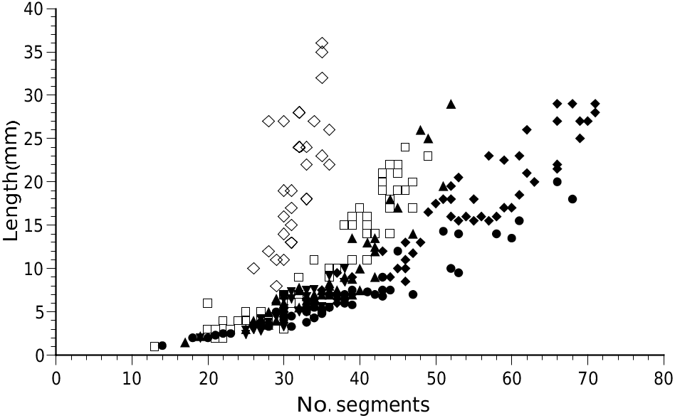
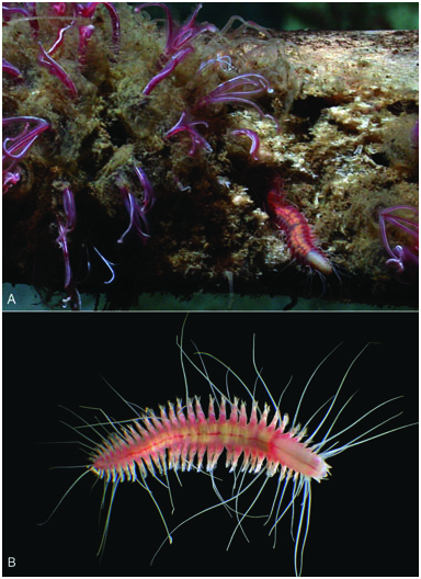
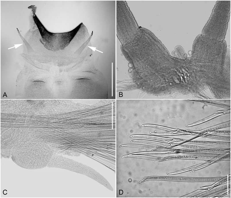
Description: Up to 36 mm long and 9 mm wide (including parapodia but excluding chaetae) for 35 segments (see Fig. 1 View Figure 1 for further measurements). Body stout, anteriorly truncate and slightly posteriorly tapered ( Fig. 2 View Figure 2 ); specimens with fewer segments nearly as wide as longer specimens but more elliptical in dorsal outline. Longitudinal ventral depression usually distinct. Prostomium rounded rectangular, much wider than long, poorly delineated postero-dorsally towards first segment; posterior incision absent ( Fig. 3A). Palpophores cylindrical, longer and stouter than palpostyles; palpostyles evenly tapered to rounded tips ( Fig. 3A). Paired antennae as long as palps, thinner, evenly tapered, without distinct antennophores ( Fig. 3B). Eyes absent. Median antenna absent, but small mediodorsal tubercle usually present middorsally on prostomium ( Fig. 3B). Nuchal organs small, as postero-lateral prostomial pits, not extending dorsally ( Fig. 3B). Peristomium only visible ventrally as lips. Distinct facial tubercle present, forming large rounded triangular elevation on specimens with everted proboscis ( Fig. 3A, B), and anterio-ventral sac-like protrusion on specimens with inverted proboscis. Lip glands forming three pairs of distinct pads surrounding mouth opening: dorsal pair (below palps and paired antennae), lateral pair and ventral pair ( Fig. 3A–C). Terminal proboscis ring densely ciliated, with ten long, digitate and distally pointed papillae ( Fig. 4A View Figure 4 ); jaws, jaw plates and teeth absent; inner lining sclerotized ( Fig. 4A View Figure 4 ). Segment 1 reduced, not dorsally visible, segment 2 fully developed. Dorsal cirri segments 1–5 reaching segments 10–15, 12–21, 10–13, 10–18 and 16–20, respectively; cirri and cirrophores not differentiated compared with following segments. Ventral cirri on segments 1–3, reaching segments 5–7, 5–7 and 5, respectively; first pair longest and third pair shortest; cirrophores cylindrical, well differentiated ( Fig. 3D). Small papilla-shaped neuropodial lobes on segment 3, with aciculae but usually without chaetae ( Fig. 4B View Figure 4 ); sometimes with few chaetae in smaller specimens ( Fig. 3D). Neuropodial lobes, neurochaetae and ventral cirri on segment 4 similar to following segments ( Fig. 3D). Notopodial lobes and notochaetae absent on all segments. Cirrophores and dorsal cirri segments 5, 8, 10, 12, 15, 17, 20, 23 and 25 slightly enlarged and elevated, although difficult to assess posterior to about segment 19 or 20. Dorsal cirrophores prominent; dorsal cirri very long ( Fig. 2 View Figure 2 ), sometimes longer than body, weakly ringed, proximal rings wider than long, distal rings longer than wide; 2–3 notoaciculae per notopodium. Neuropodial lobes of median segments with rounded, elongated preaciculary lobes, pointed aciculary lobes and asymmetrical postaciculary lobes with truncate ends. Large number of compound chaetae, +50; 2–3 neuroaciculae per neuropodium. Shafts internally distinctly camerated ( Fig. 4D View Figure 4 ). Blades unidentate with one side finely serrated, dorsal and median blades up to twice as long as ventral ones. Prolonged teeth absent. Ventral cirri subdistally inserted on neuropodium, evenly tapered ( Fig. 3E). Ventral segmental globular swellings at bases of ventral cirri and cirrophores ( Figs 3E, 4C View Figure 4 ); apparent on some specimens, possibly associated with reproduction. Pygidial cirri long, similar to dorsal cirri on posterior segments; pygidial papilla absent ( Fig. 3F). Anus dorsally situated. Some specimens mature females, eggs rounded, small, c. 60 Mm in diameter.
Colour: Live specimens yellowish to red, with parapodia and lateral parts of dorsum usually with rich supply of blood vessels. Eggs light rose-coloured.
Distribution and habitat: Known only from the type locality on a grey whale carcass at 2891 m depth in Monterey Canyon off California.
Remarks: The length/segment distribution of V. balaenophila is unusual. In one example (12 mm for 28 segments) a specimen showed obvious signs of regeneration, but this was not the case with others, and the relationship between the length and the number of ·
segments differs from other hesionids, where segments are added continually (excluding Hesionini , in which adults always have 21 segments), although at reduced rates with increasing size. For comparison with close relatives, we plotted the distribution for specimens of Hesiospina aurantiaca (Sars, 1862) , Nereimyra punctata (O.F. Muller, 1776) , Micropodarke dubia (Hessle, 1925) , Psamathe fusca Johnston, 1836 and Syllidia armata Quatefages, 1866 ( Fig. 1 View Figure 1 ). The observed pattern in V. balaenophila indicates a continuous growth of individual segments, with virtually no new ones added after c. 35 segments.
Glandular lip pads have been reported in several other hesionid group, including Gyptis ( Pleijel, 1993) and Nereimyra ( Pleijel, 1998) . The glandular nature of these structures is indicated by a large number of pores with an aperture of about 1 Mm ( Pleijel, 1998). In these taxa, however, the lip pads are situated as a single pair on each side of the mouth opening; in V. balenophila there are three pairs, which is a unique feature.
A facial tubercle has been reported from a series of hesionids by Pleijel (1998), especially prominent in taxa such as Hesione Lamarck, 1818 and Leocrates Kinberg, 1866 . It is a distinct triangular pad or protuberance that is positioned ventrally to the prostomium proper; when the proboscis is everted it becomes situated dorsally on the basal section of the proboscis, anterior to the prostomium. The term ‘facial tubercle’ has previously been used in scale worms (e.g. Rouse & Pleijel, 2001) for a structure situated in a similar position, although it is currently uncertain if the structure is homologous in the two groups.
The shape of the neuropodial lobes on segment 3 is characteristic for V. balaenophila . In some of the examined specimens they were also provided with a few compound chaetae. In all studied hesionids the juveniles have neuropodial lobes and chaetae starting at segment 2. These are then successively reduced during ontogeny until they reach the adult stage with neuropodial lobes and chaetae starting at segment 3, 4 or 5 (segment 2 in Sirsoe grasslei ). In V. balaenophila it appears that the lobes on segment 3 are partly but not fully reduced.
Several of the examined specimens are provided with conspicuous bulbous swellings at the bases of ventral cirri, possibly glandular, whereas these are absent from others. One possibility is that this feature is related to reproduction, but we were unable to confirm this from the available specimens.
| RV |
Collection of Leptospira Strains |
| T |
Tavera, Department of Geology and Geophysics |
| SMNH |
Department of Paleozoology, Swedish Museum of Natural History |
| SMNH SMNH |
Department of Paleozoology, Swedish Museum of Natural History |
| SMNH SMNH SMNH SMNH |
Department of Paleozoology, Swedish Museum of Natural History |
No known copyright restrictions apply. See Agosti, D., Egloff, W., 2009. Taxonomic information exchange and copyright: the Plazi approach. BMC Research Notes 2009, 2:53 for further explanation.