
Selizitapia, Świerczewski & Stroiński, 2021
|
publication ID |
https://doi.org/ 10.5852/ejt.2021.750.1367 |
|
publication LSID |
lsid:zoobank.org:pub:FB09F5B2-162A-48B4-9687-D611F24792DE |
|
DOI |
https://doi.org/10.5281/zenodo.5493126 |
|
persistent identifier |
https://treatment.plazi.org/id/7CB34782-C76D-43AE-808C-4911F4CB0A2E |
|
taxon LSID |
lsid:zoobank.org:act:7CB34782-C76D-43AE-808C-4911F4CB0A2E |
|
treatment provided by |
Felipe |
|
scientific name |
Selizitapia |
| status |
gen. nov. |
Selizitapia View in CoL gen. nov.
urn:lsid:zoobank.org:act:7CB34782-C76D-43AE-808C-4911F4CB0A2E
Figs 1–7 View Fig View Fig View Fig View Fig View Fig View Fig View Fig
Type species
Selizitapia pennyi View in CoL gen. et sp. nov., here designated.
Diagnosis
The new genus differs from other taxa of Selizini in Madagascar by the following characters: 1) wings subrectangular (wings strongly constricted apically in Stenocyarda Fennah, 1965 ); 2) frons with median carina not diverged (frons with Y-shaped median carina in Urana ); 3) mesonotum without gibbosities (mesonotum with four gibbosities in Urana ); 4) dorsal part of periandrium unilobate (dorsal part of periandrium bilobate in Lembakaria and trilobate in Kelyflata Świerczewski & Stroiński, 2019 ); 5) lateral split of periandrium exceeding ⅓ of its length (lateral split of periandrium reaching ⅓ of its length in Peyrierasus Stroiński & Świerczewski, 2013 ).
Etymology
The generic name is an arbitrary combination of the name of the tribe ‘Selizini’, which the new genus belongs to, and the name of the forest formation – ‘tapia’, which the insect is associated with. Gender feminine.
Description
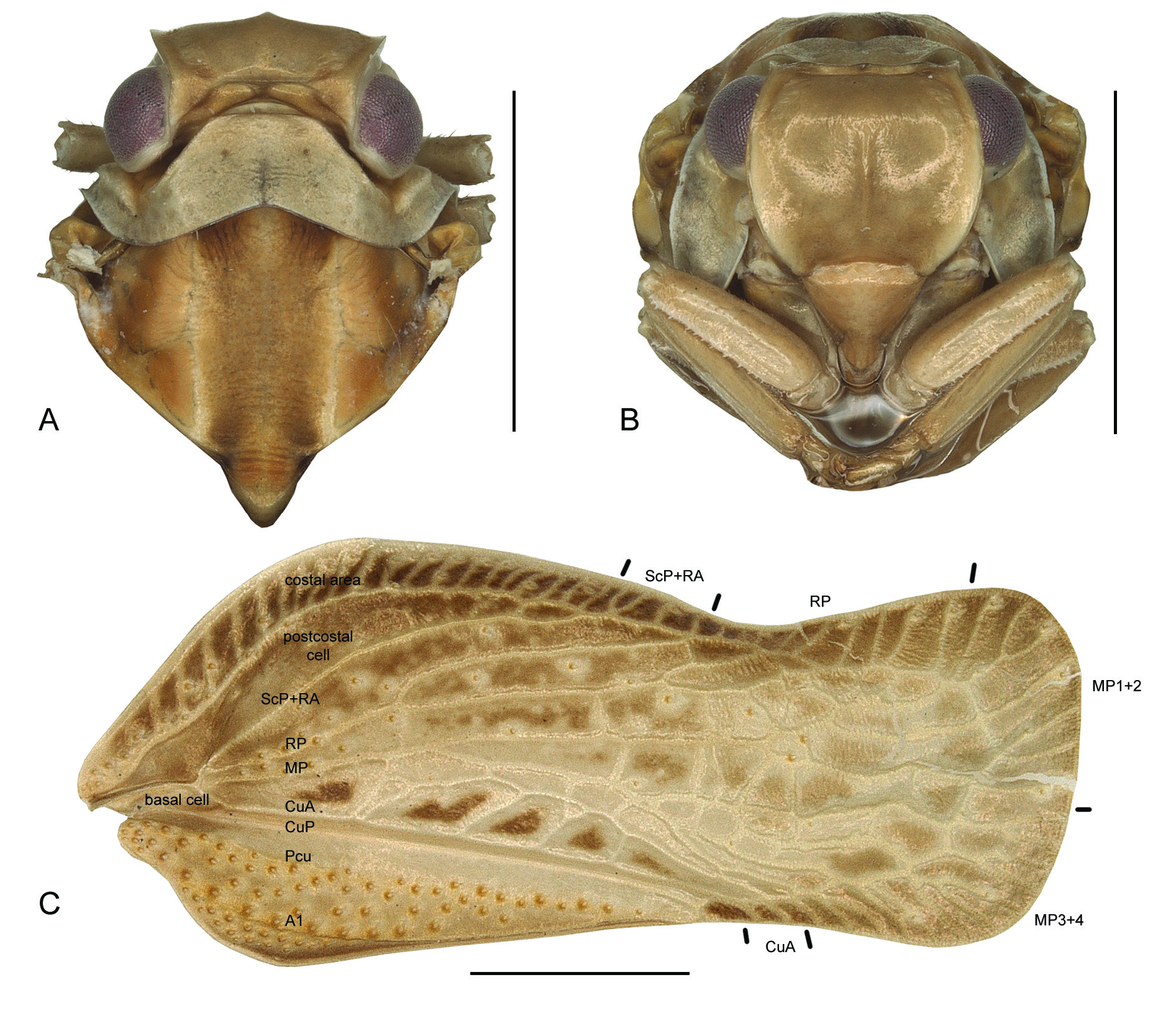
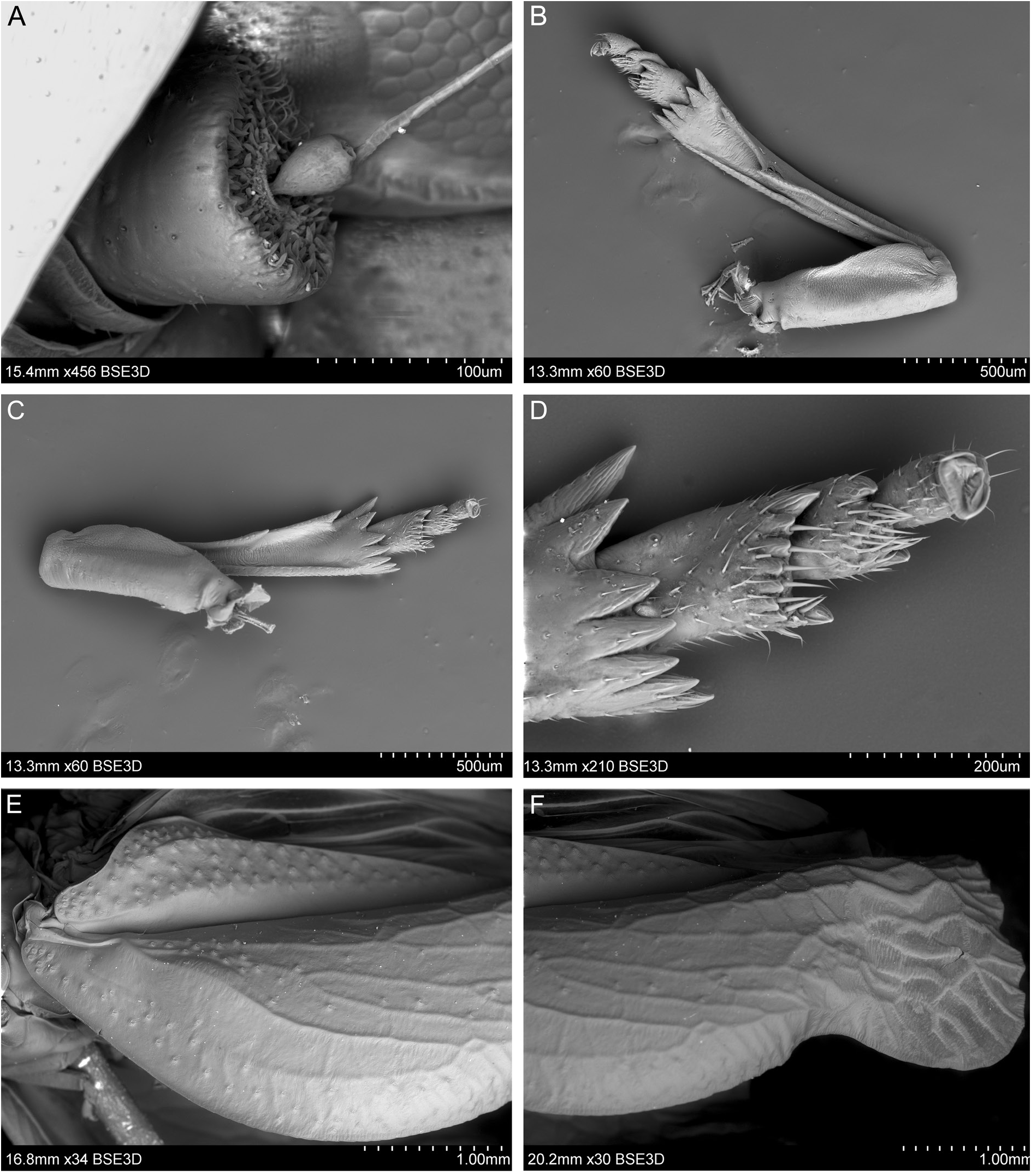
HEAD. Head with compound eyes, in dorsal view, slightly narrower than thorax. Vertex transverse, constricted in middle, medially slightly overlapped by pronotum: posterior margin carinate and strongly elevated, anterior margin carinate medially, covered by posterior margin, lateral parts obsolete; lateral margins carinate and subparallel ( Figs 1A View Fig , 2A, C–E View Fig ). Frons convex, widest at its lower third in frontal view; lateral margins carinate, arcuate and elevated, without incisions; upper margin almost straight; disc of frons with single, well-visible, median carina, laterally with obsolete ridges; frontoclypeal suture arcuate ( Figs 1B View Fig , 2A–B View Fig ). Clypeus smooth, weakly convex, without carinae ( Figs 1B View Fig , 2B View Fig ). Rostrum with apical segment shorter than subapical one, apex reaching hind coxae level. Compound eyes oval, with narrow callus at posterior margin. Lateral ocelli present. Antennae placed very close to medioventral margin of eyes; scapus small, ring-like, with single setae; pedicel shorter than diameter of eye but distinctly longer than scapus, bulbous, functional area at the top and on dorsal surface with trichoid sensilla type 1, antennal plate organs present on apical concavity and basally delimiting lateral margins of dorsal functional surface ( Figs 2F View Fig , 3A View Fig ).
THORAX. Pronotum, in dorsal view, shorter than mesonotum at midline: anterior margin arcuate with median portion almost straight, reaching anterior margin of compound eyes, posterior margin concave; disc of pronotum wrinkled, without carinae, with lateral impressions and central groove; postocular eminences conical ( Figs 1A View Fig , 2A, C–E View Fig ). Mesonotum with scutellum widely deltoid, wider than long at midline, scutellum with elevated apex; disc of mesonotum medially depressed with shallow groove; lateral carinae as ridges, only visible in posterior part and connected with posterior margin ( Figs 1A View Fig , 2E View Fig ). Tegmina longer than wide, subrectangular, with distinct venation and numerous transverse veinlets in apical part, without nodal line and with single apical line; costal margin sinuate, costal and sutural angle rounded, apical margin slightly rounded, postclaval sutural margin straight. Costal area short, with dense transverse veinlets, ending at the level of fusion of claval veins ( Figs 1C View Fig , 3E–F View Fig ). Costal cell about the same width as costal area, tapering apicad. Basal cell longer than wide. Tegmen with longitudinal veins ScP+RA and RP arising as short common stem from basal cell before bulla. Vein ScP+RA with fork distinctly after RP fork, ending on costal margin with 4 terminals; vein RP with fork before MP fork, ending on costal margin with 8–9 terminals; vein MP with fork distinctly apicad to CuA fork, ending on apical and postclaval margins; CuA with the first fork distinctly before RP fork. Apical cells subrectangular. Veins of apical half of tegmen wrinkled. Sensory and wax gland-plates concentrated on bulla and costal area, with a few scattered on the whole tegmen ( Figs 1C View Fig , 3E–F View Fig ). Clavus ending a bit before the end of costal area; Pcu and A 1 joined slightly anterior to clavus apex; A 1 slightly elevated; sensory and wax gland-plates concentrated on the area between Pcu and A 1 and basal part of the area after A 1 vein; single transverse veinlet after Pcu-A 1 connection ( Figs 1C View Fig , 3E View Fig ).
LEGS. Pro- and mesofemora slightly shorter than tibiae, subrectangular in cross section. Pro- and mesotibiae with shallow groove on external side; apical tarsomere of anterior and median legs longer than cumulative length of second and basal tarsomeres. Metatibiae longer than metafemora, triangular in cross section with two lateral spines and apical row of spines – first lateral spine placed subapically, second lateral spine placed a bit after midlength, apical spines in formula 2 longer (external) + 5 shorter (internal); basitarsomere of metatarsus a bit longer than cumulative length of second and apical tarsomeres, with apical spines lined as semicircle – 2 external spines a bit longer than 7 shorter internal spines; each internal spine bearing single, distinct seta; second segment of tarsomere with two lateral spines and median pad with setae. Metatibiotarsal formula: 2-(2+5)/(2+7)/2 ( Figs 3B–D View Fig ).
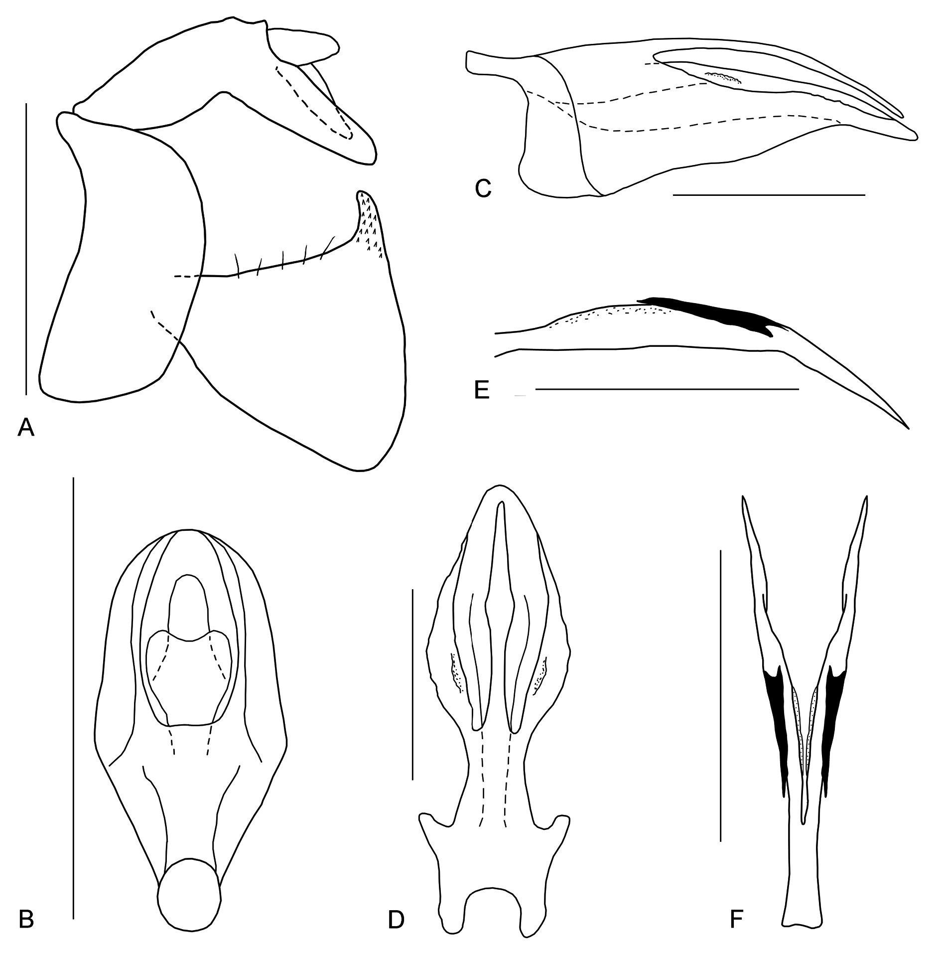
MALE TERMINALIA. Anal tube, in lateral view, elongate, with breaking point before anal opening, tapering apicad; anal opening placed a bit after midlength; basal part wider than apical part ( Figs 4A–B View Fig , 5A View Fig ); in dorsal view, rhomboid, with rounded apex ( Figs 4C–D View Fig , 5B View Fig ). Pygofer, in lateral view, with dorsal and ventral margin almost the same length, subparallel; anterior margin weakly concave, posterior margin convex. Genital style triangular, bearing short, hook-like capitulum with apex oriented anteriad ( Figs 4A–B, E View Fig , 5A View Fig ).
PHALLIC COMPLEX. Periandrium without any additional processes; in lateral view, about as long as aedeagus; lateral split reaching ⅔ of periandrium ( Fig. 5C View Fig ). Dorsal part of periandrium, in dorsal view, a bit shorter than ventral part, unilobate, smooth, spearhead-shape. Ventral part of periandrium elliptic, tapering apicad, basally with lateral lobes ( Fig. 5D View Fig ). Aedeagus, in lateral view, long and narrow, oriented ventrad, medially with acute process oriented apicad ( Fig. 5E View Fig ); in dorsal view bipartite, symmetrical, with deep median split, reaching ¾ of its length ( Fig. 5F View Fig ).
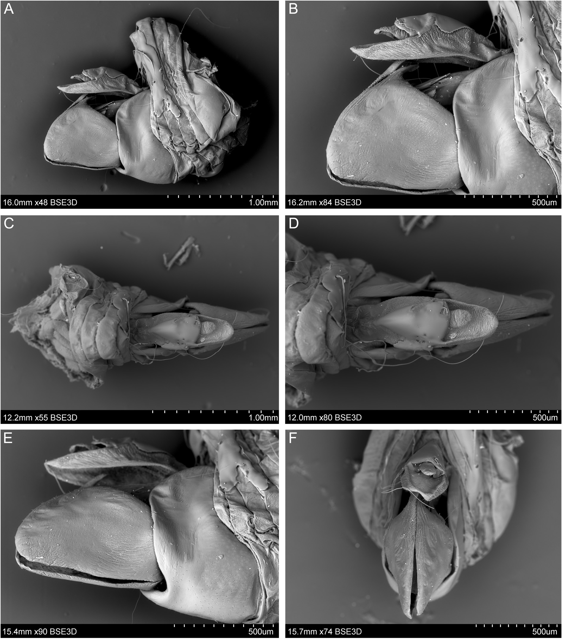
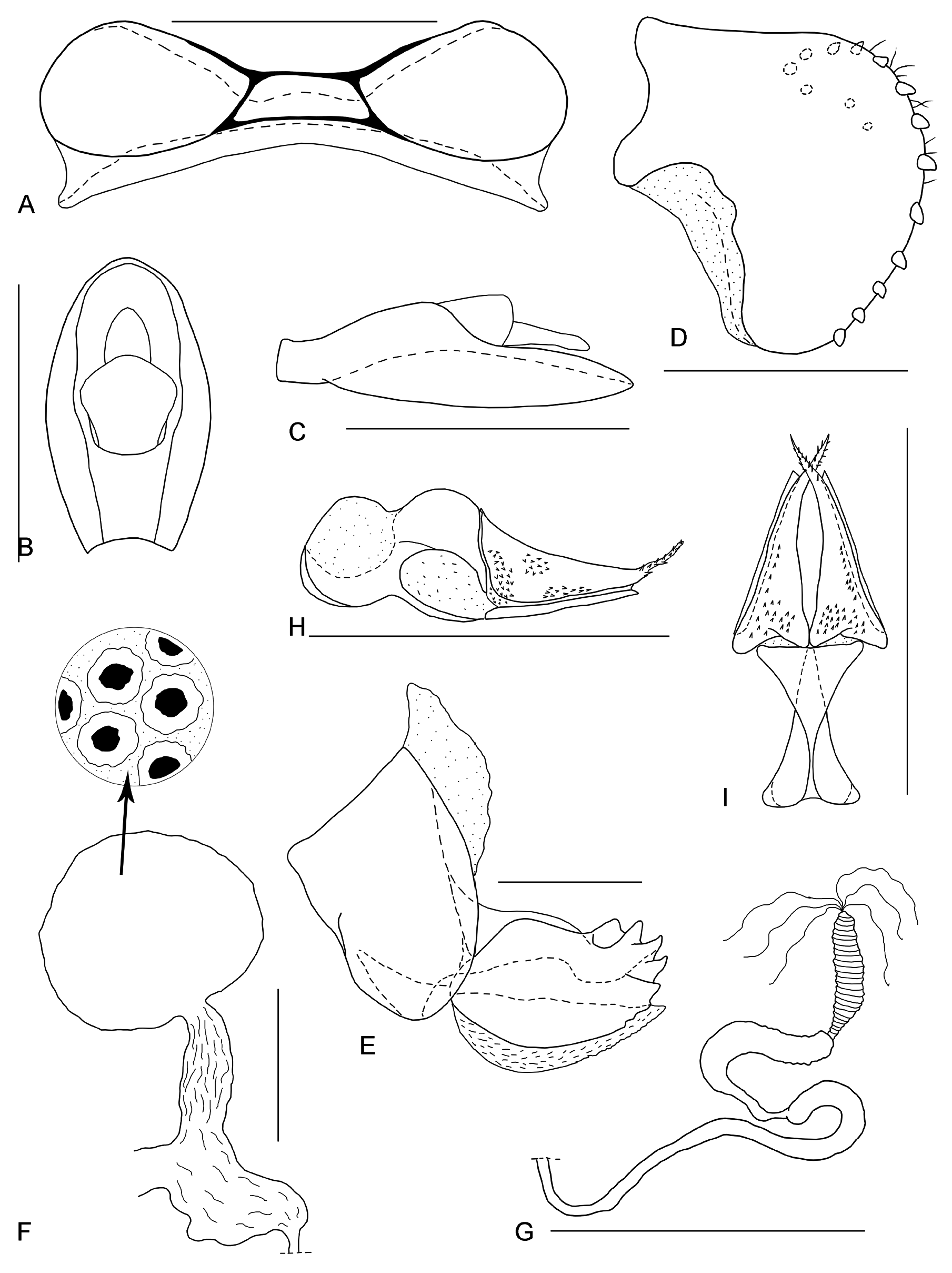
FEMALE TERMINALIA. Pregenital sternite with lateral lobes distinctly separated ( Figs 6A–B View Fig , 7A View Fig ). Anal tube, in lateral view, covering gonoplac and reaching its posterior margin ( Figs 6B View Fig , 7C View Fig ); in dorsal view, elliptic ( Figs 6C View Fig , 7B View Fig ). Gonoplac unilobate, rounded posteriorly, oriented ventrad, covering gonapophysis VIII ( Figs 6B View Fig , 7D View Fig ); posterior margin with one row of stout teeth, positioned at some distance one from another; teeth of both gonoplacs fitting together in a zip-like manner ( Figs 6E–F View Fig , 7D View Fig ). Gonapophysis VIII widely triangular, flattened, slightly oblique in respect to longitudinal body axis ( Fig. 7E View Fig ); endogonocoxal process as long as gonapophysis, wide, tapering apicad, with spiniferous microsculpture. Gonospiculum as in Fig. 7H–I View Fig . Bursa copulatrix with single pouch, rounded, cells with weakly sclerotized central areas ( Fig. 7F View Fig ). Spermatheca well developed; ductus receptaculi longer than diverticulum ductus, both parts smooth ( Fig. 7G View Fig ). Tergites of abdomen membranous in median portion ( Fig. 6C–D View Fig ).
Diversity and distribution
The genus is monotypic and contains a single species from Madagascar.


No known copyright restrictions apply. See Agosti, D., Egloff, W., 2009. Taxonomic information exchange and copyright: the Plazi approach. BMC Research Notes 2009, 2:53 for further explanation.