
Pseudostenophyma Furth, 2010
|
publication ID |
https://doi.org/ 10.5281/zenodo.199276 |
|
DOI |
https://doi.org/10.5281/zenodo.6204438 |
|
persistent identifier |
https://treatment.plazi.org/id/03E0B947-FF9B-FFB5-FF4B-FF26FDAEFCDC |
|
treatment provided by |
Plazi |
|
scientific name |
Pseudostenophyma Furth |
| status |
gen. nov. |
Pseudostenophyma Furth , new genus
Type species. Stenophyma modesta Weise, 1921 , 137: Male: Brasil: “Rio Purus”. “Amazon, Roman”, “Hyutanahã, 2/215. Hõglãnd“ [hand-written]. Stenophyma modesta m. [hand-written] (Swedish Museum of Natural History) – Herein designated as the Lectotype. Male: Brasil: “Rio Autaz”, “Amazon, Roman” (Swedish Museum of Natural History) – Herein designated as the Paralectotype. These Lectotype and Paralectotype designations are being made because no primary type was mentioned in the original description by Weise (1921), thus they are syntypes. Therefore, the designation of a primary type specimen establishes a single specimen as the unique bearer of this name and the standard for its application in the future, thus avoiding any confusion.
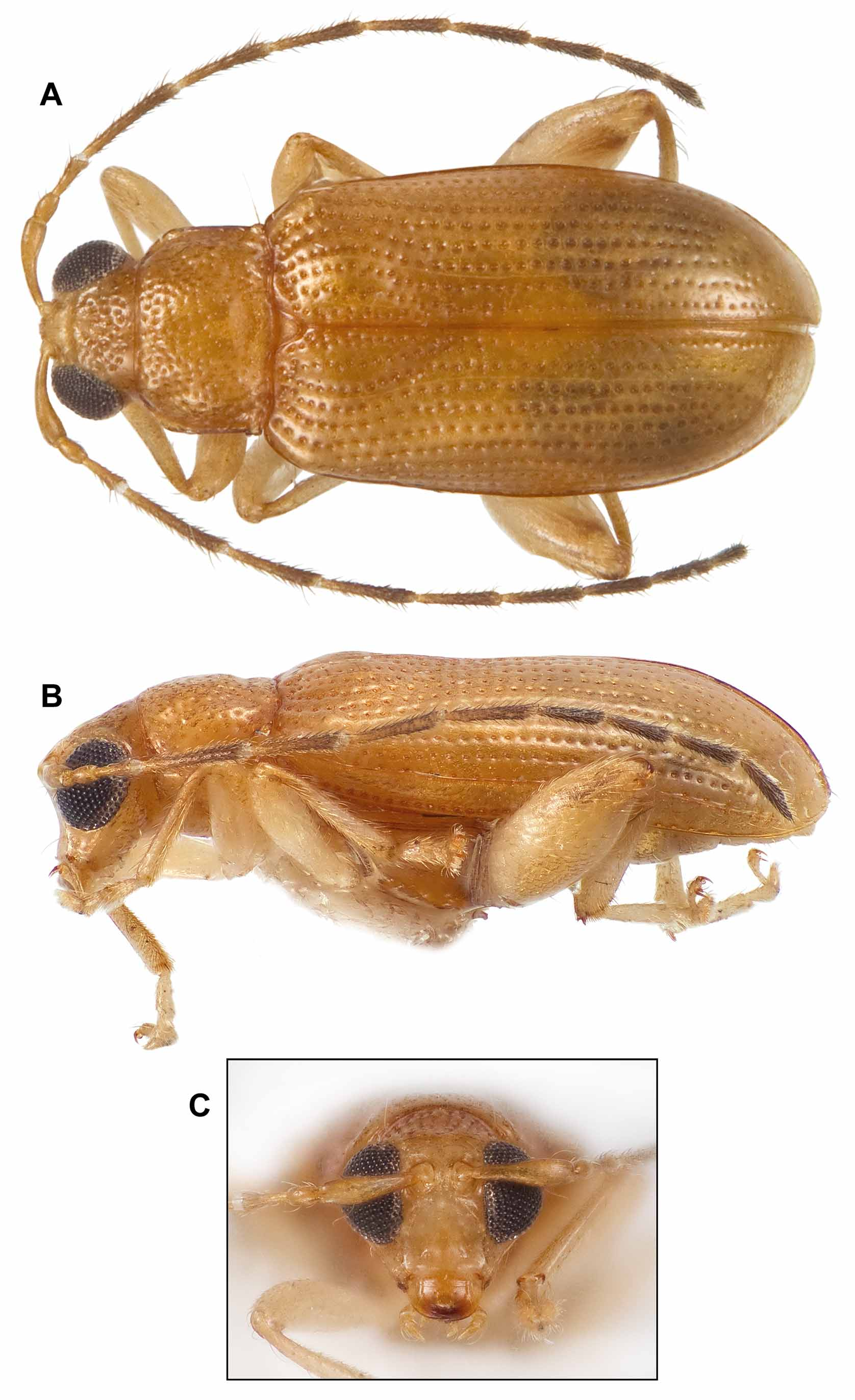
Description. Body shape elongate, narrow. Dorsum with coarse, dense punctation. Body length: 2.7– 3.05mm.
Head: ( Figs. 7 View FIGURE 7 a–c; 11a–b): Broadly connected to pronotum at base (posteriorly); vertex with coarse, dense punctation; frons narrow, smooth, narrowing ventrally, concave in lateral view, ca. at a 90° angle to vertex in lateral view; antennal calli evident, delimited posteriorly; eyes large, round, prominent, but total eye to eye width not apparently greater than pronotal anterior width; IOD narrow; antennae almost as long or longer than body, antennal segments 2 and 3 shortest, 4 distinctly longest; labrum bearing four fine setiferous punctures on dorsal surface, two on each side.
Pronotum: Quadrate/rectangular, not distinctly narrowed basally (posteriorly); laterally straight-sided, parallel; anterolateral callosities distinctly angled; punctation coarse, dense; pre-basal transverse impression deep, laterally delimited by apparent short, longitudinal, sublateral carinae.
Elytra: Parallel-sided; base somewhat wider than base of pronotum; humeri prominent; elytral bossae distinct; punctation coarse, strongly striate throughout; epipleurae parallel-sided, equal width tapered subapically laterad of elytral apex. Two basolateral patches of microspines on underside of each elytron ( Fig. 11 View FIGURE 11 c).
Ve n te r: Procoxal cavities open ( Fig. 8 View FIGURE 8 c).
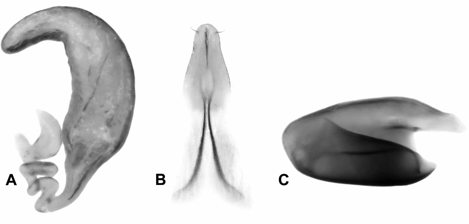
Legs: Metafemora distinctly swollen, much more so than pro- or mesofemora; metafemoral spring ( Fig. 10 View FIGURE 10 c) with dorsal lobe rather strongly arched dorsally at base with extended arm strongly depressed at apex, extended arm about 17% of total spring length, basal angle of ventral lobe obtuse, dorsal edge of ventral lobe descending sharply towards apex, recurve flange evident, most similar in shape to the Phyllotreta spring Morpho-group ( Furth & Suzuki 1998), but differs significantly from Alasia (above); tibiae with evident apical spines; metatibial apex not unusually expanded or laterally compressed, dorsal margins with only a short distance with stout spines (subequal to length of tarsomere 2); metatarsus with first tarsomere distinctly longer than in pro or mesotarsi, first metatarsomere relatively long, almost as long as tarsomeres 2–5 combined; first foretarsal segment not swollen in males; claws appendiculate.
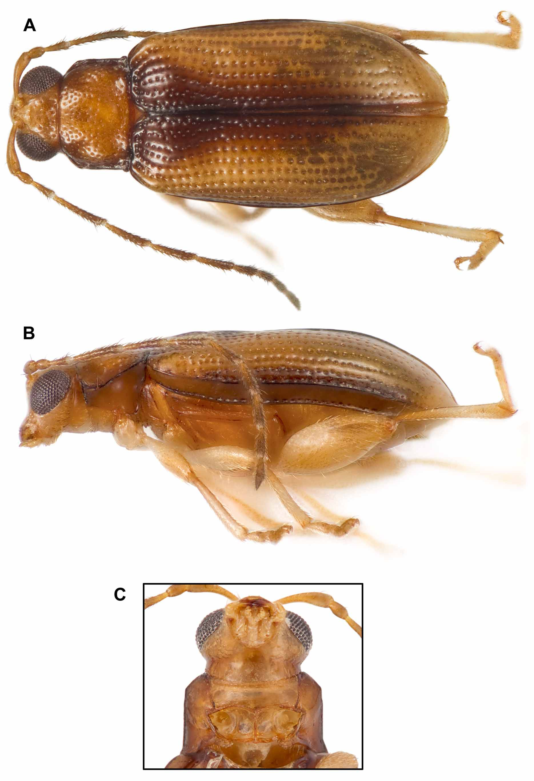
Remarks. Pseudostenophyma is similar to Alasia in general body shape, antennal length, lateral view of frons, but differs in IOD; narrow gena laterally below eye; pronotal shape, pronotal anterolateral angles; distinctly striate elytral punctation throughout; male and female genitalia, and metafemoral spring form (see also Remarks for Alasia alpina above). It is also clearly very different from Stenophyma elegans ( Fig. 12 View FIGURE 12 a–c) [see Remarks section for S. elegans ].


No known copyright restrictions apply. See Agosti, D., Egloff, W., 2009. Taxonomic information exchange and copyright: the Plazi approach. BMC Research Notes 2009, 2:53 for further explanation.
|
Kingdom |
|
|
Phylum |
|
|
Class |
|
|
Order |
|
|
Family |