
Cyrnellus, Banks, 1913
|
publication ID |
https://doi.org/10.11646/zootaxa.5082.1.2 |
|
publication LSID |
lsid:zoobank.org:pub:795488E3-DE16-4268-8968-628C9D5E3A4A |
|
DOI |
https://doi.org/10.5281/zenodo.5783142 |
|
persistent identifier |
https://treatment.plazi.org/id/03DEE90B-FFA6-451F-FF0A-FB5BFA01FE5E |
|
treatment provided by |
Plazi (2021-12-15 10:30:43, last updated 2024-11-29 15:46:45) |
|
scientific name |
Cyrnellus |
| status |
|
Key to adult males of the genus Cyrnellus View in CoL View at ENA
1. Subapicomesal spine I of each inferior appendage directed obliquely apicad with respect to longitudinal axis of inferior appendage ( Fig. 6B View FIGURE 6 )......................................................................................... 2
- Subapicomesal spine I directed perpendicularly to longitudinal axis of each inferior appendage ( Fig. 3F View FIGURE 3 )............... 4
2(1). Posterior margins of sternite IX convex and sinuous with slight undulations; subapicomesal spine I closer to apex of each inferior appendage and digitate ( Figs 6A–6D View FIGURE 6 , as).................................................... C. misionensis View in CoL
- Posterior margins of sternite IX irregular with some excisions and protuberances ( Fig. 5A View FIGURE 5 ); subapicomesal spine I of each inferior appendage in mesal or subapical position ( Fig. 5B View FIGURE 5 , as)................................................. 3
3(2). Subapicomesal spine I of each inferior appendage mesally inserted, long and digitate ( Figs 4E–4H View FIGURE 4 ).............. C. rianus View in CoL
- Subapicomesal spine I, subapically inserted, short and triangular ( Figs 5A–5D View FIGURE 5 )................................ C. risi View in CoL
4(1). Subapicomesal spine of each inferior appendage triangular, acute ( Figs 3F, 3I–3K View FIGURE 3 )................................. 5
- Subapicomesal spine rounded or mammiform ( Figs 2F View FIGURE 2 , 6F View FIGURE 6 ), or with two subapicomesal points ( Figs 1B View FIGURE 1 , 2B View FIGURE 2 , 5F View FIGURE 5 , 7B View FIGURE 7 )...... 9
5(4). Subapicomesal spine of each inferior appendage arising far from apex ( Figs 3E–3K View FIGURE 3 )...................... C. fraternus View in CoL
- Subapicomesal spine arising close to apex ( Figs 1G View FIGURE 1 , 3B View FIGURE 3 , 4B View FIGURE 4 )................................................... 6
6(5). Triangular subapicomesal spine of each inferior appendage flat ( Figs 1F–1I View FIGURE 1 )............................ C. boliviensis
- Triangular subapicomesal spine circular in cross-section ( Figs 3B View FIGURE 3 , 4B View FIGURE 4 ).......................................... 7
7(6). Subapicomesal spine of each inferior appendage with wide base, forming equilateral triangle ( Figs 3A–3D View FIGURE 3 )... C. marginalis View in CoL
- Subapicomesal spine with narrow base, forming isosceles triangle ( Fig. 4B View FIGURE 4 )....................................... 8
8(7). Subapicomesal spine of each inferior appendage conspicuously subapical, inner lobe triangular ( Figs 4A–4D View FIGURE 4 ).... C. minimus View in CoL
- Subapicomesal spine very near apex, inner lobe round ( Chamorro-Lacayo 2003, fig. 2B)................ C. zapateriensis
9(4). Subapicomesal spine II of each inferior appendage absent, single subapicomesal spine with wide, globular base ( Figs 2F View FIGURE 2 , 6F View FIGURE 6 ) .................................................................................................. 10
- Subapicomesal spines I & II present ( Figs 1B View FIGURE 1 , 2B View FIGURE 2 , 5F View FIGURE 5 , 7B View FIGURE 7 ).................................................... 11
10(9). Posterior arms of dorsal phallic sclerite slender, sinuous, and divergent ( Figs 2E–2H View FIGURE 2 )................... C. mammillatus View in CoL
- Posterior arms of dorsal phallic sclerite thick with heavily sclerotized apices ( Figs 6E–6H View FIGURE 6 )................... C. collaris View in CoL
11(9). Subapicomesal spine I of each inferior appendage very near apex ( Figs 7A–7D View FIGURE 7 )............................ C. bifidus View in CoL
- Subapicomesal spine I conspicuously preapical, with variable distance between spine and apex of inferior appendage ( Figs 1B View FIGURE 1 , 2B View FIGURE 2 , 5F View FIGURE 5 )............................................................................................ 12
12(11). Dorsal phallic sclerite complex ( Figs 1C–1E View FIGURE 1 ), its anterior arms well-developed and with long sclerotized rods projecting anterad ( Figs 1A–1E View FIGURE 1 )............................................................................ C. arotron View in CoL
- Dorsal phallic sclerite simple or indistinct ( Figs 2C View FIGURE 2 , 5H View FIGURE 5 ), with anterior arms reduced or absent...................... 13
13(12). Subapicomesal spines of each inferior appendage flat ( Fig. 2B View FIGURE 2 ); dorsal phallic sclerite with anterior arm reduced, posterior arms divergent, short, and thick ( Figs 2A–2D View FIGURE 2 )......................................................... C. guyanensis
- Subapicomesal spines circular in cross-section ( Fig. 5F View FIGURE 5 ); phallic dorsal sclerite indistinct, without anterior arm, posterior arms fused into tube ( Figs 5E–5H View FIGURE 5 )...................................................................... C. ulmeri View in CoL
Chamorro-Lacayo, M. L. (2003) Seven new species of Polycentropodidae (Trichoptera) from Nicaragua and Costa Rica. Proceedings of the Entomological Society of Washington, 105 (2), 484 - 498.
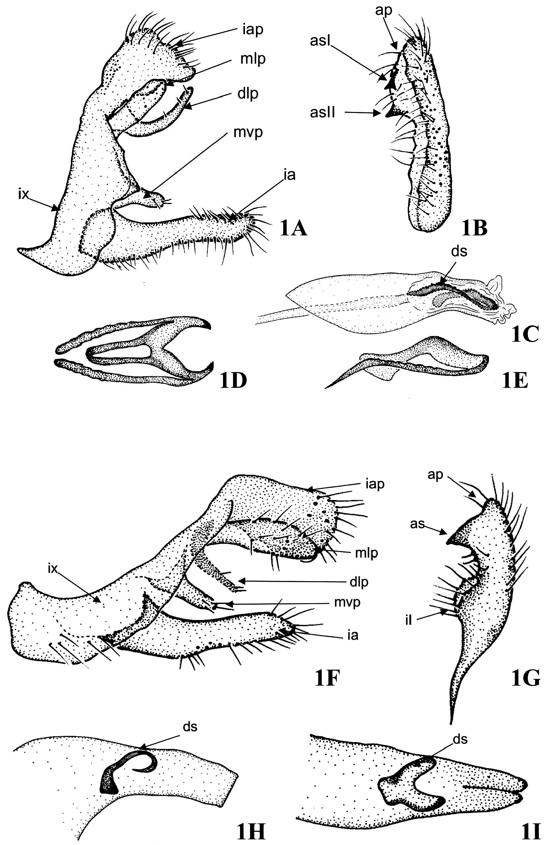
FIGURE 1. Cyrnellus spp., male genitalia. 1A–1E, Cyrnellus arotron Flint 1971: 1A, left lateral; 1B, right inferior appendage, ventral; 1C, phallus, left lateral; 1D, dorsal phallic sclerite, dorsal; 1E, dorsal phallic sclerite, left lateral. 1F–1I, Cyrnellus boliviensis sp. nov.: 1F, left lateral; 1G, right inferior appendage, ventral; 1H, phallus, left lateral; 1I, phallus, dorsal. ap = apex of an inferior appendage; asI = subapicomesal spine I; asII = subapicomesal spine II; dlp = dorsolateral process; ds = dorsal phallic sclerite; ia = inferior appendage; iap = intermediate appendage; il = internal lobe; ix = sternite IX; mlp = mesolateral process; mvp = mesoventral process.

FIGURE 2. Cyrnellus spp., male genitalia. 2A–2E, Cyrnellus guyanensis sp. nov.: 2A, left lateral; 2B, right inferior appendage, ventral; 2C, phallus, left lateral; 2D, phallus, dorsal. 2E–2H, Cyrnellus mammillatus Flint 1971: 2E, left lateral; 2F, right inferior appendage, ventral; 2G, phallus, left lateral; 2H, dorsal phallic sclerite, dorsal. ap = apex of inferior an appendage; asI = subapicomesal spine I; asII = subapicomesal spine II; dlp = dorsolateral process; ds = dorsal phallic sclerite; ia = inferior appendage; iap = intermediate appendage; ix = sternite IX; mlp = mesolateral process; mvp = mesoventral process.
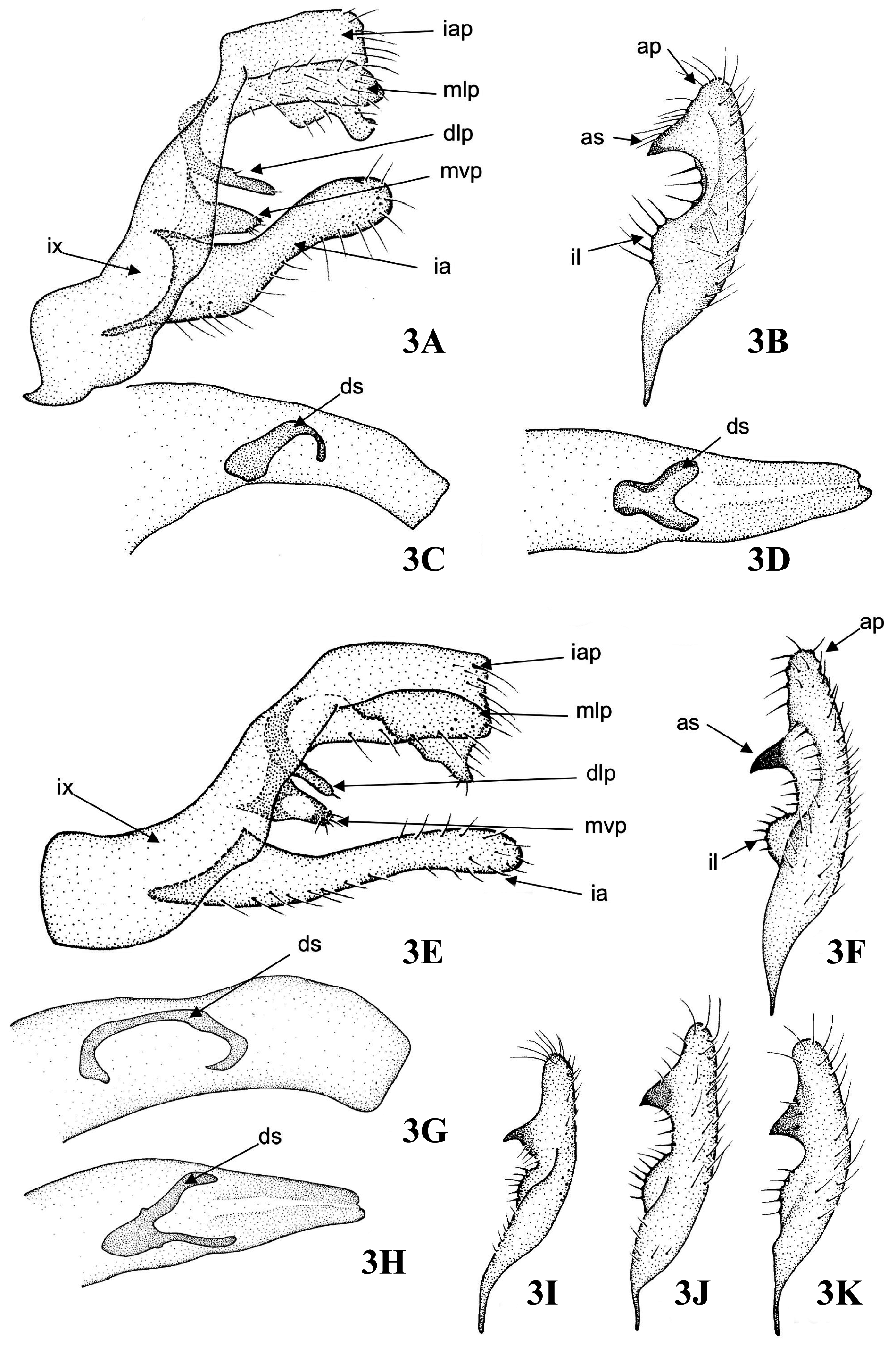
FIGURE 3. Cyrnellus spp., male genitalia. 3A–3D, Cyrnellus marginalis Banks 1930: 3A, left lateral; 3B, right inferior appendage, ventral; 3C, phallus, left lateral; 3D, phallus, dorsal. 3E–3K, Cyrnellus fraternus (Banks 1905): 3E, left lateral; 3F, right inferior appendage, ventral; 3G, phallus, left lateral; 3H, phallus, dorsal; 3I–3K, variations of the right inferior appendage, ventral. ap = apex of an inferior appendage; as = subapicomesal spine; dlp = dorsolateral process; ds = dorsal phallic sclerite; ia = inferior appendage; iap = intermediate appendage; ix = sternite IX; mlp = mesolateral process; mvp = mesoventral process.
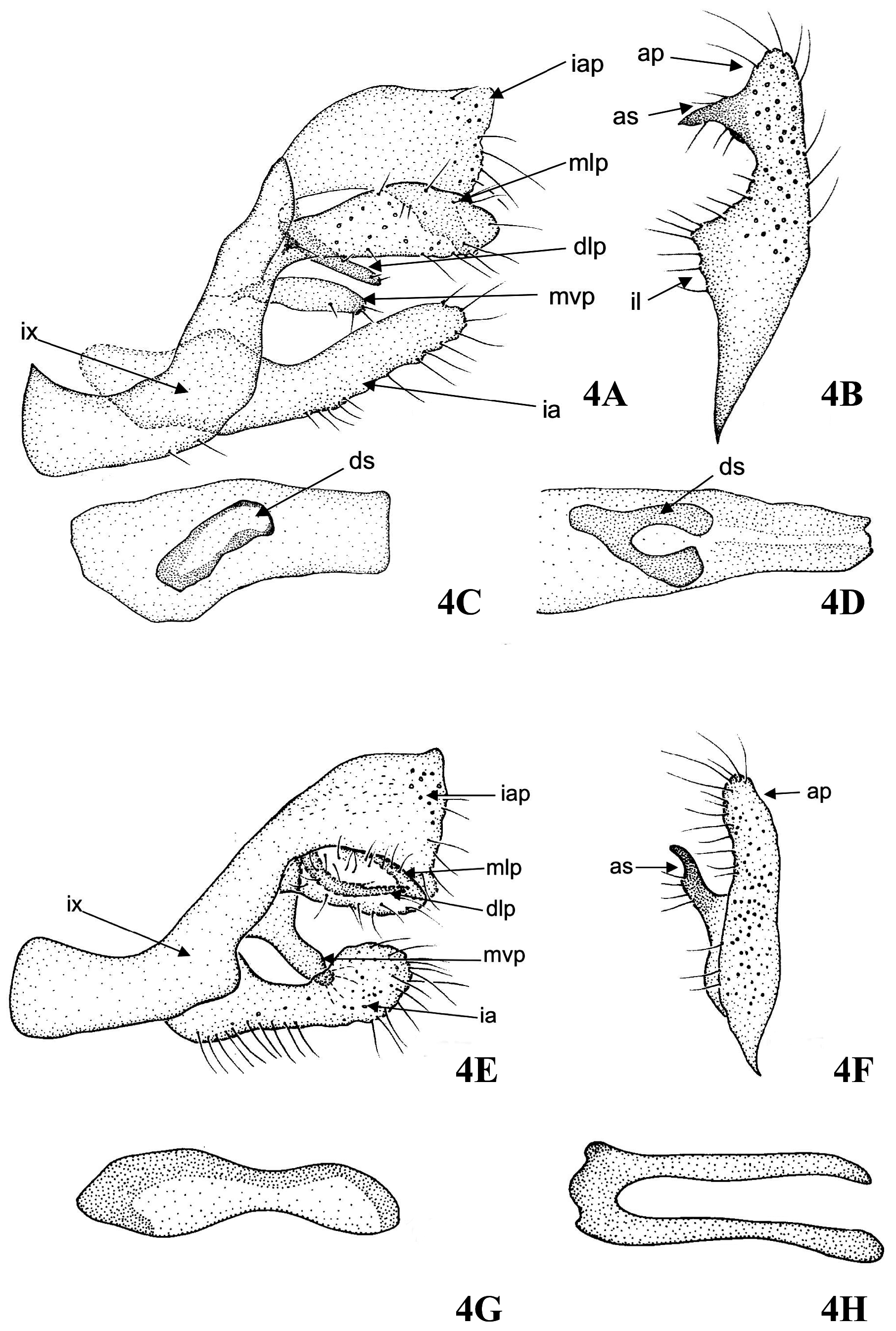
FIGURE 4. Cyrnellus spp., male genitalia. 4A–4D, Cyrnellus minimus Banks 1913: 4A, left lateral; 4B, right inferior appendage, ventral; 4C, phallus, left lateral; 4D, phallus, dorsal. 4E–4H, Cyrnellus rianus Flint 1983: 4E, left lateral; 4F, right inferior appendage, ventral; 4G, dorsal phallic sclerite, left lateral; 4H, dorsal phallic sclerite, dorsal. ap = apex of an inferior appendage; as = subapicomesal spine; dlp = dorsolateral process; ds = dorsal sclerite; ia = inferior appendage; iap = intermediate appendage; ix = sternite IX; mlp = mesolateral process; mvp = mesoventral process.

FIGURE 5. Cyrnellus spp., male genitalia. 5A–5D, Cyrnellus risi (Ulmer 1907): 5A, left lateral; 5B, right inferior appendage, ventral; 5C, dorsal phallic sclerite, left lateral; 5D, dorsal phallic sclerite, dorsal. 5E–5H, Cyrnellus ulmeri Flint 1971: 5E, left lateral; 5F, right inferior appendage, ventral; 5G, dorsal phallic sclerite, left lateral; 5H, dorsal phallic sclerite, dorsal. ap = apex of an inferior appendage; asI = subapicomesal spine I; asII = subapicomesal spine II; dlp = dorsolateral process; ia = inferior appendage; iap = intermediate appendage; ix = sternite IX; mlp = mesolateral process; mvp = mesoventral process.
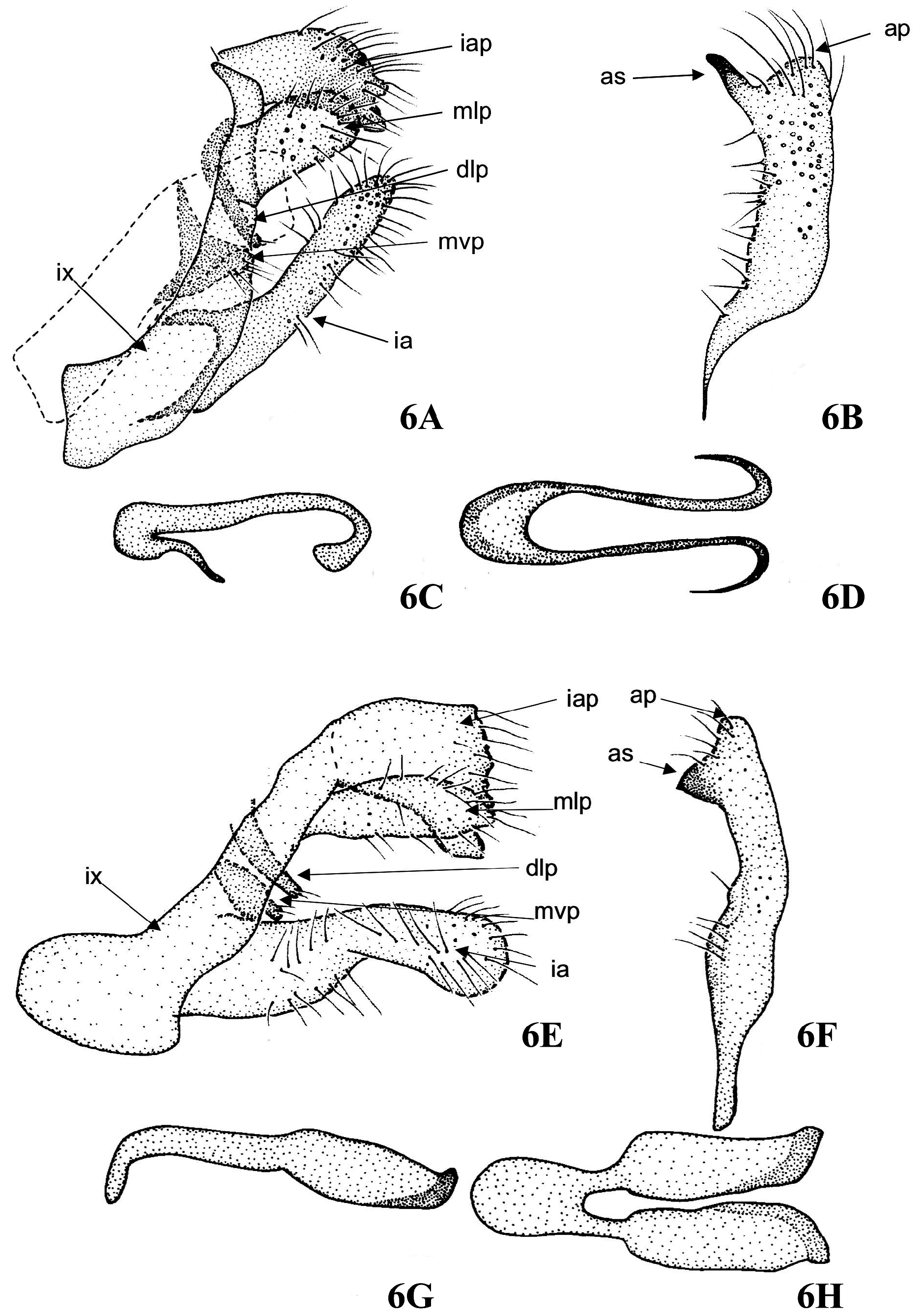
FIGURE 6. Cyrnellus spp., male genitalia. 6A–6D, Cyrnellus misionensis Flint 1983: 6A, left lateral; 6B, right inferior appendage, ventral; 6C, dorsal phallic sclerite, left lateral; 6D, dorsal phallic sclerite, dorsal. 6E–6H, Cyrnellus collaris Flint 1971: 6E, left lateral; 6F, right inferior appendage, ventral; 6G, dorsal phallic sclerite, left lateral; 6H, dorsal phallic sclerite, dorsal. ap = apex of an inferior appendage; as = subapicomesal spine; dlp = dorsolateral process; ia = inferior appendage; iap = intermediate appendage; ix = sternite IX; mlp = mesolateral process; mvp = mesoventral process.
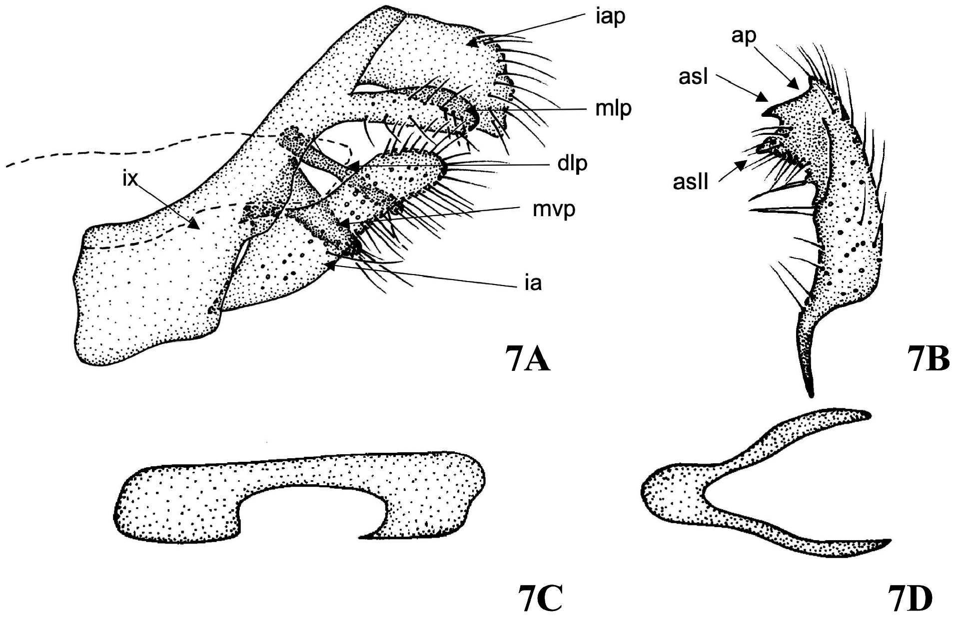
FIGURE 7. Cyrnellus bifidus Flint 1971, male genitalia. 7A, left lateral; 7B, right inferior appendage, ventral; 7C, dorsal phallic sclerite, left lateral; 7D, dorsal phallic sclerite, dorsal. ap = apex of an inferior appendage; asI = subapicomesal spine I; asII = subapicomesal spine II; dlp = dorsolateral process; ia = inferior appendage; iap = intermediate appendage; ix = sternite IX; mlp = mesolateral process; mvp = mesoventral process.
No known copyright restrictions apply. See Agosti, D., Egloff, W., 2009. Taxonomic information exchange and copyright: the Plazi approach. BMC Research Notes 2009, 2:53 for further explanation.
|
Kingdom |
|
|
Phylum |
|
|
Class |
|
|
Order |
|
|
Family |
1 (by plazi, 2021-12-15 10:30:43)
2 (by ExternalLinkService, 2021-12-15 10:38:36)
3 (by ExternalLinkService, 2021-12-15 13:34:48)
4 (by ExternalLinkService, 2021-12-15 15:00:20)
5 (by ExternalLinkService, 2022-11-11 04:52:29)
6 (by ExternalLinkService, 2023-08-13 04:31:29)
7 (by ExternalLinkService, 2023-08-13 05:01:04)