
Astrophiura caroleae, Pawson, 2018
|
publication ID |
https://doi.org/10.11646/zootaxa.4378.2.4 |
|
publication LSID |
lsid:zoobank.org:pub:C4300992-465B-49B7-A1F3-AD3DE906DD3F |
|
DOI |
https://doi.org/10.5281/zenodo.5977337 |
|
persistent identifier |
https://treatment.plazi.org/id/03DD87C4-FFE0-CC13-FF19-1C5FFEA7FE01 |
|
treatment provided by |
Plazi |
|
scientific name |
Astrophiura caroleae |
| status |
sp. nov. |
Astrophiura caroleae new species
Figures 1–3 View FIGURE1 View FIGURE 2 View FIGURE 3
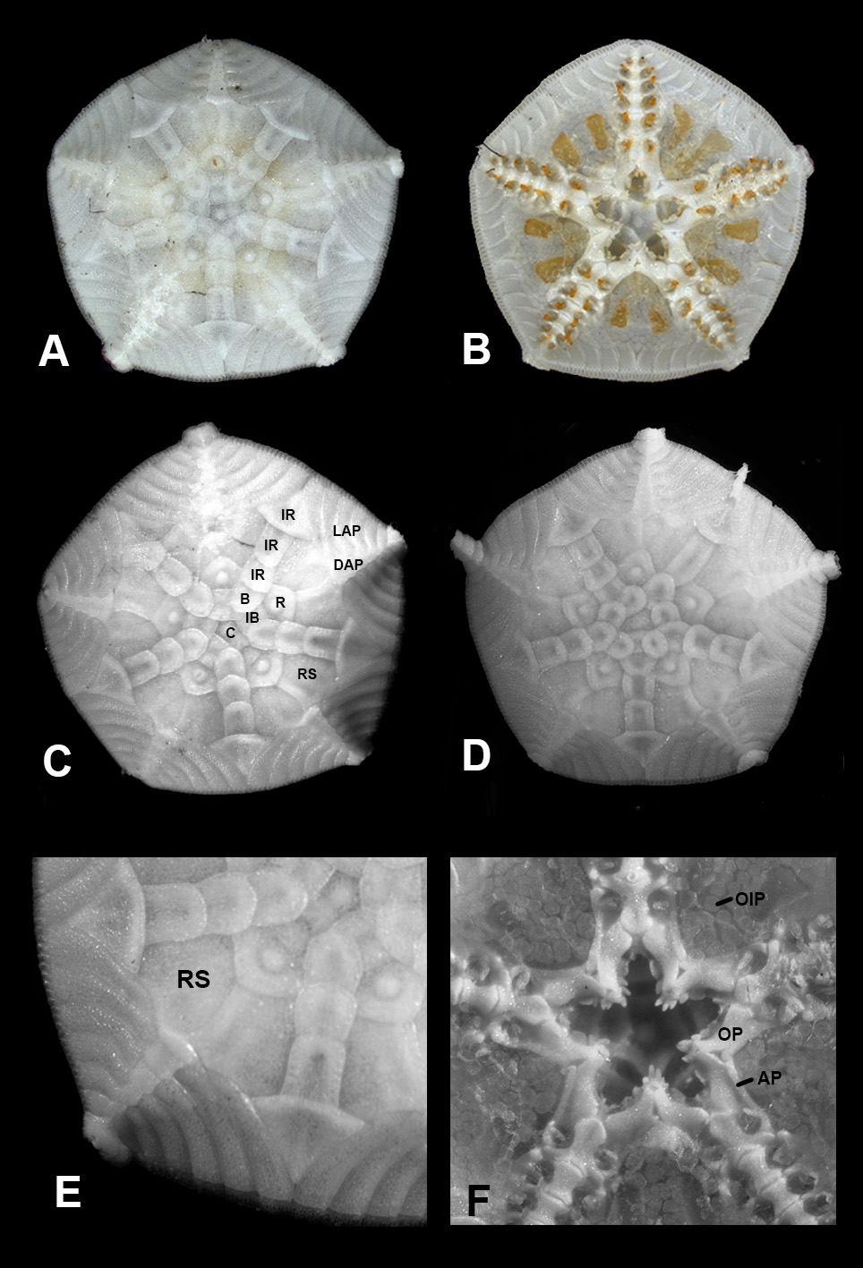
Material Examined. Holotype ( Figures 1A–C, E,F View FIGURE1 ; 2A–C), USNM 1463102, Station Number Curasub 14–15, 19 September 2014, submersible towed to Jan Thiel Bay, Curacao, 12°04′28.7″N, 68°52′57.1″W, dive depth range 152–305 meters, specimen attached to a bottle, collected at 294 meters. C. Baldwin, B. Brandt, D, Schrier. Specimen fixed and preserved in ethyl alcohol. Disc diameter of Holotype 8.5 mm.
Paratype 1 ( Figure 1D View FIGURE1 ; 3C), USNM 1463103, same collection data as Holotype. Specimen fixed and preserved in ethyl alcohol. Disc diameter of paratype 1 7.0 mm, detached arm 4.5 mm.
Paratype 2, USNM 1463104, Station Number Curasub 14–17, 23 September 2014, east of downline at Substation Curacao, 12°04′59.51″N, 68°53′56.61″W, dive depth range 215–309 meters, specimen attached to a bottle collected at 280 meters. J. Harasewych, B. van Bebber, M. McNeilus, J. Felder, Carolee. Specimen fixed and preserved in ethyl alcohol. Disc diameter of Paratype 2 (specimen broken) approximately 6 mm.
Paratypes 3 and 4, USNM E18595 View Materials , Quintana Roo, Yucatan Channel, Mexico, R/ V Pillsbury Station 587, 14 March 1968, 21°17′N, 86°13′W, 434 meters, 2 specimens (identified as Astrophiura permira by Maureen E. Downey). Specimens dried GoogleMaps . Diameter of Paratype 3, 8.4 mm; diameter of Paratype 4, 8.8 mm.on-type Material ( Photographs, taken in situ, and in the laboratory): 1. Photograph of specimen in the laboratory CURI 14106 ( Figure 2D View FIGURE 2 ), 26 June 2014, off Substation Curacao downline, 12°4′59.51″N, 68° 53′56.61″W, 235–306 meters. C. Baldwin, B. Brandt, E. Brandt, D. Schrier. 2. Photograph of specimen in situ on bottle GoogleMaps , CURI 12016 , 21 May 2012, off Substation Curacao downline, 171–309 meters. C. Baldwin, B. Brandt, D. Schrier ( Figure 3A View FIGURE 3 ). 3. Photograph of specimen in situ on automobile tire, Field Station Number 14–20, 26 September 2014, west of downline at Substation Curacao, 12°04′59.51″N, 68°53′56.61″W, 244 meters. C. Baldwin, B. Brandt, C. Castillo ( Figure 3B View FIGURE 3 ). GoogleMaps
Diagnosis. Astrophiura with disc regularly pentagonal, up to 10 mm in diameter, interradial margins convex. Free portions of arms slightly shorter than disc diameter. In dorsal disc, elongated diamond-shaped radial areas higher than interradial areas. Conspicuous ossicles in dorsal disc include central plate, infrabasals, basals, and radials, three or four interradial plates, the distalmost approximately triangular with a sharp distal point, and large radial shields, pairs of which are contiguous for their entire length. Most plates smooth, but central and radial plates have a single prominent central tubercle, and each radial shield has a more or less conspicuous prominence near the distal edge. Proximal dorsal arm plates on the disc are quadrangular, wider than long. Six or seven oral papillae on each jaw. Adoral plates elongate, visible portions at least 6 times as long as wide, the proximal ends of each pair abutting more or less in line with the apex of each jaw. Color in life variegated brownish to reddish with whitish patches, the tubercles on dorsal plates usually lighter in color than surrounding areas.
Etymology. It is a pleasure to name this species for Dr. Carole Baldwin, Chair of the Vertebrate Zoology Department, National Museum of Natural History, Smithsonian Institution. Dr. Baldwin founded the Smithsonian’s Deep Reef Observation Project (DROP) in 2011 and she has inspired its steady growth in addition to conducting her own excellent research on fishes.
Description. Holotype 8.5 mm in disc diameter, all arms broken off, disc essentially circular, with slightly convex interradial margins ( Figure 1A–C View FIGURE1 ; 2A). Disc with convex dorsal surface, ventral surface distinctly concave. Dorsal disc plates naked ( Figure 1A, C, E View FIGURE1 ), mostly unadorned, except that central plate and radial plates have a conspicuous central tubercle, and each radial shield usually carries a small, less conspicuous prominence near its distal edge. Central plate polygonal, 430µm across widest part, partly overlain by infrabasals 700 µm wide, which in turn are partly overlain by approximately polygonal basal plates 800 µm wide ( Figure 1C View FIGURE1 ). Three interradial plates present in four of five interradii; first interradial plate is rectangular with rounded corners, approximately as long as broad, second plate approximately twice as long as broad, third and distalmost plate arrow-head shaped, approximately triangular, 1.3 mm wide, with a sharp distal point ( Figure 1C View FIGURE1 ), its proximal edge overlapping adjacent radial shields. In fifth interradius first interradial plate is twice as long as broad, and second plate is arrowhead shaped, with sharp distal point. Radial shields approximately as long as wide, their radial edges broadly in contact for entire visible length of 900 µm ( Figure 1A, 1C View FIGURE1 ). Five dorsal arm plates incorporated into disc; six lateral arm plates are also incorporated into the disc, indicating that first dorsal arm plate is overgrown by radial shields, or is resorbed. Dorsal arm plates more or less rectangular ( Figure 1C View FIGURE1 ), decreasing in size distally, the first and largest plate 240 µm in length, with pointed lateral proximal extensions abutting radial shields. Lateral arm plates elongate, broader distally than proximally, each carrying four arm spines, approximately three times longer than wide ( Fig. 1A View FIGURE1 ). No conspicuous ridges and grooves on dorsal or ventral surfaces of lateral arm plates.
Six ventral arm plates within disc decreasing in size distally ( Figure 1B View FIGURE1 ), approximately rectangular, lateral edges concave. First arm plate bell-shaped, 600 µm long, second arm plate 400 µm long. Tentacle pores and tube feet large ( Figure 1B View FIGURE1 ), largest pore 200 µm in diameter, decreasing in size distally. Pores with one, occasionally two, tentacle scales. First pair of oral tentacles arise within oral slit. Interradial area covered by flat, oval to round, translucent plates ( Fig. 1F View FIGURE1 ) approximately 22 µm in diameter; covering extends from junction of adoral plates approximately 2/3 of distance to disc margin. Paired gonads visible through covering plates. Oral plates large, curved, 800 µm long, carrying six to seven oral papillae, outer papillae by far largest. Adoral plates ( Fig. 1F View FIGURE1 ) elongate, approximately 800 µm long, at least six times as long as wide, their proximal ends meeting more or less in line with junctions of oral plates. Oral shields absent, except for the shield carrying the madreporite; this shield, 400 µm in diameter, is conspicuous at juncture of two adoral plates ( Fig. 1F View FIGURE1 ).
Free arm joints absent from holotype, but present when holotype was photographed in laboratory ( Fig. 2A View FIGURE 2 ). Portions of four arms present, all with regenerating distal regions. Longest arm, with approximately 15 segments, approximately 4.6 mm long, slightly longer than one-half of the disc diameter.
Notes on paratypes: Paratype 1 (USNM 1463102). Dorsal ( Fig. 1D View FIGURE1 , 3C View FIGURE 3 ) and ventral surfaces similar in general appearance to those of holotype. Disc diameter 7.6 mm. Central plate irregular in shape ( Fig. 1D View FIGURE1 ), infrabasal plate 600 µm wide, basal plate 750 µm wide, radial 960 µm wide. Distalmost interradial plate 1.3 mm wide. Members of each pair of radial shields in broad contact radially; total length of contact area 700 µm. Six lateral arm plates in disc. On oral surface seven ventral arm plates within disc, decreasing in size distally, approximately rectangular, the lateral edges concave. First arm plate 700 µm long, second arm plate 400 µm long. Largest tentacle pore 200 µm in diameter. Width of first free arm segment 540 µm.
Paratypes 2 (USNM 1463104), 3 and 4 (USNM E18595 View Materials ). Disc diameter 6.0 mm, 7.9 mm, and 7.2 mm respectively. All paratypes with some damage to disc. Arms absent. General appearance of dorsal and ventral surfaces, and details of external morphology, similar to those in holotype.
Notes on color of type and non-type specimens: Holotype, when recently dead, with translucent ventral surface ( Fig. 2A View FIGURE 2 ), the covering of small interradial plates almost invisible. Conspicuous bright orange eggs, approximately 165 µm in diameter, packed into five pairs of gonads of female specimen, gonads occupying most of internal interradial space. Ventral surfaces of arms inside disc white, tube feet very light brown. Ventral surfaces of lateral arm plates with whitish-orange edges, central areas of plates tending towards very light brown. Arm spines light red or whitish, the red spines tending to be clumped near the center of the distal edges of the lateral arm plates. Dorsal surface of holotype in life ( Fig. 2C View FIGURE 2 ) mottled with light reddish and whitish patches, whitish areas more or less confined to: dorsal arm plates in disc, the distalmost two or three lateral arm plates, and the distalmost interradial plate. Interradial areas outside rings of primary plates reddish with whitish spots. Rings of primary plates variegated reddish-brown.
In some non-type specimens ( Figs. 2D View FIGURE 2 , 3A–B View FIGURE 3 ), dorsal surface can appear to be almost uniformly colored, light brownish-red ( Fig. 3A View FIGURE 3 ), darker centrally, or light red, with some whitish patches radially ( Fig. 3B View FIGURE 3 ). Paratype 1 ( Fig. 3C View FIGURE 3 ) has complex dorsal color patterns, with light orange radial shields and dorsal arm plates, light red lateral arm plates with whitish central patches, and a red-brown central region with whitish blotches, and conspicuous white tubercles on central and radial plates. The less conspicuous tubercles on the distal edges of the radial shields are also whitish, but less conspicuous.
Molecular data: The barcode data provided below for Astrophiura caroleae n.sp. may be the first to become available for the genus Astrophiura ; no record of the genus could be found in BOLD (Barcode of Life Database). A mtDNA COI barcode was generated from a single individual following standard Sanger sequencing protocols as outlined in Meyer (2003), and as noted in Ng & Meyer (2016). The specimen was collected from an Autonomous Reef Monitoring Structure ( ARMS; see Knowlton et al. 2010) deployed on a deep reef in Curacao.
Locality information: Catalog Number USNM 1466036 About USNM , Sample No. Curasub 15–16, BCURA _0674, Curacao, east of downline and slightly west at Substation Curacao dock, in Bapor Kibra, Curacao, 12°4′59.51″N, 68°53′56.61″W. Ophiuroid collected from ARMS at 750 feet (229 meters). Collectors Carole Baldwin, Bruce Brandt, Thomas Devine, Emily Frost GoogleMaps .
PCR primers jgLCO1490 and jgHCO2198 ( Geller et al. 2013) were used. The resulting sequence is: AACTCTTTATTTACTTTTTGGAACTTGAGCCGGAACAGTAGGAGCAGCCATGAGAAAAATTATTCGAA TAGAGCTTTCACAGCCTGGGTCCCTCATACAGAAAGATCAAATATACAATGTAATAGTAACCGCTCATG CATTTGTAATGATTTTTTTTATGGTGATGCCAATAATGATTGGAGGATTTGGAAATTGACTAATCCCATT AATGATTGGAGCTCCAGATATGGCCTTCCCACGAATGAAAAAAATGAGATTTTGACTTATCCCCCCTTC ATTTTTTCTTTTAATGGCCTCAGCAGCAAAAAAAAAGGGAGTAGGAACGGGTTGAACAATTTACCCGC CTCTCTCTGGGCACGGAGCTCACGCCGGAGGTTGCGTAGACCTAGCTATTTTTTCTCTCCATCTCGCTG GAGCTTCTTCTATAATGGCATCTATAAATTTTATAACTACCATTATCAAAATGCGAGCTCCAGGAATGGA TCTTGACCAAGCTCCACTTTTTGTTTGATCTATCCTCATCACAACTTTTCTTCTTCTACTATCCCTTCCA GTTTTAGCTGGAGCTATAACAATGTTACTAACAGATCGAAAAATTAACACTTCCTTTTTCGATCCAACA GGAGGAGGAGACCCCATTTTATTTCAACATTTATTC.
Genbank Accession Number for this sequence: MG601101 View Materials .
Preferred Habitats: It is well-known that Astrophiura prefers a hard substratum; individuals can be found attached to dead mollusk shells ( Fujita and Hendler, 2001), and rocks (Ziesenhenne, 1951; Matsumoto, 1917). A. caroleae was first observed attached to a discarded Heineken beer bottle, where its reddish coloration contrasted sharply with the green of the bottle. Other specimens were collected from miscellaneous bottles near the type locality.
Distribution: This species is known from off Curacao in the southern Caribbean, and also from off Quintana Roo, Mexico, in depths of 244 to 434 meters.
Discussion. The color of A. caroleae can vary considerably, although shades of red to reddish-brown seem always to be present. A notable feature is that the tubercles on the central and radial plates are always more or less conspicuous, being lighter-colored, usually grayish-white, than their surroundings, which are usually brownish to orange.
The fact that five or more dorsal arm plates are incorporated into the disc, while six or more lateral arm plates are present in the disc, is similar to the situation described in A. wanikawa by Fujita and Hendler (2001). The structure of the free arms is superficially similar to those described for A. marionae by Ziesenhenne (1951) and A. wanikawa by Fujita and Hendler (2001).
As Fujita and Hendler (2001) point out, descriptions of some species of Astrophiura are inadequate, rendering detailed comparisons difficult. The current new species is readily distinguished from its congeners for the following reasons: A.caroleae shares only with A. permira Sladen (1878, 1879), A. cavellae Koehler (1915) and A. chariplax Baranova (1955) the presence of distinct tubercles at the centers of primary disc plates. In A. wanikawa , the plates have “thickened and raised edges and irregular central prominences” ( Fujita and Hendler, 2001). Clark (1923) and some subsequent authors regard A. cavellae as a junior synonym of A. permira . A. permira differs from A. caroleae in possessing additional tubercles, randomly placed, on other dorsal plates. Also, as pointed out by Ziesenhenne (1951), the primary dorsal disc plates are not symmetrically arranged, the pairs of radial shields are only slightly in contact with each other, and the adoral plates are not in contact with each other proximally.
In terms of distribution of the tubercles, A. chariplax resembles A. caroleae . Baranova (1955) indicates the presence of tubercles on radial disc plates, and possibly on one radial shield, in her figure 3(1), but she does not mention these tubercles in her description. In A. chariplax , according to Baranova’s figure 3(1), the rows of dorsal interradial plates gradually increase in width distally, and the distalmost interradial plates terminate some distance from the disc margin. In contrast, in A. caroleae , the interradial plates are essentially equal in width throughout, and the distalmost interradial plate terminates quite close to the disc margin. Further, in A. chariplax , the small plates in the ventral interradii have a very restricted distribution (Baranova, 1955, figure 3(2)), whereas in A. caroleae the area of coverage is significantly more extensive. Finally, A chariplax has only five oral papillae when the disc diameter is 9.5 mm (D’yakonov, 1954; Baranova, 1955), as opposed to six or seven in A. caroleae at a disc diameter of 8.5 mm.


No known copyright restrictions apply. See Agosti, D., Egloff, W., 2009. Taxonomic information exchange and copyright: the Plazi approach. BMC Research Notes 2009, 2:53 for further explanation.
|
Kingdom |
|
|
Phylum |
|
|
Class |
|
|
Order |
|
|
Family |
|
|
Genus |