
Triplocania, Roesler, 1940
|
publication ID |
https://doi.org/10.11646/zootaxa.4336.1.1 |
|
publication LSID |
lsid:zoobank.org:pub:FA65E14F-102F-4FF1-B8D5-D7E0C9126878 |
|
DOI |
https://doi.org/10.5281/zenodo.6024736 |
|
persistent identifier |
https://treatment.plazi.org/id/03DD879B-CF47-FFA5-FF6A-EA1DFA3BF80E |
|
treatment provided by |
Plazi (2017-10-23 17:49:31, last updated 2024-11-26 02:05:38) |
|
scientific name |
Triplocania |
| status |
|
Key to the Colombian species of Triplocania View in CoL
1. Males............................................................................................... 2
- Females............................................................................................ 35
2. Forewing M3 branched ( Figs 305 View FIGURES 305 – 310 , 317 View FIGURES 317 – 322 , 323 View FIGURES 323 – 328 , 329 View FIGURES 329 – 334 , 335 View FIGURES 335 – 340 , 341 View FIGURES 341 – 346 , 347 View FIGURES 347 – 352 , 353 View FIGURES 353 – 358 and 359 View FIGURES 359 – 364 )..................................... 3
- Forewing M3 simple ( Figs 3 View FIGURES 3 – 8 , 9 View FIGURES 9 – 14 , 75 View FIGURES 75 – 80 , etc.)................................................................... 8
3. Hypandrium with only the central sclerite, this with short caudo-lateral lobular processes and one central narrow elongate process having two small apical lobes ( Fig. 326 View FIGURES 323 – 328 ); phallosome as in figure 328..................... T. lamensuraensis View in CoL n. sp.
- Hypandrium with two to four sclerites ( Figs 308 View FIGURES 305 – 310 , 338 View FIGURES 335 – 340 , 365, 365 and 367 View FIGURES 365 – 369 ); central sclerite of hypandrium and phallosome variable ( Figs 310 View FIGURES 305 – 310 , 371 View FIGURES 370 – 373 and 340 View FIGURES 335 – 340 )............................................................................. 4
4. Hypandrium of three sclerites, central sclerite with two pairs of posterior, acuminate processes, and a posterior median, elongate, acuminate process ( Figs 308 View FIGURES 305 – 310 and 365 View FIGURES 365 – 369 )................................................................. 5
- Hypandrium of two or four sclerites, the anterior-central sclerite with a pair of long acuminate, horn-shaped processes, strongly curved distally ( Figs 338 View FIGURES 335 – 340 , 366 View FIGURES 365 – 369 , and 367); posterior sclerite with an acuminate or spatulate process............... 6
5. Central sclerite of hypandrium with median, posterior, long acuminate process, almost as long as the lateral ones ( Fig. 365 View FIGURES 365 – 369 ); phallosome with phallobase almost straight proximally, posterior endophallic sclerite sinuous distally, with strongly curved tapered distal process ( Fig. 370 View FIGURES 370 – 373 )............................................................... T. furcata View in CoL New
- Central sclerite of hypandrium with median, posterior, stout process, longer than the lateral ones ( Fig. 308 View FIGURES 305 – 310 ); phallobase curved proximally toward the mesal line; posterior endophallic sclerites almost straight distally, with curved tapered apical process and two curved anteapical small teeth ( Fig. 310 View FIGURES 305 – 310 ).............................................. T. furcatoides View in CoL n. sp.
6. Hypandrium of two sclerites, acuminate processes of anterior sclerite each with a basal tooth; posterior sclerite with spatulate posterior process, dilated distally ( Fig. 338 View FIGURES 335 – 340 ); phallosome with side struts independent, each widened basally ( Fig. 340 View FIGURES 335 – 340 )........................................................................................ T. leguizamoensis View in CoL n. sp.
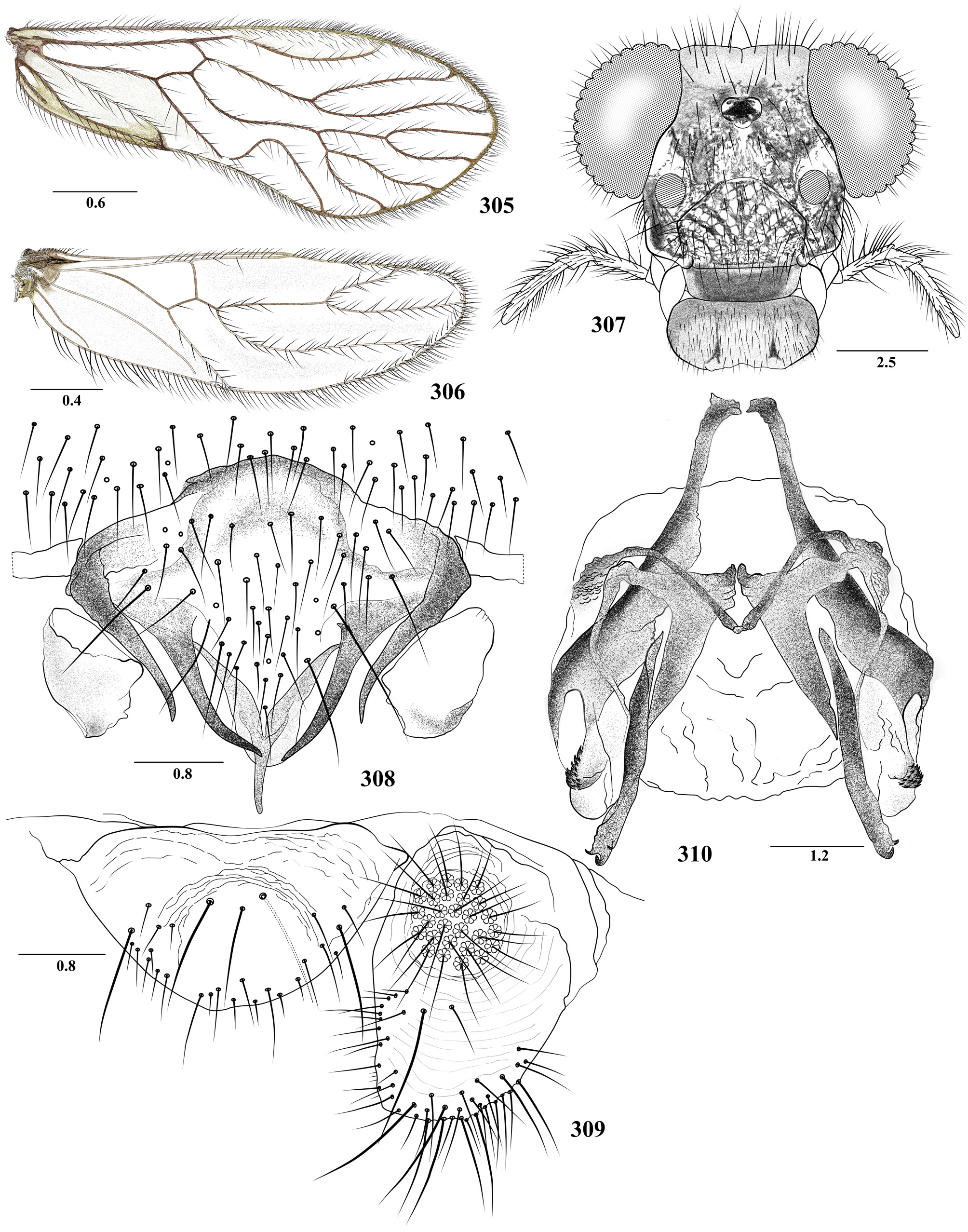
FIGURES 305 – 310. Triplocania furcatoides n. sp. Male. 305. Forewing. 306. Hindwing. 307. Front view of head. 308. Hypandrium. 309. Epiproct and right paraproct. 310. Phallosome. Scales in mm.

FIGURES 317 – 322. Triplocania huilaensis n. sp. Female. 317. Forewing. 318. Hindwing. 319. Front view of head. 320. Subgenital plate 321. Epiproct and right paraproct. 322. Ninth sternum and left gonapophyses. Scales in mm.

FIGURES 323 – 328. Triplocania lamensuraensis n. sp. Male. 323. Forewing. 324. Hindwing. 325. Front view of head. 326. Hypandrium. 327. Left paraproct and epiproct. 328. Phallosome. Scales in mm.
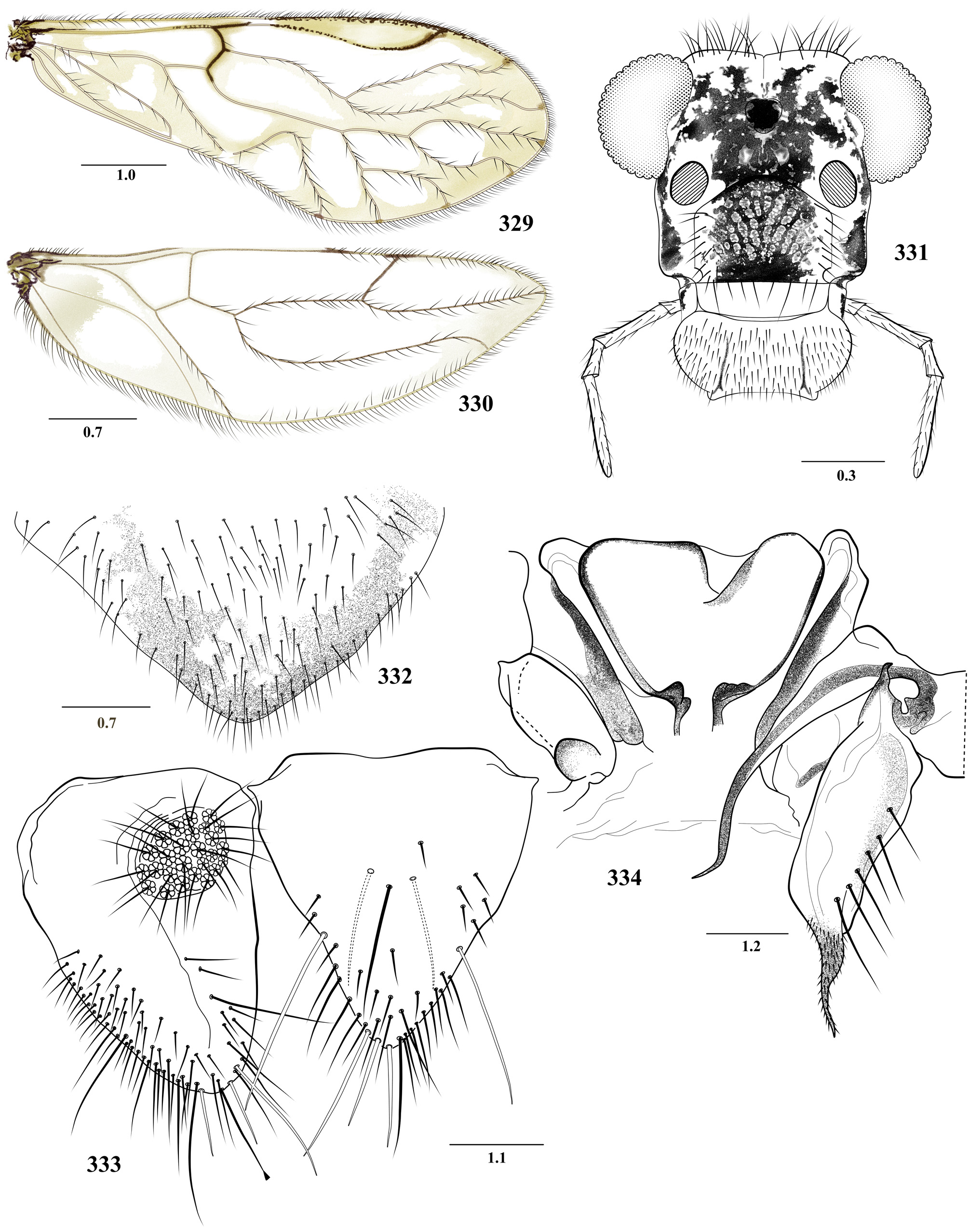
FIGURES 329 – 334. Triplocania lamensuraensis n. sp. Female. 329. Forewing. 330. Hindwing. 331. Front view of head. 332. Subgenital plate. 333. Left paraproct and epiproct. 334. Ninth sternum and left gonapophyses. Scales in mm.
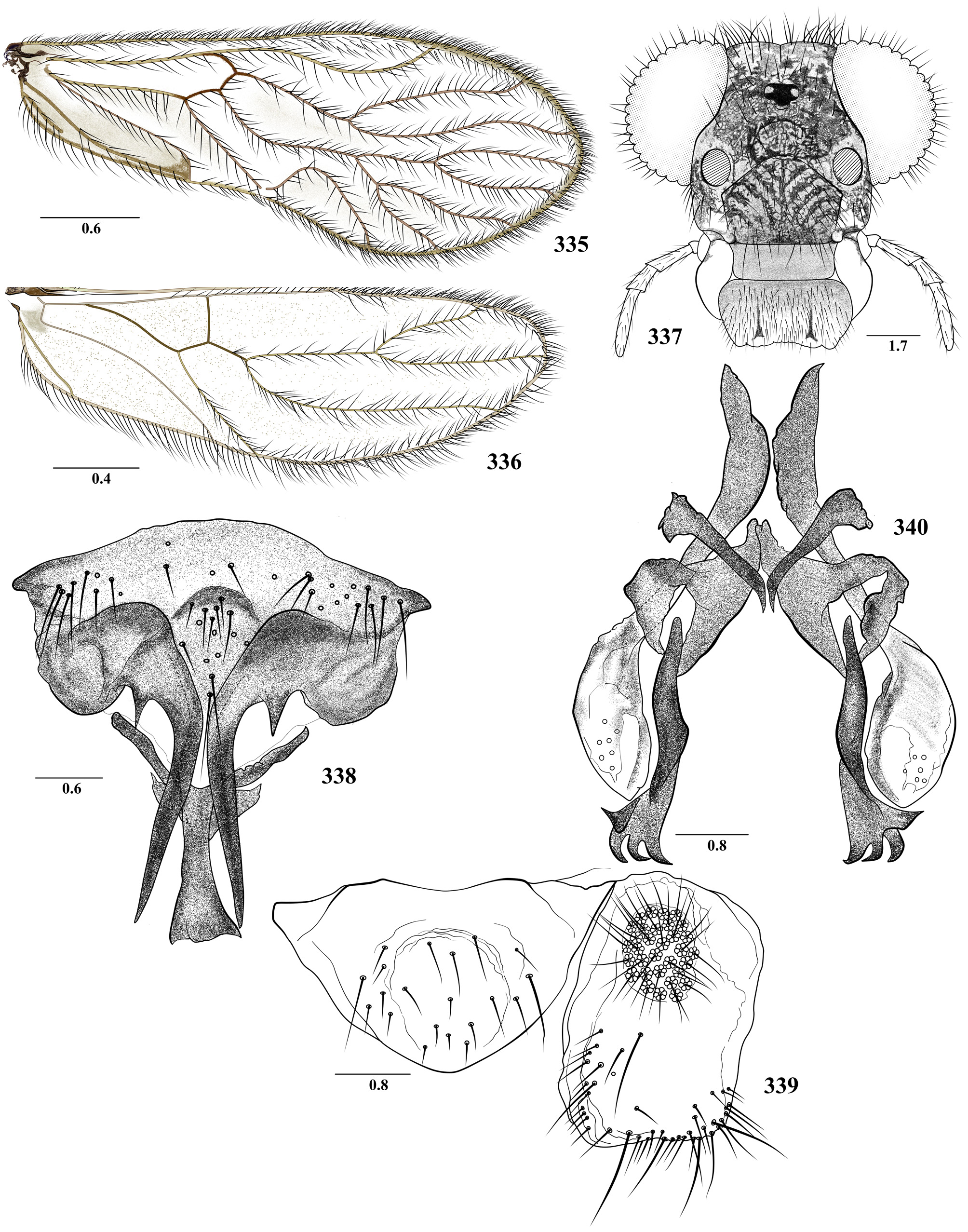
FIGURES 335 – 340. Triplocania leguizamoensis n. sp. Male. 335. Forewing. 336. Hindwing. 337. Front view of head. 338. Hypandrium. 339. Epiproct and right paraproct. 340. Phallosome. Scales in mm.

FIGURES 341 – 346. Triplocania otunquimbayaensis n. sp. Female. 341. Forewing. 342. Hindwing. 343. Front view of head. 344. Paraprocts and epiproct. 345. Subgenital plate. 346. Ninth sternum and left gonapophyses. Scales in mm.
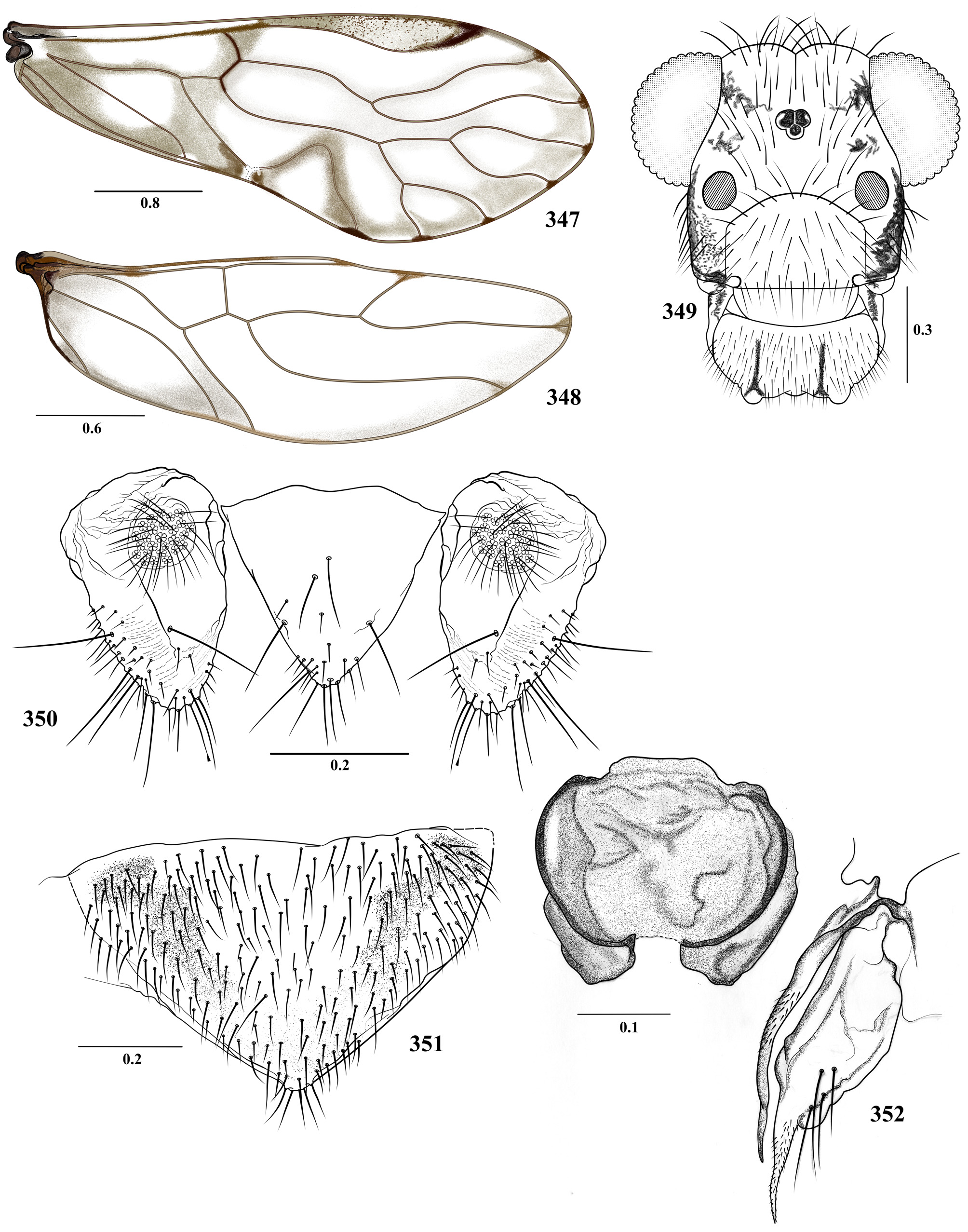
FIGURES 347 – 352. Triplocania sarriae n. sp. Female. 347. Forewing. 348. Hindwing. 349. Front view of head. 350. Paraprocts and epiproct. 351. Subgenital plate. 352. Ninth sternum and left gonapophyses. Scales in mm.

FIGURES 353 – 358. Triplocania furcata New. Female. 353. Forewing. 354. Hindwing. 355. Front view of head. 356. Subgenital plate. 357. Epiproct and right paraprocts. 358. Ninth sternum and left gonapophyses. Scales in mm.

FIGURES 359 – 364. Triplocania lamasi Silva Neto et al. Female. 359. Forewing. 360. Hindwing. 361. Front view of head. 362. Paraprocts and epiproct. 363. Subgenital plate. 364. Ninth sternum and left gonapophyses. Scales in mm.

FIGURES 3 – 8. Triplocania amacayacuensis n. sp. Male. 3. Forewing. 4. Hindwing. 5. Front view of head. 6. Epiproct and right paraproct. 7. Hypandrium. 8. Phallosome. Scales in mm.
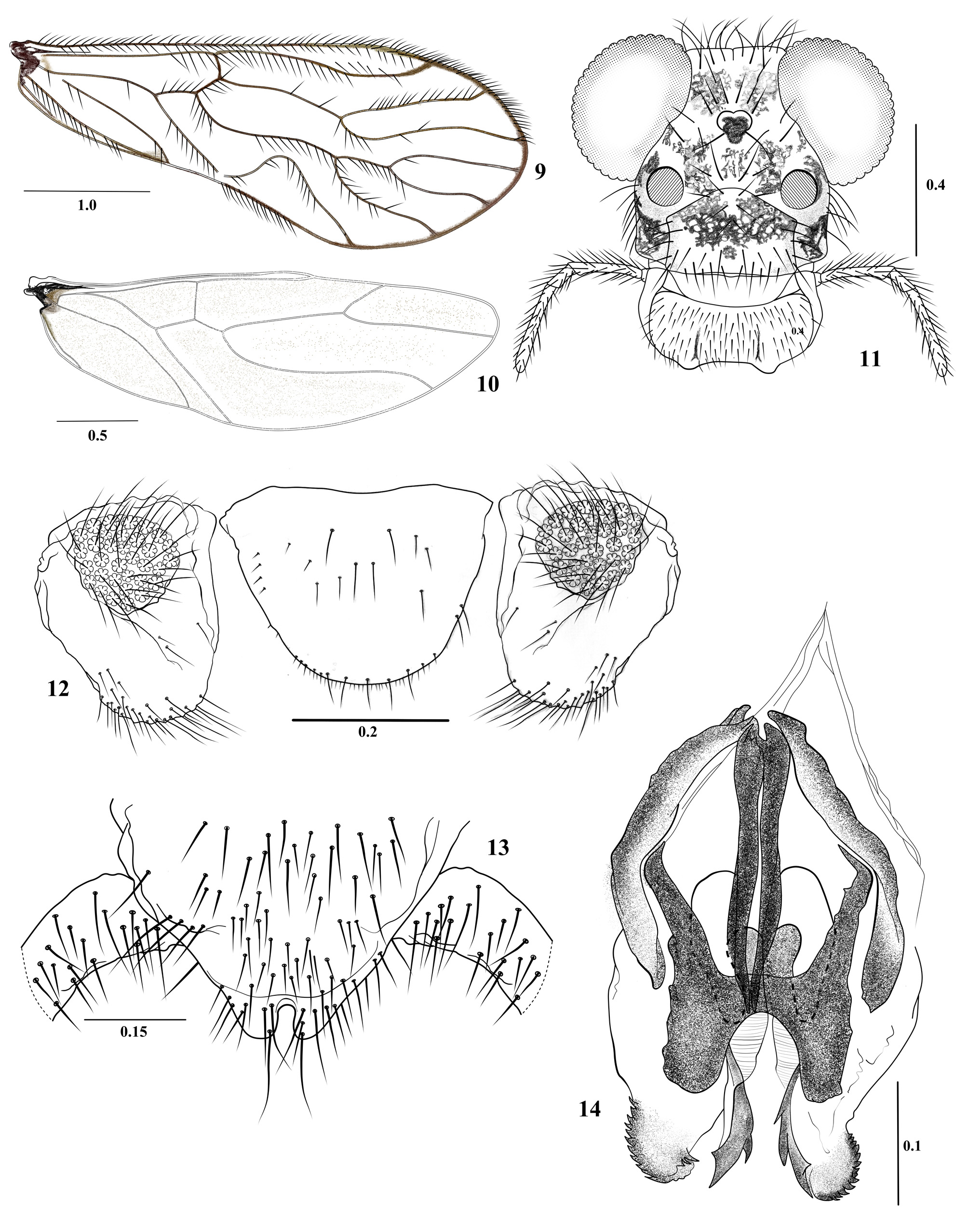
FIGURES 9 – 14. Triplocania anchicayaensis n. sp. Male. 9. Forewing. 10. Hindwing. 11. Front view of head. 12. Paraprocts and epiproct. 13. Hypandrium. 14. Phallosome. Scales in mm.
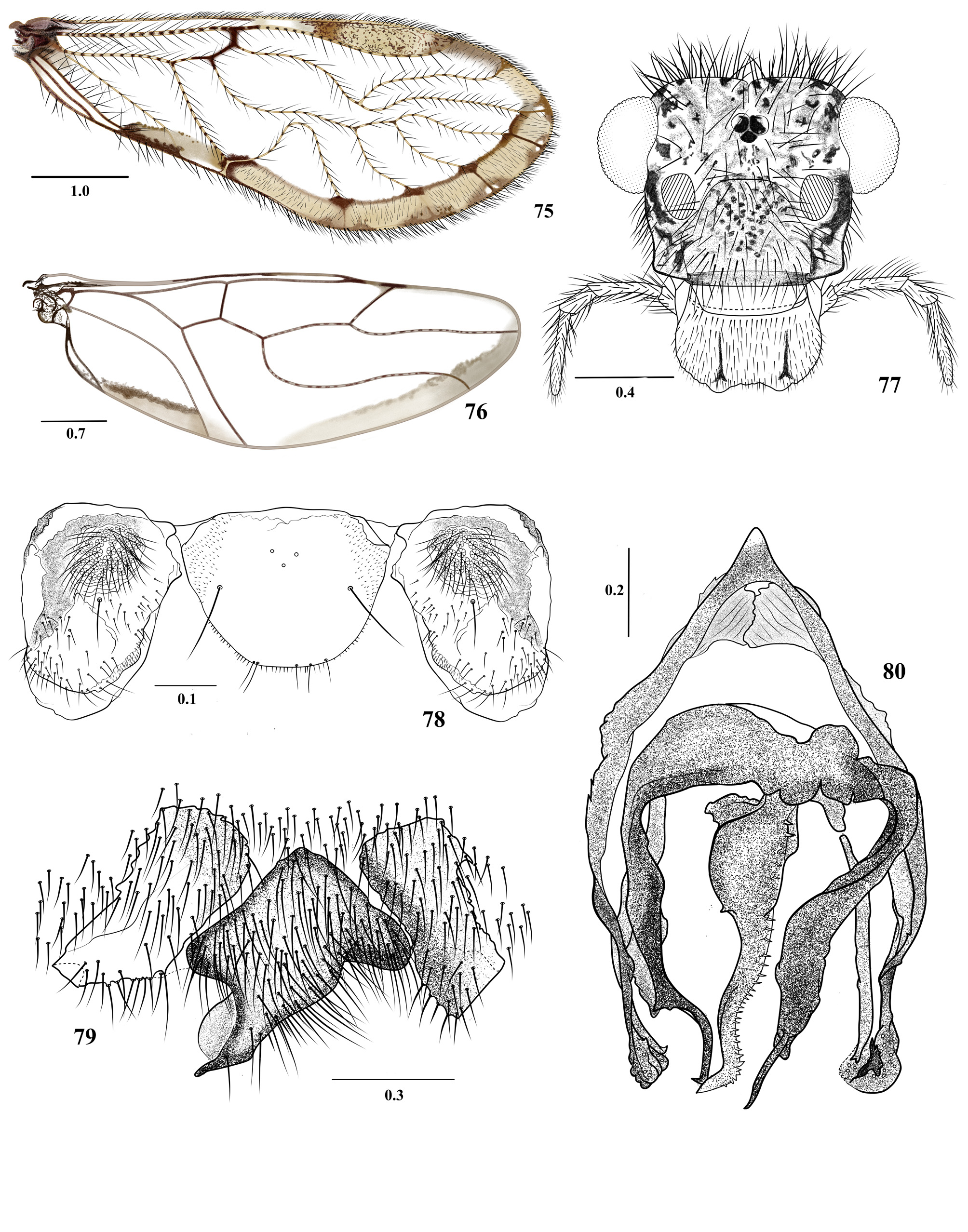
FIGURES 75 – 80. Triplocania calima n. sp. Male. 75. Forewing. 76. Hindwing. 77. Front view of head. 78. Paraprocts and epiproct. 79. Hypandrium. 80. Phallosome. Scales in mm.
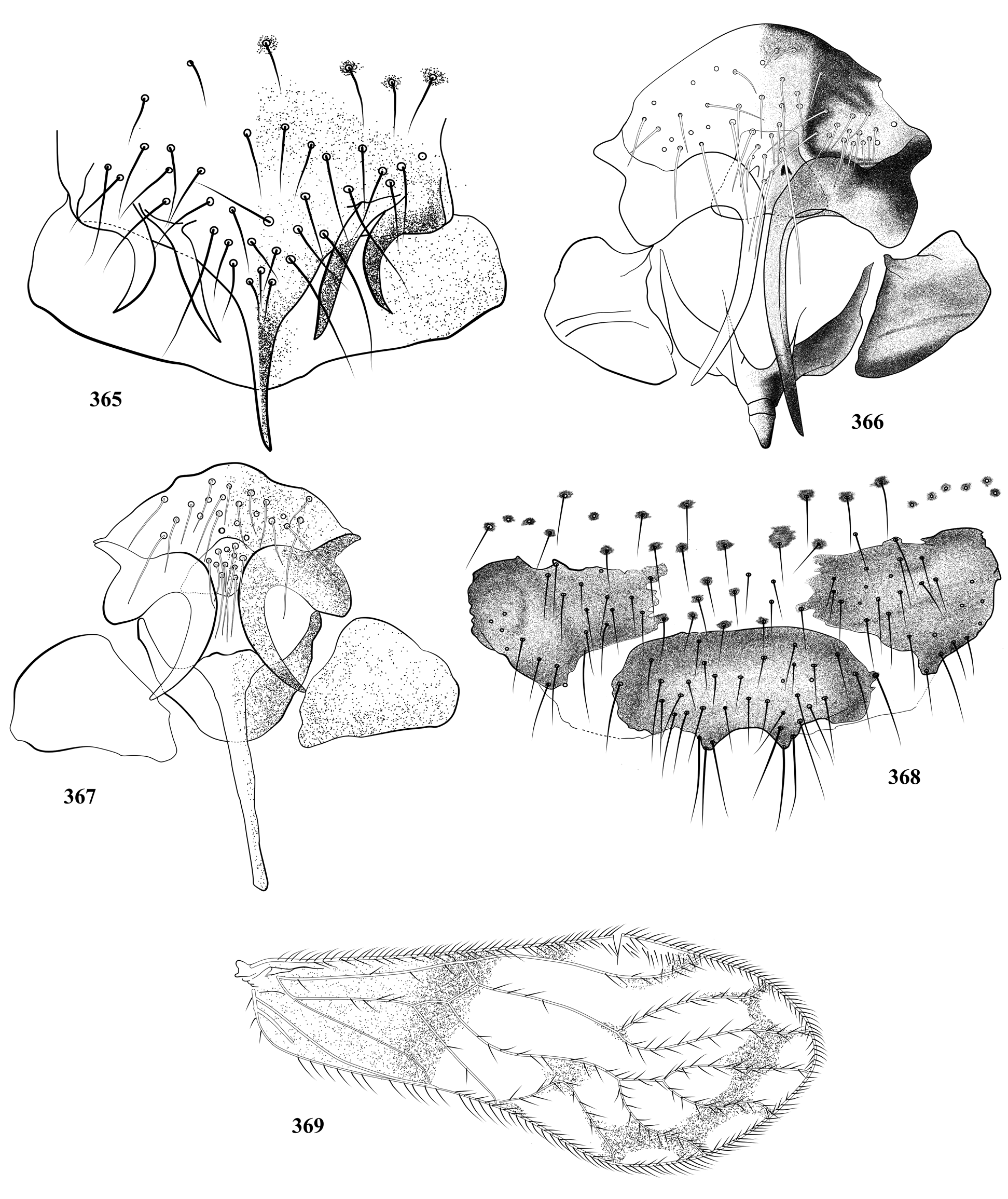
FIGURES 365 – 369. Hypandrium of Triplocania species. 365. T. furcata New. 366. T. lamasi Silva-Neto et al. 367. T. lamasoides Silva- Neto et al. 368. T. erwini Silva-Neto et al. 369. Forewing of T. colombiana García Aldrete.

No known copyright restrictions apply. See Agosti, D., Egloff, W., 2009. Taxonomic information exchange and copyright: the Plazi approach. BMC Research Notes 2009, 2:53 for further explanation.
|
Kingdom |
|
|
Phylum |
|
|
Class |
|
|
Order |
|
|
Family |
1 (by plazi, 2017-10-23 17:49:31)
2 (by ImsDioSync, 2017-10-23 18:02:23)
3 (by ImsDioSync, 2017-10-23 18:07:23)
4 (by ExternalLinkService, 2019-09-26 02:36:48)
5 (by ExternalLinkService, 2022-01-29 18:36:13)
6 (by ExternalLinkService, 2022-02-09 14:11:17)
7 (by plazi, 2023-10-28 01:49:31)