
Trichoderma dumbbelliforme T. Prameeladevi & D. Kamil, 2021
|
publication ID |
https://doi.org/ 10.11646/phytotaxa.520.3.8 |
|
persistent identifier |
https://treatment.plazi.org/id/03DC0C16-FFFC-FFB3-FF43-300E4619063C |
|
treatment provided by |
Plazi |
|
scientific name |
Trichoderma dumbbelliforme T. Prameeladevi & D. Kamil |
| status |
sp. nov. |
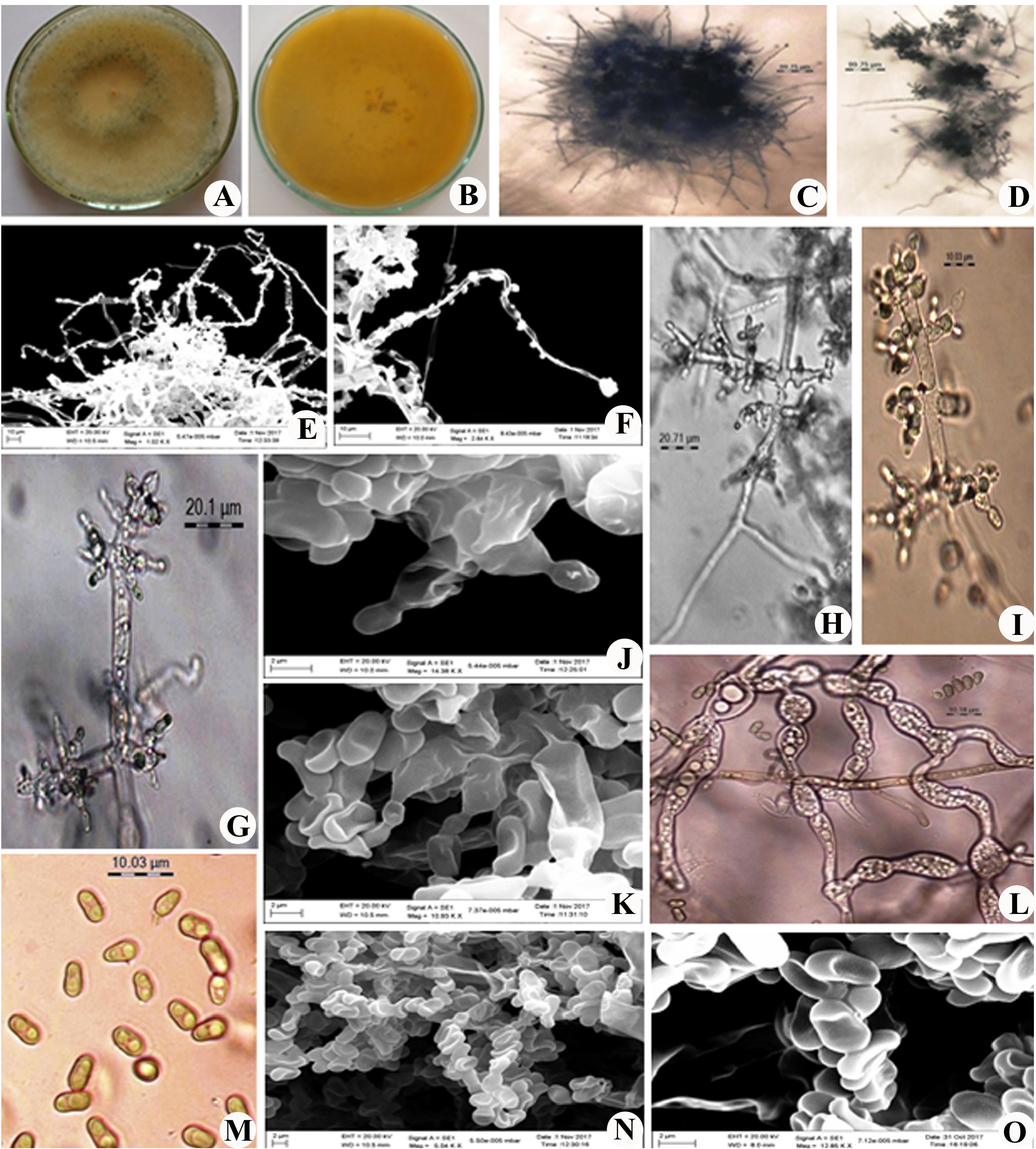
Trichoderma dumbbelliforme T. Prameeladevi & D. Kamil sp. nov. ( Fig. 2 View FIGURE 2 )
Mycobank:— MB823319
GenBank:— MG459152 View Materials
Holotype:— ITCC 8141 View Materials
Etymology:— Named after the shape of the conidia. The type strain was isolated from the soils of the virgin forest of Nagaland, India.
Host/Distribution:— soil sample collected from Dimapur, Nagaland, India.
Diagnostic characters:— Trichoderma dumbbelliforme can be distinguished from T. spirale and T. crassum by the ornamented conidiophores, shape of the conidia and presence of hyphal bulging ( Table 2). Trichoderma dumbbelliforme produced nodules on the sterile part of the conidiophores, pyriform to dumbbell shaped conidia and catenulate hyphal swellings whereas the other two species produced smooth conidiophores, globose conidia and sub-globose to obovoid chlamydospores.
Culture characteristics:— The growth of colony was observed 4.0–5.0 cm in 4 days on PDA medium at 25 0 C moderately growing. White floccose mycelium becoming glaucous to dark green ( Ridgway 1912). Cushion like flat pustules spread throughout the plate. Yellowish orange to brown soluble pigmentation was present in the medium. Reverse of the plate was brown. The conidiophores in minute pustules or effuse, branching irregularly, the apical part of the main conidiophores axis is straight or flexuous, sterile and un-branched. Long un-branched conidiophores were imbedded with smooth and round nodules with one or two phialides at the tip.The phialides were ampulliform, mainly arising in crowded whorls of 4–6 on terminal and lateral branches. Conidia was pyriform to dumbbell shaped with the size of 4.0–6.0×2.5–3.0 µm and having two big guttulae at each end, smooth and pale green Chlamydospores were smooth globose to oval with 6.0–10.0 µm diam, catenulate and branched hyphal swellings are present.
Material examined:— INDIA, Nagaland, Dimapur district, (25 o 45’24 N ” 93 o 50’26 E ”), from soil, R. Sudeep Toppo , 23 September 2016: ex-type culture deposited in Indian Type Culture Collection ( ITCC; 8141), ICAR-Indian Agricultural Research Institute, New GoogleMaps Delhi.
| R |
Departamento de Geologia, Universidad de Chile |
| ITCC |
Istituto Tecnico Stattale "Camillo Cavour" |
No known copyright restrictions apply. See Agosti, D., Egloff, W., 2009. Taxonomic information exchange and copyright: the Plazi approach. BMC Research Notes 2009, 2:53 for further explanation.
|
Kingdom |
|
|
Phylum |
|
|
Class |
|
|
Order |
|
|
Family |
|
|
Genus |